Abstract
Developmental exposure to the environmental neurotoxin β-N-methylamino-l-alanine (BMAA), a proposed risk factor for neurodegenerative disease, can induce long-term cognitive impairments and neurodegeneration in rats. While rodent studies have demonstrated a low transfer of BMAA to the adult brain, this toxin is capable to cross the placental barrier and accumulate in the fetal brain. Here, we investigated the differential susceptibility of primary neuronal cells and neural stem cells from fetal rat hippocampus to BMAA toxicity. Exposure to 250 µM BMAA induced cell death in neural stem cells through caspase-independent apoptosis, while the proliferation of primary neurons was reduced only at 3 mM BMAA. At the lowest concentrations tested (50 and 100 µM), BMAA disrupted neural stem cell differentiation and impaired neurite development in neural stem cell-derived neurons (e.g., reduced neurite length, the number of processes and branches per cell). BMAA induced no alterations of the neurite outgrowth in primary neurons. This demonstrates that neural stem cells are more susceptible to BMAA exposure than primary neurons. Importantly, the changes induced by BMAA in neural stem cells were mitotically inherited to daughter cells. The persistent nature of the BMAA-induced effects may be related to epigenetic alterations that interfere with the neural stem cell programming, as BMAA exposure reduced the global DNA methylation in the cells. These findings provide mechanistic understanding of how early-life exposure to BMAA may lead to adverse long-term consequences, and potentially predispose for neurodevelopmental disorders or neurodegenerative disease later in life.
Subject terms: Neuroscience, Stem cells, Neurological disorders
Introduction
The environmental toxin β-N-methylamino-l-alanine (BMAA) is proposed as a risk factor for neurodegenerative disease, in particular amyotrophic lateral sclerosis/parkinsonism-dementia complex (ALS/PDC)1–3. This non-proteinaceous amino acid is produced by a variety of cyanobacteria (blue-green algae) and two groups of microscopic algae, diatoms and dinoflagellates4,5. As cyanobacteria are extensively distributed in terrestrial and aquatic environments all over the world, and eutrophication of aquatic environments together with global warming are promoting a rapid increase of the algae bloom6, BMAA may be an emerging global hazard. Humans can, for example, be exposed to BMAA via drinking water, recreational water, spray-irrigated food, seafood or even through the air7–9. Recent studies have demonstrated that BMAA also can be transferred from mussel-based feed into chicken10 and accumulate in birds’ eggs11 indicating that human consumption of these products can be an additional source of BMAA exposure.
While experimental studies have demonstrated a poor transfer of the toxin into the adult brain12,13, and a low neurotoxic potential in adult rodents14, BMAA is able to cross the placental barrier, and the uptake in discrete brain regions is more efficient in rodent fetuses and neonates15. In addition, BMAA is secreted into the milk of lactating rodents and distributed to the brain of suckling pups16,17. The relatively high uptake of BMAA in the developing brain is correlated with biochemical and behavioral changes in neonatal and juvenile animals15,18,19. Neonatal exposure to BMAA can also cause cognitive impairments20,21, proteomic alterations, and progressive neurodegeneration, including neurofibrillary inclusions, in the hippocampus of adult rats22–24. Since the hippocampus is essential for learning and memory, more studies on the developmental effects of BMAA in this brain area are necessary.
During development, the central nervous system is generated from a small number of neural stem cells25, and cell division, migration, differentiation into neurons, astrocytes and oligodendrocytes, neurite outgrowth and synapse formation proceed in a well-ordered manner. Dysregulation of any of these vital processes due to either genetic causes or environmental exposures may lead to disabilities or disease later in life26. Brain development is regulated by epigenetic mechanisms such as DNA methylation, and early-life exposure to environmental contaminants may impair neural stem cells reprogramming through epigenetic alterations, which could result in long-term consequences in the adult brain27. Neural stem cell cultures are, therefore, an important tool for mechanistic studies in the field of developmental neurotoxicology28.
The aim of this study was to compare the susceptibility between hippocampal neural stem cells and primary neurons to BMAA toxicity. We examined the effects of BMAA exposure on cell proliferation, differentiation, neurite outgrowth, global DNA methylation, and investigated if the effects persist in the absence of the exposure, and are inherited from one cell generation to another.
Material and methods
Chemicals
β-N-methylamino-l-alanine hydrochloride (≥97% purity, CAS Number 16012-55-8), paraformaldehyde, 4´,6-diamidino-2-phenylindoledihydrochloride (DAPI), Triton X-100, propidium iodide (PI), DNAse-free RNAse A, 3-(4,5-dimethyl-2-yl)2,5-diphenyl-2H-tetrazolium bromide (MTT), and basic fibroblast growth factor (bFGF) were obtained from Sigma-Aldrich Co (St. Louis, MO, USA). Bovine serum, penicillin–streptomycin, Dulbecco’s phosphate-buffered saline (PBS), neurobasal medium, poliornithine, fibronectin, trypsin solution (0.05%), glutamine and B27 were obtained from Gibco (Invitrogen, Paisley, UK). The secondary antibodies Alexa fluor 555 anti-mouse IgG, 488 goat anti-rabbit IgG, 350 donkey anti-goat IgG, 647 goat anti-chicken IgG, the blocking agent (normal goat serum) and the annexin-PI kit were obtained from Molecular Probes (Invitrogen, Paisley, UK). The antibodies MAP, β III-tubulin anti-rabbit, glial fibrillary acidic protein (GFAP) anti-mouse, nestin anti-rabbit, anti-5-methylcytosine (5-mc) and the secondary antibody HRP-conjugated goat anti-rabbit and anti-mouse were obtained from Abcam (Cambridge, UK). Apoptosis-inducing factor (AIF), caspase-3, cleaved caspase 3 (5A1E), caspase 12, and cytochrome c (136F3) were obtained from Cell Signaling (Boston, MA, USA). The antibody oligo4 was obtained from Chemicon (Temecula, CA, USA). The 5-methylcytosine and cytosine DNA standards were obtained from Zymo Research (Irvine, CA, USA).
Animals and housing
Pregnant outbred Wistar rats, obtained from Charles River (Sulzfeld, Germany), were housed alone in Macrolon cages (59 × 38 × 20 cm) containing wood-chip bedding and nesting material. The dams were maintained on standard pellet food and water ad libitum. The animals were housed in a temperature and humidity-controlled environment on a 12-h light/dark cycle. All experiments were performed according to protocols approved by the Regional Animal Ethical Committee and following the Swedish Legislation on Animal experimentation (Animal welfare act SFS1998:56) and the European Union Directive on the Protection of Animals Used for Scientific Purposes (2010/63/EU).
Cell culture and BMAA exposure
Pregnant rats were euthanized by decapitation29,30 and primary neuronal cell cultures were prepared from fetal hippocampus at embryonic day 18 as previously described31. In brief, a single-cell suspension was obtained by dissociating embryonic hippocampal cells in DMEM/F12 medium and 100,000 cells/cm2 were plated on polylysine-treated 96-well plates. Neuronal cultures were kept in neurobasal medium supplemented with 2 mM glutamine and B27 for up to 24 h. The medium was then replaced and the cells were incubated for 7 days in a humid incubator at 37 °C with 5% CO2. At 8 days in vitro, the culture medium was removed and cells were treated for 24 h with 50 µM to 3 mM BMAA dissolved in neurobasal medium and used for cell viability, proliferation and morphometric assays.
Neural stem cell cultures were prepared from fetal rat hippocampus at embryonic day 15 as previously described31. In brief, 40,000 cells/cm2 were plated in 75 cm2 flasks precoated with poly-l-ornithine and fibronectin and maintained in N2 medium enriched with 10 ng/ml bFGF to keep the cells in an undifferentiated state. After 3 days in culture, cells were passaged at low density (500 cells/cm2) on plates coated with poly-l-ornithine and fibronectin, in the presence of bFGF. One day after passaging, cells were treated with 50 µM to 3 mM BMAA for 24 h. After the exposure, medium without bFGF was added to promote spontaneous differentiation of the exposed neural stem cells for 24 h (cell viability and proliferation assays) or 7 days (morphometric and differentiation assays).
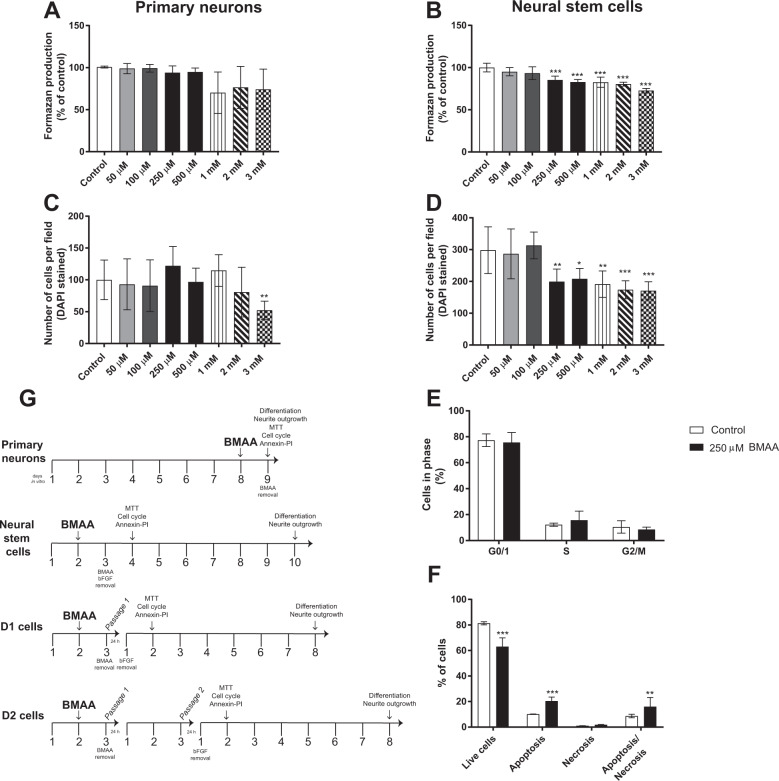
The neural stem cells were also used to study mitotically heritable effects in daughter cells. The cells were exposed to non-cytotoxic concentrations of BMAA (50 or 100 µM) for 24 h and then passaged into their daughter cells (one passage for D1 and two passages for D2) and plated at low density (500 cells/cm2) (Fig. 1G). One day after passage one (D1) or passage two (D2), bFGF was removed and the cells were allowed to differentiate for 24 h (cell viability and proliferation assays) or 7 days (morphometric and differentiation assays). All experiments were performed using six replicates and repeated three times starting from the preparation of cell cultures from new animals.
Fig. 1. Effects of BMAA on cell viability and proliferation in hippocampal primary neurons and neural stem cells.
Cell viability was determined using the MTT assay (A, B) and cell proliferation by counting the number of cells using DAPI staining (C, D) after treatment with 50 µM to 3 mM BMAA for 24 h. The cell cycle phases of neural stem cells treated with 250 µM were analyzed by flow cytometry (E), apoptotic and necrotic cells were assessed with the annexin V-PI assay (F). The experimental design used for investigating BMAA effects in hippocampal primary neurons and neural stem cells (G). Values represent mean ± SD from three independent experiments, each with six replicates. Statistically significant differences from control are indicated as follows: *p < 0.05, **p < 0.01 and ***p < 0.001 (one-way ANOVA followed by Tukey–Kramer test, or Student’s t-test when comparing only two groups).
Cell viability and proliferation analysis
3-(4,5-dimethylthiazol-2-yl)-2,5-diphenyltetrazolium bromide assay
MTT assay was performed as previously described31. Cell viability was measured after 24 h of BMAA exposure in primary neuronal and neural stem cells, and 24 h after the passage into the daughter cells. The formazan product generated was solubilized in dimethyl sulfoxide and measured at 490-630 using a SpectraMax i3 microplate reader (Molecular Devices, San Jose, CA, USA).
Annexin V-PI labeling
The apoptotic/necrotic analysis was conducted by labeling cells with the Ca2+-dependent phosphatidylserine-binding protein annexin V and PI. Neural stem cells were detached from the culture plates by 0.05% trypsin-EDTA treatment, washed once with PBS and labeled by incubation with annexin V-FITC and PI at room temperature for 15 min in the dark, according to the manufacturer’s instruction. Stained cells were analyzed (10,000 events) on a Cytoflex flow cytometer (Beckman Coulter Ltd., Brea, CA, USA).
Cell cycle analysis
Cells were processed for PI staining and flow cytometry as previously described31. Forward and light scatter data were collected in a linear mode, and fluorescence data from 10,000 cells per sample were collected in the FL3 channel on a linear scale. Side and forward light scatter parameters were used to identify the cell events and doublets cells were excluded using gating. Cells in different cell cycle phases were presented as a percentage of the total number of cells counted.
Cell death signaling analysis and neural stem cells differentiation
Western blot
Proteins involved in the regulation of apoptosis were evaluated by western blot. Cells were lysed with Laemmli lysis buffer and the protein concentration was determined by Lowry assay32. An equal amount of protein was separated by sodium dodecyl sulfate-polyacrylamide gel electrophoresis (SDS-PAGE) on a 4-20% gel and transferred to nitrocellulose membranes (Mini Trans-Blot Electrophoretic Transfer Cell; Bio-Rad, Hercules, CA, USA). The blot was then incubated in a blocking solution (TBS; 500 mM NaCl, 20 mM Trizma, pH 7.8 with defatted dry milk), followed by washes with TBS and incubated overnight in TBS containing monoclonal antibodies (apoptosis-inducing factor (AIF), caspase 3, cleaved caspase 3 (5A1E), caspase 12 and cytochrome c (136F3)) diluted 1:5000. The blots were then washed with TBS and incubated for 1 h in TBS containing peroxidase-conjugated mouse anti-rabbit or anti-mouse IgG diluted 1:10000. The blot was developed with the chemiluminescence ECL kit (Bio-rad, Hercules, CA, USA) using a charge-coupled device (CCD) imager (Thermofisher, Rockford, IL, USA), and optical density was measured using the ImageJ Software. The results were normalized by the β-tubulin content and the protein levels were expressed as a percentage of control.
Immunocytochemistry and morphometric analysis
Immunocytochemistry was performed as previously described31. In brief, cells were plated at a density of 40,000 cells/cm2 on microscope glass coverslip precoated with poly-l-ornithine and fibronectin and treated with 50 or 100 µM BMAA for 24 h for cell differentiation and AIF analysis in neural stem cells. After that, BMAA and bFGF were removed and cells were allowed to differentiate for seven days. The cells were then fixed with 4% paraformaldehyde for 30 min and permeabilized/blocked with 1% BSA/0.1% Triton X-100 in PBS for 30 min at room temperature. Cells were incubated with AIF antibody (1:500) or with β III-tubulin (1:200), anti-GFAP (1:500), anti-nestin (1:1000) and anti-oligo4 antibodies (1:1000), at room temperature, followed by washes with PBS and incubation with secondary antibodies conjugated with Alexa 488 or 555 (1:1000) for 1 h. The nucleus was stained with DAPI (0.25 mg/ml) and the cells were examined in an Olympus IX70 inverted microscope (Olympus, Tokyo, Japan). The images were collected by a CCD camera with a 20x objective using constant intensity settings and exposure time for all samples. Semiquantitative analyses of differentiated cells were conducted in five random microscopic fields and images were analyzed with the ImageJ software (Sound Vision) after the digital acquisition. Negative controls were performed by omitting the primary antibody.
For morphometric analysis, primary neurons and neural stem cells were plated at a density of 40,000/cm2 on 96-well plates, and treated with 50 µM to 3 mM BMAA for 24 h. After that, cells were stained with β III-tubulin and MAP2 antibodies as described above. Mitotically inherited effects on the neurite outgrowth were analyzed in daughter cells of neural stem cells exposed to 50 or 100 µM BMAA. Images were collected with a 10x objective in an ImageXpress Micro XLS Widefield High-Content Analysis System (Molecular Devices, Sunnyvale, CA, USA). Nine fields per well were automatically analyzed with the MetaXpress Software after digital acquisition using the Neurite outgrowth application module, based on β III-tubulin staining.
Flow cytometry
To further study effects on cell differentiation, neural stem cells exposed to 50 or 100 µM and their daughter cells were fixed in 4% paraformaldehyde, permeabilized and blocked with 0.1% Triton X-100 and 1% BSA in PBS for 15 min. Then the cells were incubated with primary antibodies (anti-β III-tubulin, anti-GFAP, anti-nestin and anti-oligo4) at 4 °C overnight. After that, cells were stained with secondary antibodies (anti-rabbit Alexa 488, anti-goat Alexa 350, anti-chicken Alexa 647 and anti-mouse Alexa 555) for 60 min at room temperature. Samples were analyzed using a Cytoflex flow cytometer (Beckman Coulter Ltd., Brea, CA, USA). The quadrants to determine the negative and positive areas were placed on unstained and single stained samples. Forward and side light scatter gates were used to exclude cell aggregates and small debris. The number of cells in each quadrant was computed and the proportion of cells stained with the antibodies was calculated.
Global DNA methylation
DNA was extracted from neural stem cells treated with 100 µM BMAA using the AllPrep DNA/RNA micro kit (Qiagen, Germany). The concentration of DNA was measured using Nanodrop 2000 spectrophotometer (Thermo Scientific, USA). The concentration of 5-methylcytosine was quantified by ELISA as previously described33. In brief, unmethylated DNA was used as negative control and the standard curve was prepared using methylated DNA. For each sample, 100 ng of DNA and PBS to reach the final volume of 100 µl were added to the PCR tubes. The DNA samples were denatured by heating at 98 °C for 5 min and then transferred immediately to ice for 10 min. The DNA samples were added to a 96-well microtiter plate, covered with aluminum foil and incubated at 37 °C for 1 h. After the incubation, the liquid content of the wells was discarded and the wells washed three times with 200 µl washing buffer (PBS with 0.2% Tween-20). The plates were blocked with a blocking buffer (Thermo Pierce, Rockford, IL, USA) at 37 °C for 30 min, and incubated overnight with anti-5-methylcytosine in PBS at 4 °C. The wells were then emptied and washed three times with washing buffer, and incubated with the HRP-conjugated secondary antibody for 30 min. After that, the wells were washed three times with washing buffer and 50 µl of 3,3′,5,5′-tetramethylbenzidine (TMB) substrate solution (Thermo Scientific Pierce, Waltham, MA, USA) was added into each well. The color reaction was terminated by the addition of 50 µl of stop solution (Thermo Scientific Pierce, Waltham, MA, USA). Optical density was measured at 450 nm using a SpectraMax i3 microplate reader (San Jose, CA, USA).
Statistical analysis
The results are presented as mean ± standard deviation (SD) for each experimental group consisting of at least three individual cell cultures, each with five to six replicates. Differences compared with the control group were analyzed by one-analysis of variance (ANOVA) followed by Tukey–Kramer multiple tests, or by Student’s t-test when comparing only two groups (cell cycle and flow cytometry analysis) using Prism 7 (Graphpad Software, San Diego, CA, USA).
Results
Effects of BMAA exposure on cell viability and proliferation in primary neurons and neural stem cells
Initially, we examined the effects of 50 µM to 3 mM BMAA on cellular viability and proliferation in hippocampal primary neurons and neural stem cells. The results showed that BMAA exposure did not affect cell viability in primary neurons when measured with the MTT assay (Fig. 1A), and the cell number was decreased only at 3 mM BMAA (Fig. 1C). However, in the neural stem cells, BMAA induced a decrease in cell viability (Fig. 1B) and cell number (Fig. 1D) from 250 µM to 3 mM BMAA.
The lowest BMAA concentration that reduced the cell viability and cell number in the neural stem cells (250 µM) was then used to analyze the effects on cell cycle and cell death mechanisms by flow cytometry. While the results revealed no effects of BMAA exposure on the cell cycle (Fig. 1E), the Annexin V-PI assay demonstrated a decrease in the number of viable cells and an increase of cells in apoptosis and apoptosis/necrosis in the neural stem cells compared with the control group (Fig. 1F).
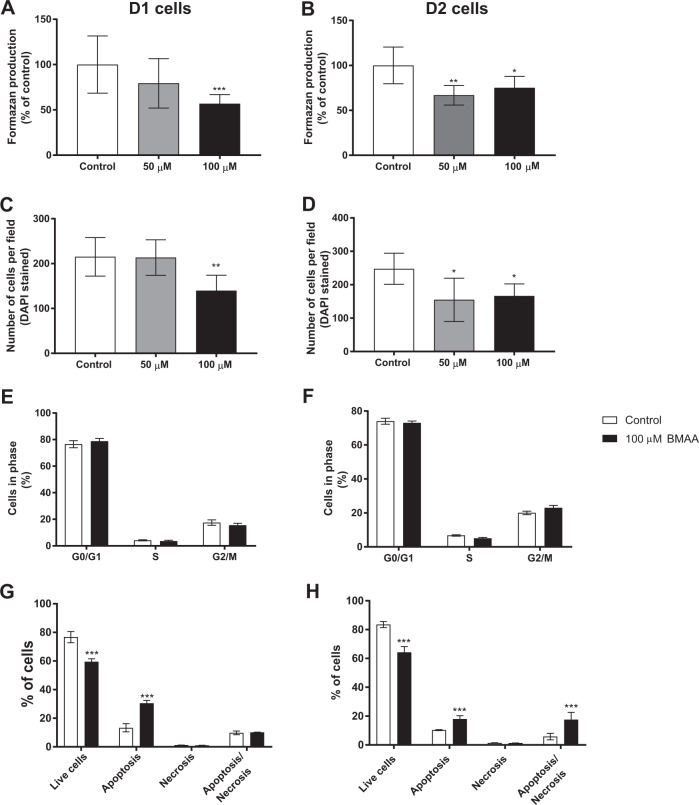
Mitotically heritable effects of BMAA exposure on cell viability and proliferation were investigated in daughter cells of neural stem cells exposed to non-cytotoxic doses of BMAA (50 and 100 µM) (Fig. 1G). In contrast to the exposed cells, the results demonstrated a decreased cell viability in D1 (100 µM) and D2 (50 and 100 µM) cells compared with their respective control groups (Fig. 2A, B). This was confirmed by the cell counting (Fig. 2C, D) illustrating that the effects on cell viability and proliferation are shown in the daughter cells at even lower concentrations than in the neural stem cells actually exposed to BMAA. Cell cycle effects and cell death mechanisms were evaluated in the daughter cells of neural stem cells exposed to 100 µM BMAA. The results demonstrated no alteration in the cell cycle phases (Fig. 2E, F). To confirm that the decreased cell proliferation in the daughter cells is due to an increase in cell death, similar to the exposed neural stem cells, we performed annexin V-PI assay also in the daughter cells. The results showed an increase in apoptosis in D1 cells (Fig. 2G), and apoptosis and apoptosis/necrosis in D2 cells (Fig. 2H).
Fig. 2. Effects of BMAA on neural stem cell viability and proliferation in daughter cells after one (D1) or two passages (D2).
Cell viability was determined using the MTT assay (A, B) and proliferation by counting the number of cells using DAPI staining (C, D) in daughter cells of neural stem cells treated with 50 or 100 µM BMAA. The cell cycle phase was analyzed by flow cytometry (E, F), apoptotic and necrotic cells were assessed with the annexin V-PI assay (G, H) in daughter cells of neural stem cells exposed to 100 µM BMAA. Values represent mean ± SD from three independent experiments, each with six replicates. Statistically significant differences from control are indicated as follows: *p < 0.05, **p < 0.01 and ***p < 0.001 (one-way ANOVA followed by Tukey–Kramer test, or Student’s t-test when comparing only two groups).
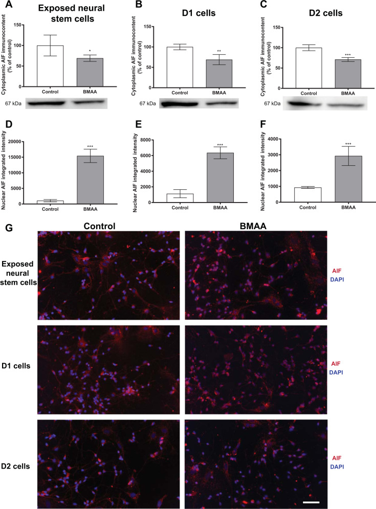
To better clarify the signaling pathways of cell death triggered by BMAA in neural stem cells, we studied proteins involved in mechanisms of apoptosis (cleaved caspase-3, caspase 12, cytochrome c and AIF) by western blot. The analysis was conducted in neural stem cells treated with 250 µM BMAA and in daughter cells of neural stem cells treated with 100 µM BMAA. The toxin induced no alterations in cytochrome c, cleaved caspase 3/caspase 3 and caspase 12 levels (Supplementary Fig.). However, the AIF levels in the cytoplasmic homogenate was decreased in the exposed neural stem cells (Fig. 3A) and in D1 and D2 cells (Fig. 3B, C). As AIF translocate from mitochondria to the nucleus when apoptosis is induced, we evaluated the effects of BMAA on AIF translocation through immunocytochemistry. The image analysis revealed an increased AIF translocation to the nuclei in the neural stem cells exposed to 250 µM BMAA (Fig. 3D) and in both D1 and D2 cells (Fig. 3E, F, respectively) derived from neural stem cells exposed to 100 µM BMAA. Representative images are shown in Fig. 3G.
Fig. 3. Involvement of the apoptosis-inducing factor (AIF) in cell death triggered by BMAA in neural stem cells.
AIF levels were evaluated by western blot in cytoplasmic homogenates from neural stem cells exposed to 250 µM BMAA (A), or D1 (B) and D2 (C) daughter cells of neural stem cells exposed to 100 µM BMAA. β-tubulin was used as a loading control. Representative blots of three experiments are shown. Nuclear AIF levels were analyzed by immunocytochemistry using ImageJ (D–F). Representative images are shown (G). Values represent mean ± SD from three independent experiments. Statistically significant differences from control are indicated as follows: *p < 0.05, **p < 0.01 and ***p < 0.001 (Student’s t-test). Scale bar: 30 µm.
BMAA exposure reduces neural stem cell differentiation
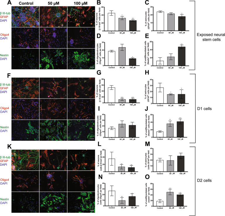
We examined the ability of BMAA to alter neural stem cell differentiation into neurons, astrocytes and oligodendrocytes in neural stem cells exposed to 50 or 100 µM BMAA and their daughter cells. Representative images of exposed cells are shown in Fig. 4A. Exposure to BMAA caused a significant decrease in the percentage of neural stem cell-derived neurons (Fig. 4B) at both concentrations, while only 100 µM BMAA caused a decrease in the percentage of astrocytes (Fig. 4C) and oligodendrocytes (Fig. 4D), as well as a significant increase in the percentage of undifferentiated cells (Fig. 4E). The effects on cell differentiation were also analyzed by flow cytometry, in neural stem cells treated with 100 µM BMAA and demonstrated a reduction in the percentage of neurons and oligodendrocytes, and an increase in the percentage of undifferentiated cells (Table 1).
Fig. 4. BMAA exposure to 50 or 100 µM alters the differentiation of neural stem cells and their daughter cells (D1 and D2).
The cells were stained with the neuronal marker, β III-tubulin; astroglia marker, GFAP; the oligodendrocyte marker, Oligo4; and undifferentiated cells were marked with nestin. Nuclei were stained with DAPI. Representative images are shown (A, F, and K). Semiquantitative analyses of differentiated neurons (B, G, L), astrocytes (C, H, M), oligodendrocytes (D, I, N), and undifferentiated cells (E, J, O) were performed according to the material and methods section. Values represent mean ± SD from three independent experiments each with six replicates. Statistically significant differences from control are indicated as follows: *p < 0.05, **p < 0.01, and ***p < 0.001 (one-way ANOVA followed by the Tukey–Kramer test). Scale bar = 30 µm.
Table 1.
Effects on differentiation in neural stem cells exposed to 100 µM BMAA and their daughter cells (D1 and D2).
| Cells | Control | BMAA |
|---|---|---|
| Exposed neural stem cells | ||
| Neurons | 43.38 ± 4.15 | 22.9 ± 1.92*** |
| Astrocytes | 24.38 ± 3.25 | 27.49 ± 13 |
| Oligodendrocytes | 10.24 ± 3.45 | 4.17 ± 1.91* |
| Undifferentiated cells | 21.26 ± 4.94 | 46.27 ± 7.73* |
| D1 daughter cells | ||
| Neurons | 36.98 ± 8.88 | 17.74 ± 7* |
| Astrocytes | 25.34 ± 3.97 | 11.31 ± 2.23* |
| Oligodendrocytes | 13.84 ± 6.85 | 11.74 ± 2.5 |
| Undifferentiated cells | 22.83 ± 12 | 59.2 ± 8** |
| D2 daughter cells | ||
| Neurons | 40.73 ± 5.52 | 22.68 ± 3.48** |
| Astrocytes | 22.67 ± 5.75 | 29.23 ± 16 |
| Oligodendrocytes | 25.28 ± | 10 ± 2.06** |
| Undifferentiated cells | 11.4 ± 5.9 | 40.02 ± 12* |
Results are expressed as percentage for total events (10,000 events).
Statistically significant differences from control are indicated as follow: ***p < 0.001; **p < 0.01, and *p < 0.05 (Student’s t-test).
The mitotically inherited effects of BMAA on cell differentiation were investigated in the daughter cells after a multitude of cell divisions. Representative images of D1 and D2 cells are shown in Fig. 4F, K, respectively. The results revealed that the reduction of the neuron differentiation was persistent in D1 and D2 cells (Fig. 4G, L, respectively) for both concentrations, while the reduction in astrocyte differentiation only persisted in D1 cells (Fig. 4H, M). A reduction in oligodendrocytes was only observed in D2 cells (Fig. 4N). The percentage of undifferentiated cells were increased in both D1 and D2 cells (Fig. 4J, O, respectively). These immunocytochemistry results were confirmed by flow cytometry analysis (Table 1).
Effects of BMAA on neuronal morphology
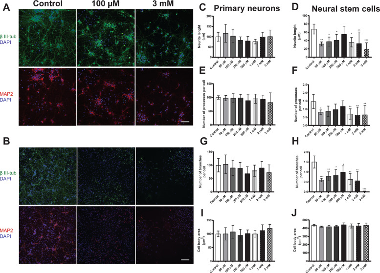
Immunocytochemical staining with anti-β III-tubulin and anti-MAP2 antibodies were performed to analyze morphological parameters of primary neurons and neurons derived from neural stem cells treated with 50 µM to 3 mM BMAA. Representative images are shown in Fig. 5A, B for 100 µM and 3 mM BMAA, respectively.
Fig. 5. BMAA causes morphometric alterations in neurons derived from hippocampal neural stem cells but not in primary neurons.
The effects on BMAA treatment with 50 µM to 3 mM BMAA was investigated directly after 24 h exposure of primary neurons, while the neural stem cells were allowed to differentiate for 7 days after the exposure. Representative images of cells immunostained with anti-β III-tubulin (green), anti-MAP2 (red), and DAPI (blue) are shown (Primary neurons, A; Exposed neural stem cells, B). Morphometric analysis was conducted using an ImageXpress Micro XLS Widefield HCA System (Molecular Devices, Sunnyvale CA, USA), where images were automatically captured and analyzed with the MetaXpress Software. Neurite length (C, D), the number of processes per cell (E, F), the number of branches per cell (G, H), and the cell body area (I, J) were determined. Values represent mean ± SD from three independent experiments, each with five to six replicates. Statistically significant differences from control are indicated as follows: *p < 0.05, **p < 0.01, and ***p < 0.001 (one-way ANOVA followed by Tukey–Kramer test). Scale bar = 50 µm.
BMAA exposure caused no morphological alterations in primary neurons at any of the concentrations (Fig. 5C, E, G, and I). In contrast, neurons derived from neural stem cells treated with 50 µM to 3 mM BMAA demonstrated a reduction in the neurite length (Fig. 5D), the number of processes per cell (Fig. 5F) and the number of branches per cell (Fig. 5H). The cell body area was not affected (Fig. 5J).
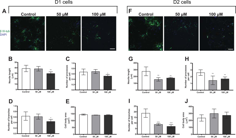
Morphological alterations were also investigated in neurons derived from daughter cells of neural stem cells treated with 50 or 100 µM BMAA, and most of the effects on neuronal morphology persisted in D1 and D2 cells. Representative images of D1 and D2 cells are shown in Fig. 6A and F, respectively. The results revealed a decrease in the neurite length (Fig. 6B), the number of processes (Fig. 6C) and the number of branches per cell (Fig. 6D) in D1 daughter cells of neural stem cells exposed to 100 µM of BMAA, while both 50 and 100 µM BMAA decreased the neurite length (Fig. 6G), the number of processes (Fig. 6H) and the number of branches per cell (Fig. 6I) in D2 cells. No alterations were observed in the cell body area of D1 or D2 cells (Fig. 6E, J, respectively).
Fig. 6. Morphometric alterations in neurons derived from daughter cells (D1 and D2) of neural stem cells treated with 50 or 100 µM BMAA.
Representative images of cells immunostained with anti-β III-tubulin (green) and DAPI (blue) (D1 cells, A; D2 cells, F). Morphometric analysis was conducted using an ImageXpress Micro XLS Widefield HCA System (Molecular Devices, Sunnyvale, CA, USA), where images were automatically captured and analyzed with the MetaXpress Software. Neurite length (B, G), the number of processes per cell (C, H), the number of branches per cell (D, I), and the cell body area (E, J) were determined. Values represent mean ± SD from three independent experiments, each with six replicates. Statistically significant differences from control are indicated as follows: *p < 0.05, **p < 0.01 and ***p < 0.001 (one-way ANOVA followed by Tukey–Kramer test). Scale bar = 50 µm.
BMAA alters global DNA methylation in hippocampal neural stem cells
As epigenetic modifications such as DNA methylation play critical roles in the regulation of neural stem cell proliferation and differentiation27, we analyzed the global DNA methylation in cells treated with 100 µM BMAA. The results revealed that BMAA decreased global DNA methylation in neural stem cells compared with the control group (Fig. 7).
Fig. 7. BMAA decreases global DNA methylation in neural stem cells exposed to 100 µM BMAA.

DNA methylation was determined by measuring total 5-methylcytosine (5-mc) with ELISA. Values represent mean ± SD from three independent experiments with six replicates each. Statistically significant differences from control are indicated as follows: *p < 0.05 (Student’s t-test).
Discussion
The development of the nervous system requires an intricate balance between proliferation of the progenitors, differentiation of the correct cell types, and the subsequent migration and connection of these cells. The importance of the regulation of neural development is demonstrated by the many neurodevelopmental disorders that can arise from defects in morphogenesis34. In this study, hippocampal neural stem cells were found to be clearly more sensitive to BMAA than hippocampal primary neurons. Exposure to 250 µM BMAA inhibited neural stem cell proliferation through induction of apoptosis, while the non-cytotoxic dose 100 µM BMAA inhibited the differentiation of neural stem cells into neurons, astrocytes and oligodendrocytes. BMAA induced a decrease in cell numbers also in primary neurons, but only at the relatively high concentration 3 mM. This adds additional evidence to previous research showing that BMAA is more neurotoxic for the developing brain14,15,22,35 and illustrate the importance to mechanistically elucidate BMAA’s effects during brain development. Importantly, the effects on neural stem cells were mitotically inherited, demonstrating that the BMAA-induced alterations can persist, and even be evident at lower concentrations, in daughter cells after a multitude of cell divisions. In line with this, we observed an altered global DNA methylation pattern in neural stem cells treated with this toxin.
The neural stem cell number is rigorously regulated during central nervous system development. More than half of the immature neurons are deleted in certain brain regions during normal development without interfering with the further development of the remaining cells. Many components of the machinery for programmed cell death are highly expressed in the developing brain, making it more susceptible to unintended activation. Impaired regulation of cell death may lead to brain malformation, impaired learning and memory, or tumor development36,37. BMAA exposure induced AIF translocation from mitochondria to the nucleus in hippocampal neural stem cells, which culminated in caspase-independent apoptosis. The mitochondrion-localized flavoprotein AIF translocates to the nucleus where it can bind to DNA and induce DNA fragmentation when apoptosis is induced38. Studies have suggested that this protein is important for regulating cell death in multiple neuronal injury pathways39, and in particular NMDA-induced neurotoxicity40 where the nuclear protein PARP1 induces AIF translocation to the nucleus41. As BMAA can induce excitotoxicity via glutamate receptors, it may also induce AIF translocation through PARP1 activation, triggering cell death by apoptosis. Intriguingly, BMAA exposure, at even lower concentration, increased caspase-independent apoptosis also as a mitotically inherited effect in daughter cells.
It is important to point out that the mechanisms by which BMAA affects cell proliferation or triggers cell death can be different depending on the brain area. Recently, we demonstrated that exposure to BMAA reduced striatal neural stem cell proliferation through apoptosis and G2/M arrest31. This may be explained by the differences in cell types between striatum and hippocampus. Moreover, the low susceptibility of primary hippocampal neurons to BMAA exposure may be related to the expression of glutamate receptors. Although studies have shown that primary rat neurons grown in culture for 8 days express glutamate receptors, they might not be fully sensitive to glutamate toxicity42,43. However, other studies have shown that primary rat neurons are sensitive to glutamate agonists such as quinolinic acid, kainic acid and glutaric acid at this stage of culture44–46. It is also possible that different neuronal populations may be more or less sensitive to BMAA exposure, which may explain the variability in the viability of primary neurons in our study.
Neural stem cells give rise to diverse populations of neurons, followed by astrocytes and oligodendrocytes, during embryogenesis47. Production of the correct number and subtype of cells during a critical development window is vital for the formation of functional neural circuitry, and defects in this process contribute to neurodevelopmental and neurological disorders48. Interestingly, non-cytotoxic BMAA concentrations altered the spontaneous neuronal, astrocytic and oligodendrocytic differentiation in both exposed neural stem cells and daughter cells, demonstrating that the impairments in neural stem cells differentiation can be mitotically inherited to daughter cells after a multitude of cell divisions. The observed effects may be related to previous studies showing that neonatal exposure to BMAA caused cognitive impairments, neuronal degeneration, cell loss, calcium deposits, and astrogliosis in the hippocampus of adult rats21,24,49.
The regulation of neurite outgrowth is another crucial process during brain development. Neurons grow out cellular processes, termed neurites, to establish synaptic connections with other neurons, forming highly specialized networks50. Abnormal neurite outgrowth and neuronal migration can cause impaired brain functions, where cognitive and motor deficits are the most frequent consequences51. Neurons derived from neural stem cells treated with BMAA demonstrated a significantly compromised neurite network even at the lowest concentration tested (50 µM). The effects on the neurite outgrowth also persisted in the daughter cells. Notably, primary neurons treated with 50 µM to 3 mM BMAA did not present any similar alteration.
Epigenetic mechanisms play a critical regulatory role in neural stem cell proliferation and differentiation, and DNA methylation is a key modulator52. DNA methylation can influence neural cell identities and altered global DNA methylation often result in cell death during differentiation53. Several neurodevelopmental disorders have been linked to proteins involved in the regulation of DNA modification and DNA methylation is closely correlated to stem cell-related diseases27. In line with previous studies demonstrating that various toxic agents can induce global DNA hypomethylation54–56, BMAA was found to decrease global DNA methylation in neural stem cells. The BMAA-induced effects on neural stem cell proliferation and differentiation, which were mitotically inherited to unexposed cells, could therefore be related to an altered DNA methylation pattern. However, more studies are necessary to identify the specific genes that are hypomethylated and potentially activated by BMAA.
In conclusion, this study provides additional evidence that neural stem cells are more susceptible to BMAA exposure than primary neurons. While BMAA only reduced cell proliferation in primary hippocampal neurons at the highest concentration 3 mM, neural stem cells were clearly affected even at the lowest concentration tested (50 μM). The heritable programming effects induced by BMAA in hippocampal neural stem cells on caspase-independent apoptosis and neural differentiation support the idea that developmental exposures can result in long-lasting brain changes, which may lead to neurodevelopmental disorders or predispose to brain disease later in life.
Supplementary information
Acknowledgements
Open Access funding provided by Stockholm University.
Conflict of interest
The authors declare that they have no conflict of interest.
Footnotes
Edited by B. Joseph
Publisher’s note Springer Nature remains neutral with regard to jurisdictional claims in published maps and institutional affiliations.
Supplementary information
Supplementary Information accompanies this paper at (10.1038/s41419-020-03093-6).
References
- 1.Cox PA, Kostrzewa RM, Guillemin GJ. BMAA and Neurodegenerative Illness. Neurotox. Res. 2018;33:178–183. doi: 10.1007/s12640-017-9753-6. [DOI] [PubMed] [Google Scholar]
- 2.Cox PA, et al. Diverse taxa of cyanobacteria produce beta-N-methylamino-L-alanine, a neurotoxic amino acid. Proc. Natl Acad. Sci. USA. 2005;102:5074–5078. doi: 10.1073/pnas.0501526102. [DOI] [PMC free article] [PubMed] [Google Scholar]
- 3.Cox, P. A., Davis, D. A., Mash, D. C., Metcalf, J. S. & Banack, S. A. Dietary exposure to an environmental toxin triggers neurofibrillary tangles and amyloid deposits in the brain. Proceedings. 283 (Biological sciences/The Royal Society, 2016). [DOI] [PMC free article] [PubMed]
- 4.Jiang L, et al. Diatoms: a novel source for the neurotoxin BMAA in aquatic environments. PLoS ONE. 2014;9:e84578. doi: 10.1371/journal.pone.0084578. [DOI] [PMC free article] [PubMed] [Google Scholar]
- 5.Lage S, et al. BMAA in shellfish from two Portuguese transitional water bodies suggests the marine dinoflagellate Gymnodinium catenatum as a potential BMAA source. Aquat. Toxicol. 2014;152:131–138. doi: 10.1016/j.aquatox.2014.03.029. [DOI] [PubMed] [Google Scholar]
- 6.Paerl H. Nutrient and other environmental controls of harmful cyanobacterial blooms along the freshwater-marine continuum. Adv. Exp. Med. Biol. 2008;619:217–237. doi: 10.1007/978-0-387-75865-7_10. [DOI] [PubMed] [Google Scholar]
- 7.Codd GA, Morrison LF, Metcalf JS. Cyanobacterial toxins: risk management for health protection. Toxicol. Appl. Pharm. 2005;203:264–272. doi: 10.1016/j.taap.2004.02.016. [DOI] [PubMed] [Google Scholar]
- 8.Falconer IR, Dornbusch M, Moran G, Yeung SK. Effect of the cyanobacterial (blue-green algal) toxins from Microcystis aeruginosa on isolated enterocytes from the chicken small intestine. Toxicon. 1992;30:790–793. doi: 10.1016/0041-0101(92)90016-X. [DOI] [PubMed] [Google Scholar]
- 9.Banack SA, et al. Detection of cyanotoxins, beta-N-methylamino-L-alanine and microcystins, from a lake surrounded by cases of amyotrophic lateral sclerosis. Toxins (Basel) 2015;7:322–336. doi: 10.3390/toxins7020322. [DOI] [PMC free article] [PubMed] [Google Scholar]
- 10.Kim, S. Y. & Rydberg, S. Transfer of the Neurotoxin beta-N-methylamino-l-alanine (BMAA) in the agro-aqua cycle. Mar. Drugs18, 244 (2020). [DOI] [PMC free article] [PubMed]
- 11.Andersson M, Karlsson M, Brandt I. The environmental neurotoxin β-N-methylamino-l-alanine (l-BMAA) is deposited into birds’ eggs. Ecotoxicol. Environ. Saf. 2018;147:720–724. doi: 10.1016/j.ecoenv.2017.09.032. [DOI] [PubMed] [Google Scholar]
- 12.Karlsson O, Berg C, Brittebo EB, Lindquist NG. Retention of the cyanobacterial neurotoxin beta-N-methylamino-l-alanine in melanin and neuromelanin-containing cells-a possible link between Parkinson-dementia complex and pigmentary retinopathy. Pigment Cell Melanoma Res. 2009;22:120–130. doi: 10.1111/j.1755-148X.2008.00508.x. [DOI] [PubMed] [Google Scholar]
- 13.Smith QR, Nagura H, Takada Y, Duncan MW. Facilitated transport of the neurotoxin, beta-N-methylamino-L-alanine, across the blood-brain barrier. J. Neurochem. 1992;58:1330–1337. doi: 10.1111/j.1471-4159.1992.tb11346.x. [DOI] [PubMed] [Google Scholar]
- 14.Perry TL, Bergeron C, Biro AJ, Hansen S. Beta-N-methylamino-L-alanine. Chronic oral administration is not neurotoxic to mice. J. Neurol. Sci. 1989;94:173–180. doi: 10.1016/0022-510X(89)90227-X. [DOI] [PubMed] [Google Scholar]
- 15.Karlsson O, Lindquist NG, Brittebo EB, Roman E. Selective brain uptake and behavioral effects of the cyanobacterial toxin BMAA (beta-N-methylamino-L-alanine) following neonatal administration to rodents. Toxicol. Sci. 2009;109:286–295. doi: 10.1093/toxsci/kfp062. [DOI] [PubMed] [Google Scholar]
- 16.Andersson M, Karlsson O, Bergstrom U, Brittebo EB, Brandt I. Maternal transfer of the cyanobacterial neurotoxin beta-N-methylamino-L-alanine (BMAA) via milk to suckling offspring. PLoS ONE. 2013;8:e78133. doi: 10.1371/journal.pone.0078133. [DOI] [PMC free article] [PubMed] [Google Scholar]
- 17.Andersson M, Karlsson O, Banack SA, Brandt I. Transfer of developmental neurotoxin beta-N-methylamino-l-alanine (BMAA) via milk to nursed offspring: Studies by mass spectrometry and image analysis. Toxicol. Lett. 2016;258:108–114. doi: 10.1016/j.toxlet.2016.06.015. [DOI] [PubMed] [Google Scholar]
- 18.Engskog MK, et al. The cyanobacterial amino acid beta-N-methylamino-l-alanine perturbs the intermediary metabolism in neonatal rats. Toxicology. 2013;312C:6–11. doi: 10.1016/j.tox.2013.07.010. [DOI] [PubMed] [Google Scholar]
- 19.Karlsson O, et al. Neurotoxin-induced neuropeptide perturbations in striatum of neonatal rats. J. Proteome Res. 2013;12:1678–1690. doi: 10.1021/pr3010265. [DOI] [PubMed] [Google Scholar]
- 20.Karlsson O, Roman E, Brittebo EB. Long-term cognitive impairments in adult rats treated neonatally with beta-N-Methylamino-L-Alanine. Toxicol. Sci. 2009;112:185–195. doi: 10.1093/toxsci/kfp196. [DOI] [PubMed] [Google Scholar]
- 21.Karlsson O, Roman E, Berg AL, Brittebo EB. Early hippocampal cell death, and late learning and memory deficits in rats exposed to the environmental toxin BMAA (beta-N-methylamino-L-alanine) during the neonatal period. Behav. Brain Res. 2011;219:310–320. doi: 10.1016/j.bbr.2011.01.056. [DOI] [PubMed] [Google Scholar]
- 22.Karlsson O, et al. Intracellular fibril formation, calcification, and enrichment of chaperones, cytoskeletal, and intermediate filament proteins in the adult hippocampus CA1 following neonatal exposure to the nonprotein amino acid BMAA. Arch. Toxicol. 2015;89:423–436. doi: 10.1007/s00204-014-1262-2. [DOI] [PMC free article] [PubMed] [Google Scholar]
- 23.Karlsson O, Michno W, Ransome Y, Hanrieder J. MALDI imaging delineates hippocampal glycosphingolipid changes associated with neurotoxin induced proteopathy following neonatal BMAA exposure. Biochim. Biophys. Acta Proteins Proteom. 2017;1865:740–746. doi: 10.1016/j.bbapap.2016.12.004. [DOI] [PMC free article] [PubMed] [Google Scholar]
- 24.Karlsson O, et al. Neonatal exposure to the cyanobacterial toxin BMAA induces changes in protein expression and neurodegeneration in adult hippocampus. Toxicol. Sci. 2012;130:391–404. doi: 10.1093/toxsci/kfs241. [DOI] [PMC free article] [PubMed] [Google Scholar]
- 25.Kriegstein A, Alvarez-Buylla A. The glial nature of embryonic and adult neural stem cells. Annu. Rev. Neurosci. 2009;32:149–184. doi: 10.1146/annurev.neuro.051508.135600. [DOI] [PMC free article] [PubMed] [Google Scholar]
- 26.Huang L, Wang G. The effects of different factors on the behavior of neural stem cells. Stem Cells Int. 2017;2017:9497325. doi: 10.1155/2017/9497325. [DOI] [PMC free article] [PubMed] [Google Scholar]
- 27.Zhou H, Wang B, Sun H, Xu X, Wang Y. Epigenetic regulations in neural stem cells and neurological diseases. Stem Cells Int. 2018;2018:6087143. doi: 10.1155/2018/6087143. [DOI] [PMC free article] [PubMed] [Google Scholar]
- 28.Tofighi R, Moors M, Bose R, Ibrahim WN, Ceccatelli S. Neural stem cells for developmental neurotoxicity studies. Methods Mol. Biol. 2011;758:67–80. doi: 10.1007/978-1-61779-170-3_5. [DOI] [PubMed] [Google Scholar]
- 29.Pierozan P, Jerneren F, Ransome Y, Karlsson O. The choice of euthanasia method affects metabolic serum biomarkers. Basic Clin. Pharm. Toxicol. 2017;121:113–118. doi: 10.1111/bcpt.12774. [DOI] [PMC free article] [PubMed] [Google Scholar]
- 30.Jerneren F, Soderquist M, Karlsson O. Post-sampling release of free fatty acids-effects of heat stabilization and methods of euthanasia. J. Pharm. Toxicol. Methods. 2015;71:13–20. doi: 10.1016/j.vascn.2014.11.001. [DOI] [PubMed] [Google Scholar]
- 31.Pierozan P, Karlsson O. Mitotically heritable effects of BMAA on striatal neural stem cell proliferation and differentiation. Cell Death Dis. 2019;10:478. doi: 10.1038/s41419-019-1710-2. [DOI] [PMC free article] [PubMed] [Google Scholar]
- 32.Lowry OH, Rosebrough NJ, Farr AL, Randall RJ. Protein measurement with the Folin phenol reagent. J. Biol. Chem. 1951;193:265–275. [PubMed] [Google Scholar]
- 33.Pierozan, P., Cattani, D. & Karlsson, O. Perfluorooctane sulfonate (PFOS) and perfluorooctanoic acid (PFOA) induce epigenetic alterations and promote human breast cell carcinogenesis in vitro. Arch. Toxicol. (2020). 10.1007/s00204-020-02848-6. [DOI] [PMC free article] [PubMed]
- 34.Long KR, Huttner WB. How the extracellular matrix shapes neural development. Open Biol. 2019;9:180216. doi: 10.1098/rsob.180216. [DOI] [PMC free article] [PubMed] [Google Scholar]
- 35.Scott, L. L. & Downing, T. G. A single neonatal exposure to BMAA in a rat model produces neuropathology consistent with neurodegenerative diseases. Toxins (Basel)10, 1–26 (2017). [DOI] [PMC free article] [PubMed]
- 36.Blomgren K, Leist M, Groc L. Pathological apoptosis in the developing brain. Apoptosis. 2007;12:993–1010. doi: 10.1007/s10495-007-0754-4. [DOI] [PubMed] [Google Scholar]
- 37.Bergstrom T, Forsberg-Nilsson K. Neural stem cells: brain building blocks and beyond. Ups. J. Med. Sci. 2012;117:132–142. doi: 10.3109/03009734.2012.665096. [DOI] [PMC free article] [PubMed] [Google Scholar]
- 38.Candé C, Vahsen N, Garrido C, Kroemer G. Apoptosis-inducing factor (AIF): caspase-independent after all. Cell Death Differ. 2004;11:591–595. doi: 10.1038/sj.cdd.4401400. [DOI] [PubMed] [Google Scholar]
- 39.Klein JA, et al. The harlequin mouse mutation downregulates apoptosis-inducing factor. Nature. 2002;419:367–374. doi: 10.1038/nature01034. [DOI] [PubMed] [Google Scholar]
- 40.Yuan J, Lipinski M, Degterev A. Diversity in the mechanisms of neuronal cell death. Neuron. 2003;40:401–413. doi: 10.1016/S0896-6273(03)00601-9. [DOI] [PubMed] [Google Scholar]
- 41.Yu SW, et al. Mediation of poly(ADP-ribose) polymerase-1-dependent cell death by apoptosis-inducing factor. Science. 2002;297:259–263. doi: 10.1126/science.1072221. [DOI] [PubMed] [Google Scholar]
- 42.Janssens N, Lesage AS. Glutamate receptor subunit expression in primary neuronal and secondary glial cultures. J. Neurochem. 2001;77:1457–1474. doi: 10.1046/j.1471-4159.2001.00369.x. [DOI] [PubMed] [Google Scholar]
- 43.Mizuta I, Katayama M, Watanabe M, Mishina M, Ishii K. Developmental expression of NMDA receptor subunits and the emergence of glutamate neurotoxicity in primary cultures of murine cerebral cortical neurons. Cell Mol. Life Sci. 1998;54:721–725. doi: 10.1007/s000180050199. [DOI] [PMC free article] [PubMed] [Google Scholar]
- 44.Smialowska M, et al. Selective mGluR1 antagonist EMQMCM inhibits the kainate-induced excitotoxicity in primary neuronal cultures and in the rat hippocampus. Neurotox. Res. 2012;21:379–392. doi: 10.1007/s12640-011-9293-4. [DOI] [PMC free article] [PubMed] [Google Scholar]
- 45.Pierozan P, Ferreira F, de Lima BO, Pessoa-Pureur R. Quinolinic acid induces disrupts cytoskeletal homeostasis in striatal neurons. Protective role of astrocyte–neuron interaction. J. Neurosci. Res. 2015;93:268–284. doi: 10.1002/jnr.23494. [DOI] [PubMed] [Google Scholar]
- 46.Pierozan P, et al. Toxic synergism between quinolinic acid and glutaric acid in neuronal cells is mediated by oxidative stress: insights to a new toxic model. Mol. Neurobiol. 2018;55:5362–5376. doi: 10.1007/s12035-017-0761-6. [DOI] [PubMed] [Google Scholar]
- 47.Jabaudon D. Fate and freedom in developing neocortical circuits. Nat. Commun. 2017;8:16042. doi: 10.1038/ncomms16042. [DOI] [PMC free article] [PubMed] [Google Scholar]
- 48.Navarro Quiroz, E. et al. Cell signaling in neuronal stem cells. Cells7, 1–26 (2018). [DOI] [PMC free article] [PubMed]
- 49.Scott L, Downing T. Dose-dependent adult neurodegeneration in a rat model after neonatal exposure to beta-N-methylamino-L-alanine. Neurotox. Res. 2019;35:711–723. doi: 10.1007/s12640-019-9996-5. [DOI] [PubMed] [Google Scholar]
- 50.Okujeni, S. & Egert, U. Self-organization of modular network architecture by activity-dependent neuronal migration and outgrowth. Elife8, 1–29 (2019). [DOI] [PMC free article] [PubMed]
- 51.Guerrini R, Dobyns WB, Barkovich AJ. Abnormal development of the human cerebral cortex: genetics, functional consequences and treatment options. Trends Neurosci. 2008;31:154–162. doi: 10.1016/j.tins.2007.12.004. [DOI] [PubMed] [Google Scholar]
- 52.Challen GA, et al. Dnmt3a is essential for hematopoietic stem cell differentiation. Nat. Genet. 2011;44:23–31. doi: 10.1038/ng.1009. [DOI] [PMC free article] [PubMed] [Google Scholar]
- 53.Stricker SH, Gotz M. DNA-methylation: master or slave of neural fate decisions? Front. Neurosci. 2018;12:5. doi: 10.3389/fnins.2018.00005. [DOI] [PMC free article] [PubMed] [Google Scholar]
- 54.Lin N, et al. Homocysteine induces cytotoxicity and proliferation inhibition in neural stem cells via DNA methylation in vitro. FEBS J. 2014;281:2088–2096. doi: 10.1111/febs.12764. [DOI] [PubMed] [Google Scholar]
- 55.Bose R, Onishchenko N, Edoff K, Janson Lang AM, Ceccatelli S. Inherited effects of low-dose exposure to methylmercury in neural stem cells. Toxicol. Sci. 2012;130:383–390. doi: 10.1093/toxsci/kfs257. [DOI] [PubMed] [Google Scholar]
- 56.Zhou FC, et al. Alcohol alters DNA methylation patterns and inhibits neural stem cell differentiation. Alcohol Clin. Exp. Res. 2011;35:735–746. doi: 10.1111/j.1530-0277.2010.01391.x. [DOI] [PMC free article] [PubMed] [Google Scholar]
Associated Data
This section collects any data citations, data availability statements, or supplementary materials included in this article.


