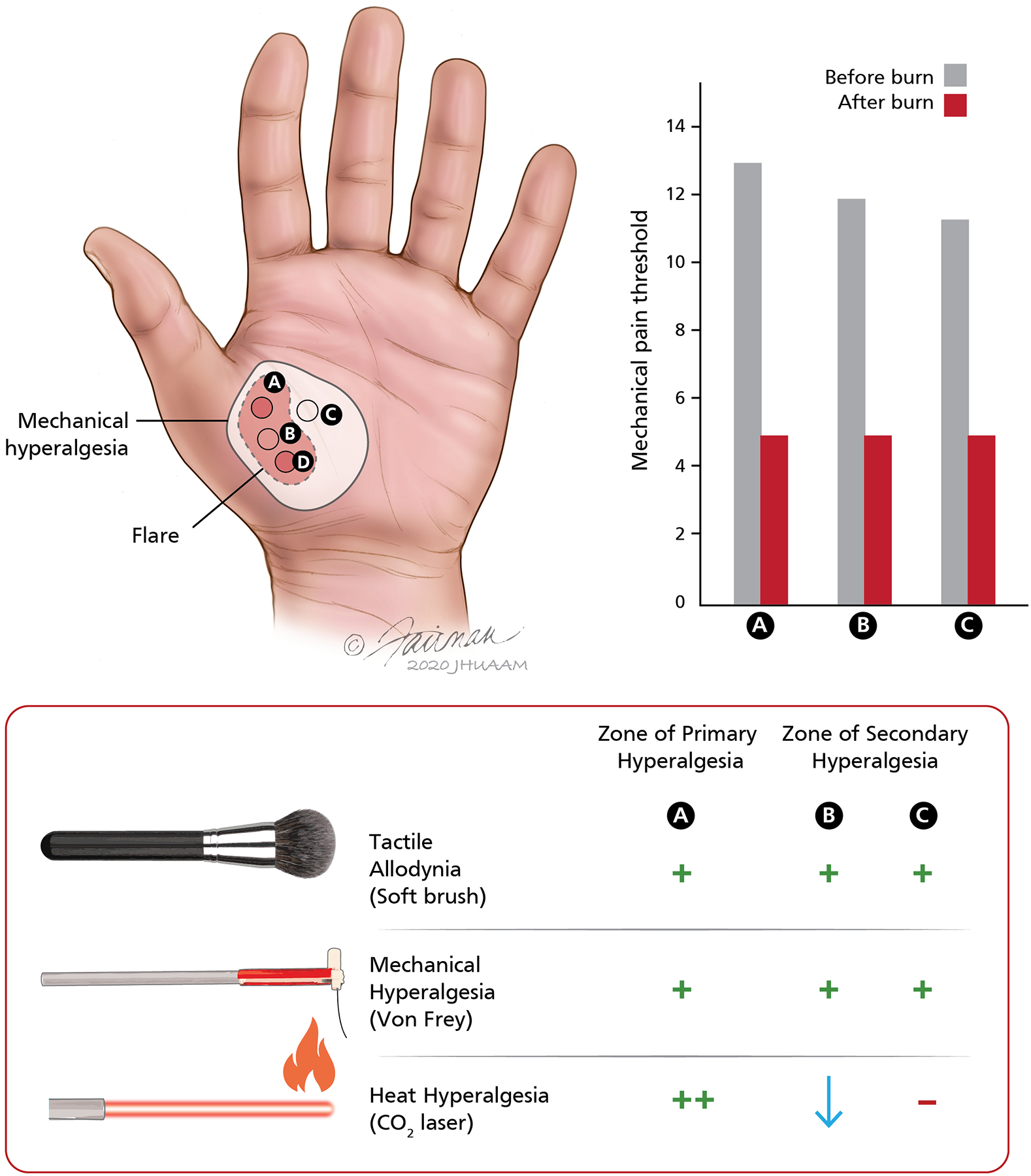
Figure 2.

Evidence for different mechanisms of primary and secondary hyperalgesia after cutaneous burn injuries in human subjects. (Top left) The illustration shows the relative location of the stimulus sites on the glabrous skin of the hand and the area of flare and mechanical hyperalgesia that develops after cutaneous burn injuries at sites A and D. The heat injury consisted of two burns (53°C, 30 s) applied over an area 7.5 mm in diameter separated (center to center) by a 2 cm interval. The area of mechanical hyperalgesia was larger than the flare region, and mechanical hyperalgesia persisted even after the flare disappeared. Mechanical thresholds for pain, assessed by von Frey hairs, and ratings of pain to controlled laser thermal stimuli (41–49°C, 10 stimuli at 1°C increments) were recorded at sites A, B, and C before and after the burns at sites A and D. (Top right) Mean pain thresholds to mechanical stimuli at the primary site (site A) and secondary hyperalgesia sites (B, C) before and after the burns at A and D. The mechanical pain threshold was significantly reduced after the cutaneous injury and was similar at the regions of primary and secondary hyperalgesia. (Bottom) Schematic representation of sensory changes in the zones of primary and secondary hyperalgesia. In the region of primary hyperalgesia (site A), allodynia to brush stimuli and hyperalgesia to von Frey hairs were observed. Pain ratings to heat stimuli were increased more than four-fold at this site (thermal hyperalgesia). Although pain in response to mechanical stimuli was enhanced at both secondary hyperalgesia sites (B, C), the pain to thermal stimuli was either unchanged (site C) or decreased (site B). Thus, thermal hyperalgesia was observed only at the injury site, whereas hypoalgesia to heat was observed at the site between the two burn injuries. Adapted from Raja et al. [115].
