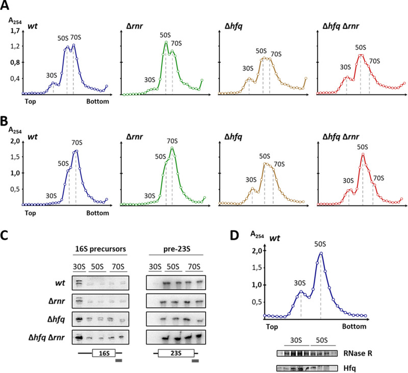
FIG 5.
Ribosome assembly defects arise upon double inactivation of Hfq and RNase R. (A and B) Fifteen to 50% sucrose density gradient analysis of ribosomal particles extracted from exponential-phase (A) and stationary-phase (B) cells analyzed under associative conditions (10 mM Mg2+) to promote subunit association into 70S ribosomes. Gradient fractions were collected, and absorbance at 254 nm was measured and plotted. Top and bottom denote the lower and higher sucrose concentrations in the gradient, respectively. Gradient analyses shown are representative of at least three independent experiments. (C) RNA was isolated from representative gradient fractions of each ribosomal particle (30S, 50S, and 70S), and Northern blot analysis was performed to assess the presence of 17S and pre-23S precursor rRNAs (left and right, respectively). Each set of wild-type and single and double mutant strains belongs to the same blotted membrane using a specific probe for the 3′ end extension of either the 17S or pre23S rRNA, as depicted below. (D) Ten to 30% sucrose density gradient of wild-type ribosomal particles extracted from stationary-phase cells, analyzed under dissociative conditions (0.1 mM Mg2+), to promote the complete dissociation of 70S ribosomes into the free 30S and 50S subunits. The protein content of representative gradient fractions was isolated and probed by Western blotting for the presence of RNase R and Hfq using specific antibodies. Ribosomal particles are indicated on top of the analyzed fractions and of each peak.

