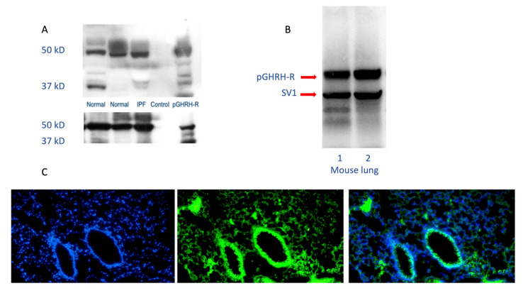
Figure 1.
Human and mouse lung westerns (upper panels A and B) and immunofluorescence staining (lower panel C) demonstrate growth hormone-releasing hormone-receptor (GHRH-R) protein. As shown in the upper panels, Western blotting confirms the presence of pituitary-type GHRH-R (pGHRH-R) and splice variant (SV1) in both normal and IPF human lung tissues (upper left panel). Likewise, the pGHRH-R is abundant in lung tissue protein from normal C57BL/6J mice (upper right panel). GHRH-R was detected using a rabbit polyclonal IgG primary antibody (Origene Technologies, Inc., Rockville, MD, USA). As shown in the lower panel, immunofluorescent staining for GHRH-R protein demonstrates prominent expression of the GHRH-R protein in the bronchial epithelium, as well as in alveolar parenchymal cells. (Left, DAPI staining; middle, immunofluorescent antibody to GHRH-R; right, merged images).

