Abstract
Objective:
Vertebral artery injury (VAI) after cervical spine trauma often remains undiagnosed. Despite various clinical studies suggesting simultaneous occurrence of VAI with cervical spine trauma, guidelines regarding screening and management of posttraumatic VAI are yet to be formulated. The primary objective of the current study was to formulate a low-cost screening protocol for posttraumatic VAI, thereby reducing the incidence of missed VAI in developing countries.
Materials and Methods:
This was a single-center prospective study performed on 61 patients using plain magnetic resonance imaging (MRI) as a screening tool to assess the frequency of VAI and routine X-ray to detect morphological fracture patterns associated with the VAI in posttraumatic cervical spine cases. If the MRI study showed any evidence of vascular disruption, then further investigation in the form of computed tomography angiography was done to confirm the diagnosis.
Results:
This study showed the incidence of VAI was 14.75% (9/61). Of 61 patients, 16 had supraaxial, and 45 patients sustained subaxial cervical spine fractures. In the cohort of nine cases of VAI, eight patients had subaxial cervical spine injuries, of which seven were due to flexion-distraction injury. C5–C6 flexion-distraction injury was most commonly associated with VAI (4 cases). Of the nine cases, five succumbed to injury (mortality 55.55%), and 19 patients from the non-VAI group succumbed to injury (mortality 36.53%). From surviving four cases with VAI, two had improvement in the American Spinal Injury Association scale by Grade 1.
Conclusion:
VAI in cervical spine trauma is an underrecognized phenomenon. Plain MRI axial imaging sequence can be an instrumental low-cost screening tool in resource-deficient parts of the world. VAI has tendency to occur with high-velocity trauma like bi-facetal dislocation, which has a high mortality and poor neurological recovery.
Keywords: Cervical spine, computed tomography angiography, vertebral artery injury
Introduction
Cervical spine fractures are inherently life-threatening injuries which can get further complicated by associated injury to the vertebral arteries. The incidence of this association has been reported from 25% to 46% in the literature.[1] The symptoms of vertebral artery injury (VAI) often go unnoticed due to poor general condition of these patients. VAI is common in its second segment where it traverses the subaxial spine due to rigid fixation within foramen transversarium. Hence, this segment is supposedly most susceptible to injury to the vertebral artery (VA).[1,2] Unilateral VAI may not necessarily cause vertebrobasilar insufficiency due to the adequacy of contralateral VA circulation. It is no surprise that ischemia caused by bilateral VAI can prove fatal for life.
The prevalence of undetected VAI following cervical spine trauma is statistically significant.[3,4,5,6] Considering both factors of increased detection rate of VAI with modern imaging techniques and adverse impact on functional outcome,[7] it becomes prudent for the treating clinician to be aware of this entity. In a resource-constrained setup, it is important to have a low-cost readily available screening protocol to detect the VAI. Magnetic resonance imaging (MRI) is the standard of care in cervical spine injuries to know the status of cord and other soft-tissue structures. We have used a fat-suppressed T1-weighted (T1W) and T2-weighted (T2W) sequences, acquired in the axial or oblique plane in a 1.5T MRI as a screening tool to detect VAI.[8,9] This study is a systematic effort to analyze the prevalence of VAI using plain MRI and study patterns of cervical spine injury leading to VAI along with functional outcome assessment.
Materials and Methods
This study was conducted at a tertiary care center in Mumbai, India. Institutional Review Board approval was taken before the commencement of the study. None of the authors have any conflicts of interest to declare. We recruited patients with established diagnosis of cervical spine injury for our study in a period ranging from March 2014 to April 2017. After initial stabilization as per the Advanced Trauma Life Support protocol, we performed detailed imaging, including X-ray and MRI. Inclusion criteria consisted of age between 15 and 60, cervical spine fractures/subluxations occurring between C1 and C7 irrespective of neurological status. Exclusion criteria consisted of patients with preexisting progressive neurological disorders and the presence of head injury. After a detailed analysis of X-ray and MRI, all patients of cervical spine injuries were classified according to the Allen and Ferguson classification[10] for subaxial spine, and supraaxial injury was classified as per specific bony component injury.
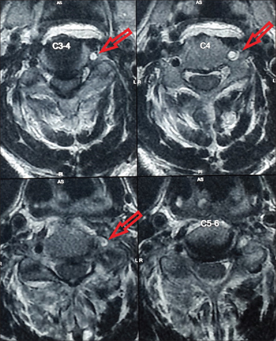
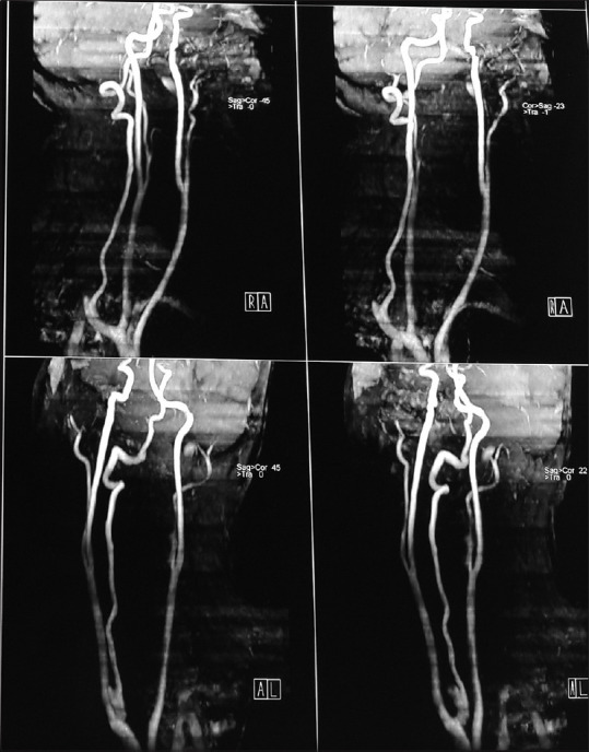
All MRI scans were scrutinized for signal changes on the axial T1 fat-saturation sequence with 1.5 T using T1W (TR/TE of 600/10 m) sequences and T2W (TR/TE of 4000/90 m). The T1W sequence with fat saturation along with T2 axial sequences enables visualization of the VAI (intramural hematoma/thrombosis/dissection) in the V2 and V3 segments of VA with good sensitivity and specificity.[9] In contrast to contralateral VA, these intramural pathologies appear hyperintense with a “crescent” shape that is eccentric into circular hyperintense residual arterial lumen due to flowing blood in T1 fat-suppressed sequence.[8] On the T2 sequence, it appears as crescentic hyperintensity involving the wall of VA with complete loss of flow void [Figure 1]. If the MRI study showed any evidence of vascular disruption, then further investigation in the form of computed tomography (CT) angiography was ordered to confirm the diagnosis [Figure 2]. There was no further attempt to subclassify vascular injuries of VA. Neurological examination was performed with help of the American Spinal Injury Association (ASIA) impairment scale[11] at the time of admission and at the 6-month follow-up. Medical records of all patients were screened for vertebrobasilar insufficiency in the form of occurrence of posttraumatic giddiness, headache, ataxia, blurred vision, ipsilateral facial numbness, or MRI evidence of cerebellar infarction.[12]
Figure 1.

Crescentic hyperintensity involving wall of the vertebral artery with complete loss of flow void in T2 axial sequence
Figure 2.

Computed tomography angiography showing absent flow in the left vertebral artery
Results
We considered 62 patients in a period ranging from March 2014 to April 2017, with established diagnosis of cervical spine injury for our study. Sixty-one patients satisfied the eligibility requirements of the study. The mean age was 33.5 years, with 46 males and 15 females in our study group. We had an average follow-up of 19 months following injury. Incidence VAI in the current study was 14.75% (9/61). Sixteen cases had supraaxial fractures, and 45 had subaxial fractures. Of 45 subaxial injuries, seven had flexion compression injury, two had vertical compression injury, and the rest 36 cases had flexion-distraction injury pattern [Table 1]. Nine patients had MRI evidence of VAI, which was confirmed with CT angiography. One had supraaxial vertebral V3 artery injury and eight had subaxial V2 level VA injury. Four had C5–C6 flexion-distraction injury with bilateral facetal dislocation, two had C4–C5 flexion-distraction injury with bilateral facetal dislocation, one had C6–C7 flexion distraction with unilateral facetal subluxation, one had flexion compression injury at C5 level, and one had posttraumatic atlantoaxial dislocation due to dens fracture (type 2) [Table 2].
Table 1.
Injury pattern in cervical spine trauma
| Pattern of injury | VAI (%) | Non-VAI (%) |
|---|---|---|
| Flexion distraction | 7 (87.5) | 29 (78.37) |
| Stage 1 | 0 | 3 |
| Stage 2 | 1 | 14 |
| Stage 3 | 5 | 9 |
| Stage 4 | 1 | 3 |
| Flexion compression | 1 (12.5) | 6 (16.21) |
| Vertical compression | 0 | 2 (5.4) |
| Supraaxial injury | 1 (6.25) | 15 (93.75) |
VAI – Vertebral artery injury
Table 2.
Level of injury in cervical spine trauma
| Level of injury | VAI (%) | Non-VAI (%) |
|---|---|---|
| Supraaxial | 1 (11.11) | 15 (28.34) |
| Subaxial C3-C4 | 0 | 4 (7.54) |
| C4-C5 | 3 (33.33) | 9 (18.86) |
| C5-C6 | 4 (44.44) | 19 (35.84) |
| C6-C7 | 1 (11.11) | 5 (9.43) |
VAI – Vertebral artery injury
Of nine VAI cases, five succumbed to the injury (mortality 55.55%), and 19 patients from the non-VAI group succumbed to injury (mortality 36.53%) although it is statistically insignificant as P = 0.28091. Out of four cases with VAI, two had improvement in neurology according to the ASIA scale. One patient who had incomplete quadriparesis improved from Asia B to C and could walk with assisted ambulation at 6-month follow-up, and another patient improved from Asia C to D with walking without support on a 3-month follow-up. Twenty-two patients from the non-VAI group showed improvement, which is again statistically insignificant with P = 0.2547 [Table 3]. None of the patients showed signs of vertebrobasilar ischemia such as dizziness, vertigo, ataxia, blurred vision, and facial numbness.
Table 3.
Mortality and improvement comparison
| VAI (%) | Non-VAI (%) | P | |
|---|---|---|---|
| Mortality | 5 (55.55) | 19 (36.53) | 0.2809 |
| Improvement | 2 (22.22) | 22 (42.30) | 0.2547 |
VAI – Vertebral artery injury
Discussion
VAI after cervical spine trauma often remains undiagnosed in limited resources setup due to the unavailability of advanced imaging modalities. In spite of various clinical studies suggesting the simultaneous occurrence of VAI with cervical spine trauma, specific guidelines regarding screening and management of posttraumatic VAI are yet to be formulated. The primary objective of the current study was to formulate a low-cost screening protocol for posttraumatic VAI, thereby reducing the incidence of missed VAI in developing countries. Secondarily, the study focuses on morphological fracture patterns associated with the VAI and functional outcomes after the VAI.
Incidence VAI in the current study was 14.75%, which is in accordance with the findings of multiple clinical studies showing the frequency of VAI from 17.2% to 26%[5,13,14,15] Miller et al. and Biffl et al.[3,6] also presented the high incidence of VAI in blunt cervical spine trauma ranging from 33% to 39% VAI incidence. On analysis of fracture pattern in VAI, we found 87.5% of patients showing flexion distraction type of injuries followed by 12.5% patients showing flexion compression type of injuries. Supraaxial fractures contribute 11.11% in all VAI patients. A study done by Gupta et al.[9] showed the subaxial VAI incidence of 37.5% in translational and rotational injuries, followed by compression in 27.5% and distraction in 22.5% of patients.
Cothren et al. showed[4] 37% of VAI frequency when patients screened with specific fracture types such as transverse foramen fracture, subluxation, and upper cervical spine trauma.
VAI, followed by cervical spine trauma, commonly occurs at the second segment of VA running through the transverse foramen of C6–C2. The fixation of the VA within the transverse foramen makes the second segment more prone to injury caused by cervical spine fractures and/or dislocations.[1,2] Among the study group of 61 patients, nine patients showed changes in axial T1 fat-saturation sequence with 1.5 T using T1W (TR/TE of 600/10 m) sequences and T2W (TR/TE of 4000/90 m). On further, CT angiography imaging, all of them showed VAI injury. This highlights the high specificity of plain MRI in VAI as a primary low-cost screening modality in suspected cases of VAI. This is similar to the observations made by Gupta et al.,[9] who made a presumptive diagnosis of VAI on the basis of axial T2 W MRI images.
The unilateral VAI, specifically the nondominant VA, may not cause ischemic symptoms, which is similar to our findings where none of the patients of VAI showed signs of vertebrobasilar ischemia such as dizziness, vertigo, ataxia, blurred vision, and facial numbness. Therefore, the injury may be underestimated in asymptomatic patients.[16,17] Many clinical studies have reported that VAI, after cervical spine trauma, occurs more frequently than the previously mentioned literature.[3,4,5,6] Functional outcome after VAI is dependent on contralateral VA for perfusion. If the collateral flow is inadequate, an occlusion may cause vertebrobasilar ischemia. Fifteen percent of patients have hypoplastic VA in whom contralateral artery remains the main perfusion artery.[18] VAI, although often initially asymptomatic, may have fatal consequences. However, we did not encounter a unilateral hypoplastic VA.
The C5–C6 level was most commonly associated with VAI, followed by C4–C5 level. This might be attributable with the most mobile junction of the cervical spine column being C5–C6.[19] This is similar to the findings of the study conducted by Giacobetti et al. and Vaccaro et al.[5,15] Flexion-distraction injury with bi-facetal dislocation accounts for a larger load of VAI possibly due to the involvement of foramen transversarium. Mortality in non-VAI patients was 36.53% as compared to 55.55% in the VAI group, the difference is statistically insignificant. This high mortality, although statistically insignificant, could probably be explained by VAI being commonly associated with high-velocity trauma like bi-facetal dislocation (85.7%). Non-VAI group (42.20%) showed higher neurological improvement compared to the VAI group (22.22%) that is statistically insignificant. Hence, the author feels that other factors such as mode of trauma, velocity of trauma, and level of trauma are more determining than VAI itself.
Due to the limitation of sample size, management of VAI, role of antiplatelet in management of VAI, and superiority of CT angiogram over MRA are still beyond the scope of the study, and further studies are warranted in this regard. While managing patients with VAI following cervical spine trauma, knowing the status of VA will help the operating surgeon to do risk stratification and take due precaution to avoid catastrophic complications.
Conclusion
VAI in cervical spine trauma is an underrecognized phenomenon. Plain MRI axial imaging sequence can be instrumental low-cost screening tool in resource-deficient developing parts of the world. VAI has tendency to occur with high-velocity trauma like bi-facetal dislocation, leading to high mortality and poor neurological recovery.
Financial support and sponsorship
Nil.
Conflicts of interest
There are no conflicts of interest.
References
- 1.Willis BK, Greiner F, Orrison WW, Benzel EC. The incidence of vertebral artery injury after midcervical Spine fracture or subluxation. Neurosurg. 1994;34:435–41. doi: 10.1227/00006123-199403000-00008. [DOI] [PubMed] [Google Scholar]
- 2.Prabhu V, Kizer J, Patil A, Hellbusch L, Taylon C, Leibrock L. Vertebrobasilar thrombosis associated with nonpenetrating cervical Spine trauma. J Trauma. 1996;40:130–7. doi: 10.1097/00005373-199601000-00027. [DOI] [PubMed] [Google Scholar]
- 3.Biffl WL, Moore EE, Elliott JP, Ray C, Offner PJ, Franciose RJ, et al. The devastating potential of blunt vertebral arterial injuries. Ann Surg. 2000;231:672–81. doi: 10.1097/00000658-200005000-00007. [DOI] [PMC free article] [PubMed] [Google Scholar]
- 4.Cothren CC, Moore EE, Biffl WL, Ciesla DJ, Ray CE, Jr, Johnson JL, et al. Cervical Spine fracture patterns predictive of blunt vertebral artery injury. J Trauma. 2003;55:811–3. doi: 10.1097/01.TA.0000092700.92587.32. [DOI] [PubMed] [Google Scholar]
- 5.Giacobetti FB, Vaccaro AR, Giacobetti MA, Deeley DM, Albert TJ, Farmer JC, et al. Vertebral artery occlusion associated with cervical Spine trauma. A prospective analysis. Spine (Phila Pa 1976) 1997;22:188–92. doi: 10.1097/00007632-199701150-00011. [DOI] [PubMed] [Google Scholar]
- 6.Miller PR, Fabian TC, Croce MA, Cagiannos C, Williams JS, Vang M, et al. Prospective screening for blunt cerebrovascular injuries: Analysis of diagnostic modalities and outcomes. Ann Surg. 2002;236:386–93. doi: 10.1097/01.SLA.0000027174.01008.A0. [DOI] [PMC free article] [PubMed] [Google Scholar]
- 7.Parent AD, Harkey HL, Touchstone DA, Smith EE, Smith RR. Lateral cervical spine dislocation and vertebral artery injury. Neurosurg. 1992;31:501–9. doi: 10.1227/00006123-199209000-00012. [DOI] [PubMed] [Google Scholar]
- 8.Provenzale JM, Morgenlander JC, Gress D. Spontaneous vertebral dissection: Clinical, conventional angiographic, CT, and MR findings. J Comput Assist Tomogr. 1996;20:185–93. doi: 10.1097/00004728-199603000-00004. [DOI] [PubMed] [Google Scholar]
- 9.Gupta P, Kumar A, Gamangatti S. Mechanism and patterns of cervical spine fractures-dislocations in vertebral artery injury. J Craniovertebr Junction Spine. 2012;3:11–5. doi: 10.4103/0974-8237.110118. [DOI] [PMC free article] [PubMed] [Google Scholar]
- 10.Allen BL, Jr, Ferguson RL, Lehmann TR, O'Brien RP. A mechanistic classification of closed, indirect fractures and dislocations of the lower cervical spine. Spine (Phila Pa 1976) 1982;7:1–27. doi: 10.1097/00007632-198200710-00001. [DOI] [PubMed] [Google Scholar]
- 11. [Last accessed on 2020 May 02]. Available from: https//www.asia-spinalinjury.org .
- 12.Tulyapronchote R, Selhorst JB, Malkoff MD, Gomez CR. Delayed sequelae of vertebral artery dissection and occult cervical fractures. Neurol. 1994;44:1397–9. doi: 10.1212/wnl.44.8.1397. [DOI] [PubMed] [Google Scholar]
- 13.Parbhoo AH, Govender S, Corr P. Vertebral artery injury in cervical spine trauma. Inj. 2001;32:565–8. doi: 10.1016/s0020-1383(00)00232-1. [DOI] [PubMed] [Google Scholar]
- 14.Taneichi H, Suda K, Kajino T, Kaneda K. Traumatically induced vertebral artery occlusion associated with cervical spine injuries: Prospective study using magnetic resonance angiography. Spine. 2005;30:1955–62. doi: 10.1097/01.brs.0000176186.64276.d4. [DOI] [PubMed] [Google Scholar]
- 15.Vaccaro AR, Klein GR, Flanders AE, Albert TJ, Balderston RA, Cotler JM. Long-term evaluation of vertebral artery injuries following cervical spine trauma using magnetic resonance angiography. Spine. 1998;23:789–94. doi: 10.1097/00007632-199804010-00009. [DOI] [PubMed] [Google Scholar]
- 16.Sim E, Vaccaro AR, Berzlanovich A, Pienaar S. The effects of staged static cervical flexion-distraction deformities on the patency of the vertebral arterial vasculature. Spine. 2000;25:2180–6. doi: 10.1097/00007632-200009010-00007. [DOI] [PubMed] [Google Scholar]
- 17.Veras LM, Gutierrez SP, Castellanos J, Capellades J, Casamitjana J, Canellas AR. Vertebral artery occlusion after acute cervical Spine trauma. Spine. 2000;25:1171–7. doi: 10.1097/00007632-200005010-00019. [DOI] [PubMed] [Google Scholar]
- 18.Louw JA, Mafoyane NA, Small B, Neser CP. Occlusion of the vertebral artery in cervical spine dislocations. J Bone Joint Surg Br. 1990;72:679–81. doi: 10.1302/0301-620X.72B4.2380226. [DOI] [PubMed] [Google Scholar]
- 19.Cothren CC, Moore EE, Ray CE, Jr, Johnson JL, Moore B, Burch JM. Cervical spine fracture patterns mandating screening to rule out blunt cerebrovascular injury. Surg. 2001;141:76–82. doi: 10.1016/j.surg.2006.04.005. [DOI] [PubMed] [Google Scholar]


