eLife logoLink to eLife
. 2020 Oct 27;9:e56151. doi: 10.7554/eLife.56151

Distinct hierarchical alterations of intrinsic neural timescales account for different manifestations of psychosis

Kenneth Wengler 1,2,, Andrew T Goldberg 2, George Chahine 3, Guillermo Horga 1,2,
Editors: Michael J Frank4, Michael J Frank5
PMCID: PMC7591251  PMID: 33107431

Abstract

Hierarchical perceptual-inference models of psychosis may provide a holistic framework for understanding psychosis in schizophrenia including heterogeneity in clinical presentations. Particularly, hypothesized alterations at distinct levels of the perceptual-inference hierarchy may explain why hallucinations and delusions tend to cluster together yet sometimes manifest in isolation. To test this, we used a recently developed resting-state fMRI measure of intrinsic neural timescale (INT), which reflects the time window of neural integration and captures hierarchical brain gradients. In analyses examining extended sensory hierarchies that we first validated, we found distinct hierarchical INT alterations for hallucinations versus delusions in the auditory and somatosensory systems, thus providing support for hierarchical perceptual-inference models of psychosis. Simulations using a large-scale biophysical model suggested local elevations of excitation-inhibition ratio at different hierarchical levels as a potential mechanism. More generally, our work highlights the robustness and utility of INT for studying hierarchical processes relevant to basic and clinical neuroscience.

Research organism: Human

Introduction

Hallucinations and delusions are burdensome symptoms that typically manifest together as the psychotic syndrome of schizophrenia. Perceptual-inference models of psychosis suggest that these symptoms result from alterations in the updating of internal models of the environment that are used to make inferences about external sensory events and their causes (Adams et al., 2013; Horga and Abi-Dargham, 2019; Sterzer et al., 2018). These models are receiving increasing empirical support (Adams et al., 2018; Baker et al., 2019; Cassidy et al., 2018; Davies et al., 2018; Powers et al., 2017; Teufel et al., 2015), yet current theories do not provide a satisfactory explanation for how hallucinations and delusions tend to co-occur but sometimes manifest in isolation. This suggests that these psychotic symptoms may share a common neurobiological mechanism while simultaneously depending on symptom-specific pathways.

We and others have proposed that this apparent tension may be resolved in the context of hierarchical perceptual-inference models (Adams et al., 2013; Baker et al., 2019; Corlett et al., 2009; Corlett et al., 2019; Fletcher and Frith, 2009; Sterzer et al., 2018). One possibility is that alterations at higher levels—supporting inferences on abstract hidden states like someone’s intentions—may drive delusions, and alterations at lower levels—supporting inferences about lower level features of stimuli such as stimulus presence or absence—may drive hallucinations (Baker et al., 2019; Davies et al., 2018; Horga and Abi-Dargham, 2019; Powers et al., 2017). In addition to these symptom-specific pathways, alterations at any level may naturally propagate throughout the interdependent levels of the hierarchy (Chaudhuri et al., 2015), potentially explaining symptom co-occurrence. Importantly, neural systems supporting inference are thought to feature a hierarchical architecture of timescales that mirrors the hierarchical temporal dynamics of natural environments, where rapidly changing events are typically nested within slower changing contexts (Kiebel et al., 2008; Kiebel, 2009). Thus, higher level inferences pertaining to slower changing contexts require neural systems with the ability to integrate information over longer periods, an ability consistent with the persistent neuronal activity that characterizes higher level regions (Major and Tank, 2004; Mazurek et al., 2003).

A hierarchy of neural timescales is observed in both single-neuron recordings in non-human primates (Murray et al., 2014) and functional magnetic resonance imaging (fMRI) recordings in humans (Hasson et al., 2015; Hasson et al., 2008; Honey et al., 2012; Lerner et al., 2011; Stephens et al., 2013), and is recapitulated by a large-scale biophysical model (Chaudhuri et al., 2015). Furthermore, a newly developed method based on resting-state fMRI that was validated against electroencephalography (EEG) similarly captures a hierarchy of intrinsic neural timescales (INT), as well as alterations in psychopathology (Watanabe et al., 2019). Here, we specifically applied this fMRI measure to test whether hallucinations and delusions are associated with distinct changes of INT at low and high levels of neural hierarchies, respectively. We hypothesized that INT at these respective levels would increase with more severe symptoms, potentially reflecting increased neural integration of prior information (Glaze et al., 2015; Mante et al., 2013; Mazurek et al., 2003), in line with behavioral findings in hallucinations and delusions (Baker et al., 2019; Cassidy et al., 2018; Powers et al., 2017). If present, these INT changes should manifest as symptom-specific differences in hierarchical gradients.

Results

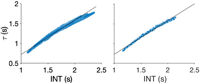
INT maps were estimated as previously described (Watanabe et al., 2019) (Materials and methods). Briefly, the autocorrelation function of the fMRI signal at each voxel (or vertex) was estimated and the sum of the autocorrelation coefficients during the initial positive period was calculated. This initial positive period included all timepoints from the first lag until the timepoint immediately preceding the first lagged timepoint with a non-positive autocorrelation coefficient. To adjust for differences in temporal resolution, the sum was multiplied by the repetition time (TR) of the fMRI data. This product was used as an index for INT (note that values are similar to those from an exponential fit [Murray et al., 2014; Figure 1—figure supplement 1]). INT maps were parcellated using the HCP-multimodal parcellation (Glasser et al., 2016) to facilitate further analysis. Additionally, T1w/T2w (myelin) and cortical-thickness maps were obtained from high-resolution structural scans from the HCP database. Both of these structural measures have previously been shown to capture an underlying brain-wide hierarchy (Burt et al., 2018; Fischl et al., 2008; Wagstyl et al., 2015), consistent with the classic use of myeloarchitecture and cytoarchitecture for cortical parcellation (Brodmann, 1909; Sarkissov et al., 1955; Vogt, 1911; Von Economo, 1929). In particular, Burt et al., 2018 validated T1w/T2w in macaques by showing strong agreement with a gold-standard tract-tracing measure of hierarchy. Establishing T1w/T2w and cortical thickness as structural indices of hierarchy in humans, Burt et al. validated these MRI measures against human postmortem gene-expression data (Hawrylycz et al., 2012)—specifically using granular layer-IV-specific gene expression as a proxy for cytoarchitecture structural type.

Selection and multimodal validation of neural hierarchies

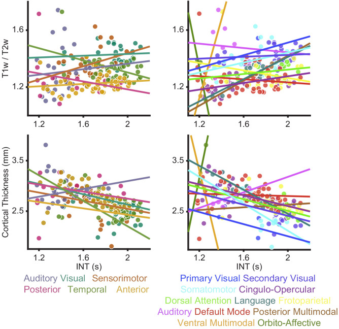
Our hypothesis of symptom-specific INT differences in hierarchical gradients was agnostic to the specific neural hierarchies involved in psychosis, as the involvement of most sensory modalities has been reported (Lewandowski et al., 2009; Postmes et al., 2014). Consistent with prior empirical and theoretical work (Chaudhuri et al., 2015; Vázquez-Rodríguez et al., 2019), in a subset of 100 unrelated young and healthy subjects from the HCP dataset, we did not observe a systematic relationship across the whole brain between INT (Figure 1A)—an index of functional hierarchy (Chaudhuri et al., 2015; Murray et al., 2014)—and the two indices of structural hierarchy (T1w/T2w and cortical thickness) (Burt et al., 2018; Fischl et al., 2008; Wagstyl et al., 2015Figure 1—figure supplement 2). We thus decided to focus on specific, well-studied hierarchies of the auditory, visual, and somatosensory systems that have been parcellated in humans, and where the functional and structural indices of hierarchy appeared better aligned.

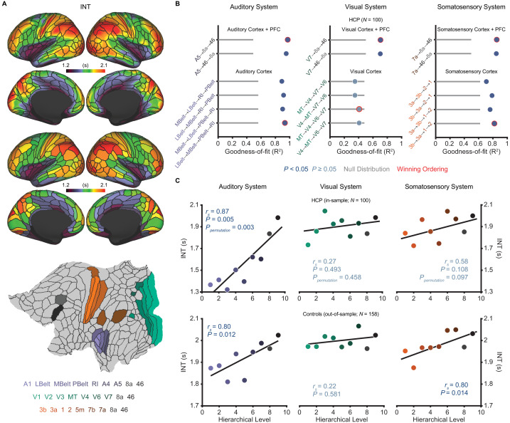
Figure 1. Model comparison to determine the hierarchical orderings of the auditory, visual, and somatosensory systems.

(A) Group-averaged intrinsic neural timescale (INT) map from the Human Connectome Project (HCP) dataset (N = 100; top), parcellated group-averaged INT map (middle), and flattened cortex showing the parcels in the auditory, visual, and somatosensory hierarchies (winning hierarchies underneath; bottom). Color coding of parcels indicates their anatomical and hierarchical location. (B) Goodness of fit (R2) of linear mixed-effects models predicting different hierarchical orderings of the auditory system (left), visual system (middle), and somatosensory system (right) from T1w/T2w and cortical thickness values in the HCP dataset (Materials and methods). First, the winning ordering (i.e. the model with the best goodness of fit) for each system was determined for the seven sensory cortex regions (bottom four models). Then, the winning ordering was determined for extended models with two downstream prefrontal cortex regions added to the respective winning models for the sensory cortex (top two models). Note that, for each of the four considered orderings within the sensory cortex for each system, only the four regions whose order is varied (out of 7 regions) are shown to delineate the models. For the auditory cortex, A1 was always the lowest order region while A4 and A5 were always the two highest order regions. For the visual cortex, V1, V2, and V3 were always the three lowest order regions. For the somatosensory cortex, 5m, 7b, and 7a were always the three highest order regions. Null distributions were generated by randomly permuting the hierarchical ordering across all regions in a given hierarchy (0th – 95th percentiles shown). (C) Scatterplots showing INT values plotted as a function of hierarchical level for the PFC-extended winning models in B (red outline) for the HCP dataset (top) and the healthy control group in the schizophrenia combined dataset (N = 158; bottom). LBelt, lateral belt; MBelt, medial belt; PBelt, parabelt; RI, retroinsular cortex; MT, middle temproal area.

Figure 1—source data 1. Data and code to reproduce Figure 1.

Figure 1.

Figure 1—figure supplement 1. Comparison of different methods for neural timescale estimation: sum of initial positive period vs exponential fit.

Figure 1—figure supplement 1.

Scatterplots showing the relationship between neural timescales estimated using the sum of the initial positive period (INT, x-axis) and an exponential fit (τ, y-axis) to the autocorrelation function. Results for vertices (left) and parcels (right). Lines of best-fit are shown for comparison. The INT method of Watanabe et al., 2019 is similar to estimating the decay rate of an exponential fit to the autocorrelation function (Murray et al., 2014). The former method was used in our study to maintain consistency with the previous study, which validated the technique against INT measured by electroencephalography (Watanabe et al., 2019). Additionally, this method avoids the need for nonlinear fitting—which is known to be challenging (Transtrum et al., 2010) and computationally expensive—and may be more robust. The difference between timescales estimated with the two methods was assessed in the 100 HCP subjects by comparing the group averaged timescale maps using Spearman correlation. The correlation between the two methods was almost perfect (vertices: rs = 0.9986, p<<0.01; parcels: rs = 0.9985, p<<0.01) but there is an upwards bias in values estimated using the method of Watanabe et al., 2019.
Figure 1—figure supplement 2. Relationships between intrinsic neural timescale and either T1w/T2w (myelin) or cortical thickness.

Figure 1—figure supplement 2.

As reported in the Main Text, we did not observe a clear parcel-wide relationship between structural markers of hierarchy and intrinsic neural timescale (INT). These relationships were investigated using two parcel-groupings; those described by Glasser et al. (Glasser et al., 2016; left) and the Cole-Anticevic networks described by Ji et al. (Ji et al., 2019; right). To better characterize the relationship between myelin (top) or cortical thickness (bottom) and INT, intercepts for each parcel group (visual, auditory, sensorimotor, posterior, anterior, and temporal) or network (primary visual, secondary visual, somatomotor, cingulo-opercular, dorsal attention, language, frontoparietal, auditory, default mode, posterior multimodal, ventral multimodal, and orbito-affective) were introduced to allow for overall differences in INT-by-parcel-group (or INT-by-network) given the apparent differences observed in the data. Parcel-group-by-T1w/T2w-value (or network-by-T1w/T2w-value) interactions and parcel-group-by-cortical-thickness (or network-by-cortical-thickness) were also introduced to allow for differences in the relationship between T1w/T2w (or cortical thickness) and INT values across parcel groups (or networks). We found that an extended model with parcel-group intercepts and interactions of parcel-group-by-T1w/T2w explained significantly more variance in INT than the reduced model (no parcel-group intercepts or interactions): parcel groups differed in INT (model with parcel-group intercepts versus reduced model: F10,167 = 12.03, p<10−14) and their relationship between T1w/T2w and INT (model with parcel-group intercepts and interactions of parcel-group-by-T1w/T2w versus a model with parcel-group intercepts but no interaction terms: F5,167 = 5.55, p<10−4). Similar results were observed for the Cole-Anticevic Networks, where we found that an extended model with network intercepts and interactions of network-by-T1w/T2w explained significantly more variance in INT than the reduced model (no network intercepts or interactions): networks differed in INT (model with network intercepts versus reduced model: F22,155 = 4.62, p<10−8) and their relationship between T1w/T2w and INT (model with network intercepts and interactions of network-by-T1w/T2w versus model with network intercepts but no interactions: F11,155 = 2.15, p=0.020). For cortical thickness, we found that an extended model with parcel-group intercepts and interactions of parcel-group-by-cortical-thickness explained significantly more variance in INT than the reduced model (no parcel-group intercepts or interactions): parcel groups differed in INT (model with parcel-group intercepts versus reduced model: F10,167 = 8.59, p<10−10) and their relationship between cortical-thickness and INT (model with parcel-group intercepts and interactions of parcel-group-by-cortical-thickness versus a model with parcel-group intercepts but no interaction terms: F5,167 = 5.05, p<10−3). Similar results were observed for the Cole-Anticevic Networks, where we found that an extended model with network intercepts and interactions of network-by-cortical-thickness explained significantly more variance in INT than the reduced model (no network intercepts or interactions): networks differed in INT (model with network intercepts versus reduced model: F22,155 = 3.58, p<10−5) and their relationship between cortical-thickness and INT (model with network intercepts and interactions of network-by-cortical-thickness versus a model with network intercepts but no interactions: F11,155 = 1.96, p=0.036). These results suggest that the relationship between structural hierarchies (T1w/T2w and cortical thickness) and functional hierarchy (INT) is not constant across the whole brain but rather changes for different parcel groups and networks.
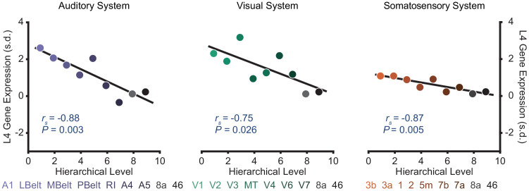
Figure 1—figure supplement 3. Selected sensory hierarchies demonstrate a hierarchical gradient of granular layer IV (L4) gene expression.

Figure 1—figure supplement 3.

Scatter plots showing L4 gene expression plotted as a function of hierarchical level for the winning orderings. Expression is plotted in units of standard deviation (s.d.; σ) from the mean. Gene expression data are from the Allen Human Brain Atlas as compiled by Burt et al., 2018.
Figure 1—figure supplement 4. Effect of framewise displacement on estimated intrinsic neural timescales.

Figure 1—figure supplement 4.

To determine the effect of head motion during resting-state fMRI data acquisition on estimated intrinsic neural timescale (INT) values, we used a linear-regression model to predict INT as a function of mean framewise displacement (FD) while controlling for age and gender. Because average FD represents a summary measure of head motion during the entire acquisition, this regression does not provide information regarding differential effects of sustained small levels of head motion versus infrequent but large levels of head motion. (A) t-statistic values from a regression model predicting INT from the average FD during fMRI data acquisition plotted as a function of the INT rank for 100 HCP subjects. Each datapoint represents one parcel. 181 out of 188 parcels showed significantly shorter INT with greater FD (Ppermutation = 0.01). (B) t-statistic values from parcels within the winning orderings of the auditory hierarchy (left), visual hierarchy (middle), and somatosensory hierarchy (right) plotted as a function of hierarchical level. No hierarchical-gradient effects of FD were observed.
Figure 1—figure supplement 5. Reliability of intrinsic neural timescale estimation.

Figure 1—figure supplement 5.

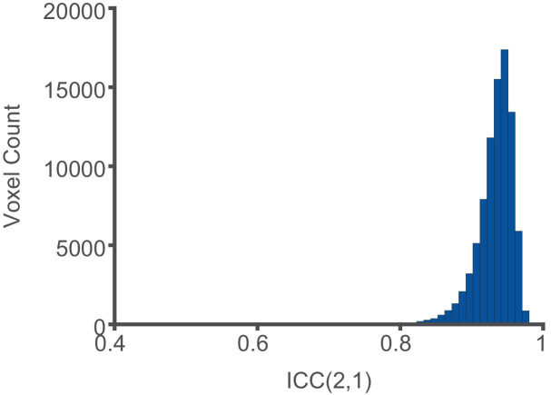
Intrinsic neural timescale (INT) maps for 100 HCP subjects were estimated in volume space and the values at each voxel falling within the 188 parcels of the HCP-MMP1.0 were compared when INT was estimated from the first 5 min or the last 5 min of fMRI acquisition. The INT maps showed excellent reliability based on the ICC(2,1) between the first and last 5 min of the fMRI acquisition (median ICC ±interquartile range across voxels: 0.94 ± 0.03).
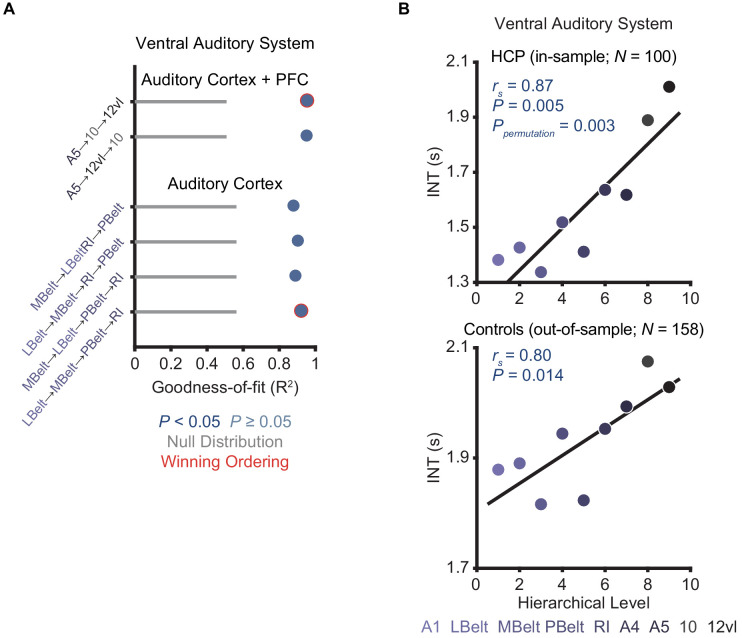
Figure 1—figure supplement 6. Model comparison to determine the hierarchical ordering of the ventral auditory pathway.

Figure 1—figure supplement 6.

(A) Goodness of fit (R2) of linear mixed-effects models predicting different hierarchical orderings of the ventral auditory system from T1w/T2w and cortical thickness values in the HCP dataset (N = 100). First, the winning ordering (i.e. the model with the best goodness of fit) was determined for the seven auditory cortex regions (bottom four models); then, the winning ordering was determined for an extended model with two ventral downstream prefrontal cortex regions (areas 10 and 12vl) added to the winning model for the auditory cortex (top two models). Note that, for each of the four considered orderings within the auditory cortex, only the four regions whose order is varied (out of seven regions) are shown to denote the models. For the auditory cortex, A1 was always the lowest-order region while A4 and A5 were always the two highest-order regions. Null distributions were generated by randomly permuting the hierarchical ordering (0th – 95th percentiles shown). (B) Scatterplots showing intrinsic neural timescale (INT) values plotted as a function of hierarchical level for the PFC-extended winning model in B (red outline) for the HCP dataset (top) and the healthy control group in the schizophrenia combined dataset (N = 158; bottom).

Despite ample anatomical investigation in primates, ambiguities in the definition of anatomical hierarchies in these sensory systems remain (Hilgetag et al., 1996; Kaas and Hackett, 2000) and have not been fully addressed in human MRI work. To address this issue, we used an anatomically informed, data-driven approach to determine the most suitable hierarchical orderings of each sensory system. First, using the HCP dataset, we determined the hierarchical orderings of the sensory cortex parcels (auditory, visual, or somatosensory) by selecting the ordering that was best predicted by T1w/T2w and cortical thickness parcel-wise values for each system (i.e. the ordering for which these values explained the most variance). To enhance robustness, we specifically constrained this comparison to the four most plausible hierarchical orderings of each system based on previous anatomical studies (Felleman and Van Essen, 1991; Galaburda and Pandya, 1983; Hyvärinen and Poranen, 1978; Morel et al., 1993). The winning orderings were A1 → lateral belt (LBelt) → medial belt (MBelt) → parabelt (PBelt) → retroinsular cortex (RI) → A4 → A5 for the auditory cortex, V1 → V2 → V3 → middle temporal area (MT) → V4 → V6 → V7 for the visual cortex, and 3b → 3a → 1 → 2 → 5m → 7b → 7a for the somatosensory cortex (Figure 1B). Using the same approach to build upon these winning orderings and capture the broadest possible range of the hierarchies, we then determined the hierarchical position of two additional prefrontal cortex (PFC) regions known to be downstream projections of the auditory, visual, and somatosensory cortices: area 8a and area 46 (Felleman and Van Essen, 1991; Kaas and Hackett, 2000). For all three sensory systems, area 46 was selected as the highest hierarchical level (Figure 1B). Notably, each of the PFC-extended winning models explained more variance than chance based on a null distribution of 10,000 random orderings (auditory system: Ppermutation < 10−4; visual system: Ppermutation = 0.003; somatosensory system: Ppermutation = 0.001).

We then evaluated whether these winning hierarchies—selected solely based on structural measures of hierarchy—were able to capture functional variability in the INT measure, such that higher levels exhibit longer INT. Within the HCP dataset, hierarchical position significantly correlated with INT in the auditory system (Spearman correlation rs = 0.87, p=0.005; Figure 1C), and this correlation was above chance level based on a null distribution of 10,000 random orderings (Ppermutation = 0.003). The hierarchical ordering was further validated in an out-of-sample group of 158 healthy controls from the schizophrenia combined dataset (Materials and methods), where hierarchy similarly correlated with INT in the auditory system (rs = 0.80, p=0.01; Figure 1C). Positive but non-significant correlations were observed in the visual system (in-sample: rs = 0.27, p=0.49, Ppermutation = 0.47; out-of-sample: rs = 0.22, p=0.58; Figure 1C). Stronger positive correlations were observed in the somatosensory system that reached significance in the out-of-sample group (in-sample: rs = 0.58, p=0.108, Ppermutation = 0.097; out-of-sample: rs = 0.80, p=0.014; Figure 1C). Despite the non-significant effects in the visual system (which surprisingly seemed to reflect less pronounced hierarchical gradients on all MRI measures, as suggested by the structural MRI gradients for all four tested orderings of the visual cortex falling within the null distribution; Figure 1B), these data showed that the winning hierarchies captured functional INT gradients, at least in the auditory and somatosensory systems. As a third independent test of our winning hierarchies, we tested their ability to capture variability in cytoarchitecture structural type using human postmortem gene-expression data from the Allen Human Brain Atlas (Hawrylycz et al., 2012). Following prior work (Burt et al., 2018), we focused on the average expression of five genes preferentially expressed in granular layer IV, a cytoarchitectural marker that is more prominent in lower hierarchical levels. Consistent with this, expression of granular layer IV genes showed strong, negative correlations with hierarchical level in all three winning hierarchies (auditory: rs = −0.88, p=0.003; visual: rs = −0.75, p=0.026; somatosensory: rs = −0.87, p=0.005; Figure 1—figure supplement 3). Thus, we empirically validated extended sensory hierarchies that captured variability in hierarchical indices across three independent datasets, although this was generally less clear for the visual system.

Assessment of robustness and reliability in the HCP dataset

Next, we set out to determine the robustness and reliability of INT. We focused on head motion, a common source of artifacts in fMRI data (Power et al., 2012). Head motion during data acquisition was associated with decreased INT values (181 out of 188 parcels, Ppermutation = 0.01; Figure 1—figure supplement 4). Yet, these effects were comparable across hierarchical levels (auditory system: rs = −0.23, Ppermutation = 0.805; visual system: rs = −0.38, Ppermutation = 0.649; somatosensory system: rs = −0.88, Ppermutation = 0.054; Figure 1—figure supplement 4). No effects were observed for gender or age (all Ppermutation > 0.174). Finally, INT maps showed excellent reliability between the first and last 5 min of the fMRI acquisition (median ICC(2,1) ± interquartile range across voxels: 0.94 ± 0.03; Figure 1—figure supplement 5).

Exploratory analyses of INT in schizophrenia versus health

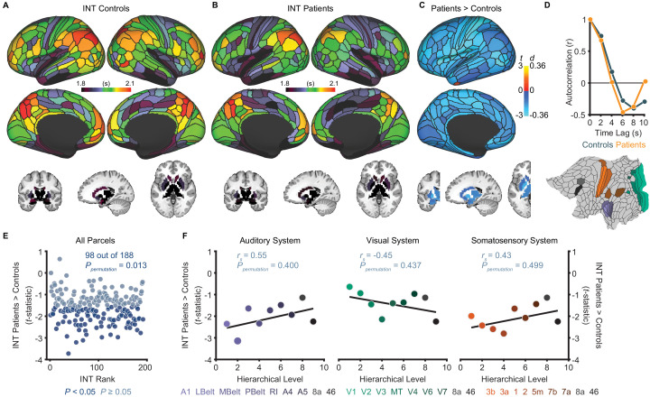
Although our primary hypothesis dealt with hierarchical differences between hallucinations and delusions, we first present exploratory analyses of diagnosis effects on INT. Table 1 lists the participant characteristics. Compared to controls (N = 158), patients (N = 127) exhibited a small-to-moderate, but widespread, reduction of INT (98 out of 188 parcels, Ppermutation = 0.013; Figure 2E). A voxelwise analysis observed a similar result (Figure 2—figure supplement 1). However, the INT reductions in patients were comparable across hierarchical levels (all Ppermutation > 0.40; Figure 2F). In silico simulations using a large-scale biophysical model (Chaudhuri et al., 2015) suggested that the global INT reduction in patients could be neuronally implemented by globally reduced excitation-inhibition (E/I) ratio (Figure 2—figure supplement 2).

Table 1. Participant characteristics.

Table 1—source data 1. Raw data for each individual subject in Table 1.
Variable Healthy controls Patients with schizophrenia
BGS COBRE NMCH UCLA All BGS COBRE NMCH UCLA All
N 24 42 25 67 158 40 31 26 30 127
Age, mean
(SD), y
36.0
(13.1)
33.9
(10.4)
29.8
(7.2)
32.3
(8.5)
32.9
(9.7)
31.1
(12.6)
30.5
(11.9)
30.5
(6.2)
35.0
(8.9)
31.8
(10.6)
Male sex, No. (%) 22 (92) 34 (81) 16 (64) 54 (81) 126 (80) 38 (95) 26 (84) 19 (73) 22 (73) 105 (83)
Framewise displacement*, mean (SD), mm 0.15
(0.04)
0.14
(0.05)
0.14
(0.11)
0.10
(0.04)
0.13
(0.06)
0.16
(0.06)
0.17
(0.06)
0.12
(0.06)
0.13
(0.04)
0.15
(0.06)
Delusion score, mean (SD) NA NA NA NA NA 2.3
(1.6)
1.7
(1.5)
3.2
(1.9)
2.5
(1.4)
2.4
(1.7)
Hallucination score, mean (SD) NA NA NA NA NA 2.1
(1.6)
1.7
(1.4)
2.9
(2.0)
2.2
(1.6)
2.2
(1.7)
Conceptual disorganization score, mean (SD) NA NA NA NA NA 0.9
(1.3)
0.6
(1.0)
2.0
(1.6)
1.4
(1.4)
1.2
(1.4)
Emotional withdrawal score, mean (SD) NA NA NA NA NA 1.8
(1.2)
1.2
(1.3)
3.4
(1.7)
2.3
(1.5)
2.1
(1.6)
Social withdrawal score, mean (SD) NA NA NA NA NA 1.8
(1.4)
1.3
(1.4)
3.2
(1.7)
2.7
(1.6)
2.2
(1.6)
Blunted affect score, mean (SD) NA NA NA NA NA 1.8
(1.6)
1.6
(1.5)
3.3
(1.6)
1.1
(1.1)
1.9
(1.6)
Alogia score, mean (SD) NA NA NA NA NA 1.3
(1.6)
1.2
(1.4)
2.1
(1.7)
0.9
(1.6)
1.3
(1.6)

*Framewise displacement values were estimated after motion censoring.

BGS, BrainGluSchi; NMCH, NMorphCH; SD, standard deviation.

Figure 2. Exploratory analyses show that patients with schizophrenia exhibit widespread reductions of intrinsic neural timescales compared to healthy controls.

(A) Parcellated group-averaged intrinsic neural timescale (INT) map for healthy controls (N = 158). (B) Parcellated group-averaged INT map for patients with schizophrenia (N = 127). (C) t-statistic (Cohen’s d) map showing the contrast of patients greater than controls. Across most parcels, INT is shorter in patients than controls in a regression model (M1exploratory; Materials and methods) controlling for age, gender, mean framewise displacement, and sample-acquisition site (overall effect of diagnosis: 98 out of 188 parcels, Ppermutation = 0.013). Only the left hemisphere is shown because statistical analyses were performed after averaging the values in each parcel across the left and right hemispheres. (D) To illustrate the effect of reduced INT in patients with schizophrenia, the group-averaged, whole-brain-averaged autocorrelation functions were estimated from subjects with fMRI data acquired with the same repetition time (top; controls: N = 132; patients: N = 101). The group-averaged autocorrelation function for patients crosses the zero point on the y-axis (i.e. autocorrelation coefficient = 0) sooner than in controls, demonstrating the global reduction of INT in patients. The flattened cortex shows the parcels in the auditory, visual, and somatosensory hierarchies for reference (bottom). (E) Scatterplot showing t-statistic values for group differences from the regression model (M1exploratory), plotted as a function of the INT rank (determined from the group-averaged INT map from HCP subjects). Each datapoint represents one parcel. (F) Scatterplots showing t-statistic values from parcels within the auditory (left), visual (middle), and somatosensory (right) hierarchies plotted as a function of hierarchical level. No hierarchical-gradient effects of schizophrenia diagnosis were observed. LBelt, lateral belt; MBelt, medial belt; PBelt, parabelt; RI, retroinsular cortex; MT, middle temproal area.

Figure 2—source data 1. Data and code to reproduce Figure 2.

Figure 2.

Figure 2—figure supplement 1. Voxelwise analysis of intrinsic neural timescales in schizophrenia versus health.
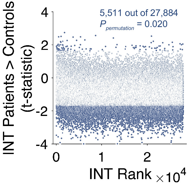
Figure 2—figure supplement 1.

Scatterplot showing t-statistic values from a regression model predicting intrinsic neural timescale (INT) controlling for age, gender, mean framewise displacement, and data collection site, plotted as a function of the INT rank. Each datapoint represents one voxel. We repeated the exploratory analysis of INT in patients with schizophrenia versus healthy controls at the level of individual voxels. Only voxels that fell within the 188 parcels were included in this analysis. Similar results were observed as reported in the Main Text (Figure 3E) where we investigated effects at the level of parcels. Relative to controls, patients with schizophrenia exhibited a small-to-moderate but widespread reduction of INT (overall effect of diagnosis: 5,511 out of 27,884 voxels, Ppermutation = 0.020;). A non-significant number of voxels also showed significantly longer INT in patients with schizophrenia compared to healthy controls (93 out of 27,884 voxels, Ppermutation = 0.49).
Figure 2—figure supplement 2. Biophysical model simulation of reduced intrinsic neural timescales in schizophrenia versus health.
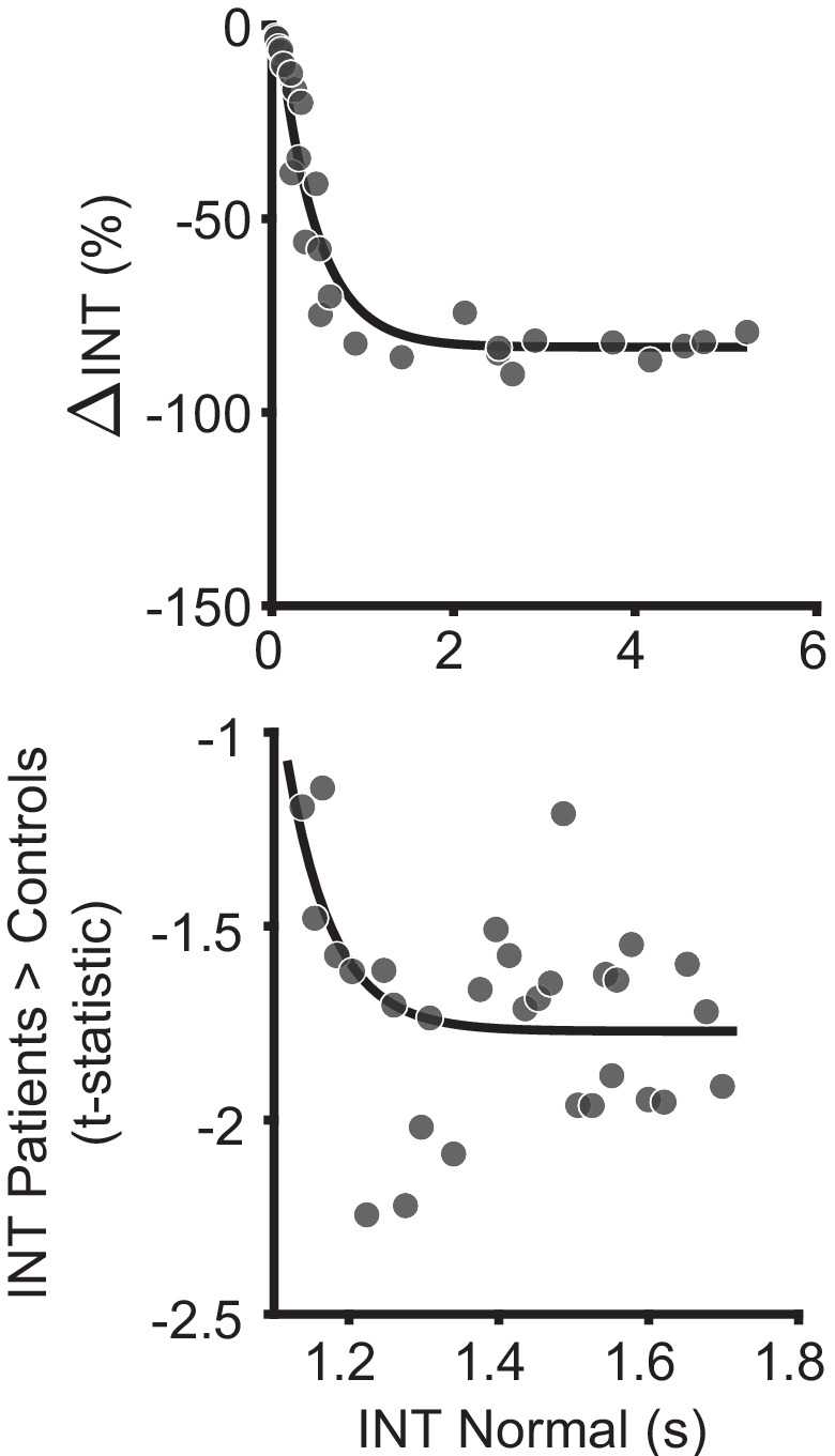
Figure 2—figure supplement 2.

An in silico simulation using a large-scale biophysical model suggested that the globally reduced intrinsic neural timescales (INT) observed in patients with schizophrenia could be neuronally implemented by globally reduced E/I ratio. Scatterplot showing the difference in INT values estimated for the reference (healthy) simulated scenario and the globally reduced excitatory-to-excitatory recurrent connection strength (reduced E/I ratio); each data point denotes 1 of the 29 nodes (top). Relative to the reference scenario, reduced excitatory-to-excitatory recurrent connection strength lead to a global reduction of INT values with the effect becoming more pronounced for regions with longer timescales in the healthy condition. The data are well described by an exponential model (R2 = 0.94). Scatterplot showin the t-statistic values from a regression model (M1exploratory) predicting INT group differences controlling for age, gender, mean framewise displacement, and data collection site, plotted as a function of the INT value in healthy subjects (determined from group-averaged INT map from HCP subjects to reduce circularity); each data point denotes the average of six parcels along the INT hierarchy for a similar number of data points as the biophysical model results (bottom). Similar to the biophysical model simulation, the data are better fit by an exponential model (Matlab fitnlm: F2,28 = 10.3, P = 0.003; BIC = 4.8), than a linear model (Matlab fitnlm: F1,29 = 1.4, P = 0.25; BIC = 9.3). Similar results are observed when no binning of the data is performed (exponential model: F2,185 = 8.3, P = 0.005; BIC = 366.4; linear model: F1,186 = 0.8, P = 0.38; BIC = 368.6). The findings are similar to a recent study that recapitulated the behavior of macaque monkeys on an evidence-varying decision-making task following intramuscular administration of a subanesthetic dose of ketamine by reducing the strength of the couplings to the excitatory population from the local excitatory population (wEE) by 1.75% to achieve a global reduction of E/I ratio (Cavanagh et al., 2019). This is consistent with the NMDA-receptor hypofunction hypothesis of schizophrenia (Corlett et al., 2011; Kehrer et al., 2008; Krystal et al., 2003; Lisman et al., 2008; Olney and Farber, 1995), and the use of ketamine as a model of schizophrenia (Becker et al., 2003; Corlett et al., 2011; Frohlich and Van Horn, 2014; Krystal et al., 1994). Furthermore, our biophysical model results support recent evidence of how global changes can preferentially affect higher order brain regions (Yang et al., 2016); and our in vivo results lend additional (although preliminary) evidence.

Hierarchical differences in INT between hallucinations and delusions

Our a priori hypothesis was that hallucinations and delusions are associated with alterations of INT at different hierarchical levels, leading to distinct changes in hierarchical gradients. To test this, we determined the unique variance associated with the effect of interindividual variability in hallucination and delusion severity on INT (M1primary; Materials and methods). As expected, severity of hallucinations and delusions in our sample were correlated (rs = 0.62, p<0.01) but had sufficient unique variance [(1 – R2)=0.62] to evaluate their independent contributions. The severity of these symptoms was uncorrelated with antipsychotic dose among the 109 patients with available data (chlorpromazine equivalents: both p>0.86), making medication an unlikely confound (Figure 3—figure supplement 1).

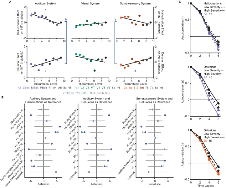
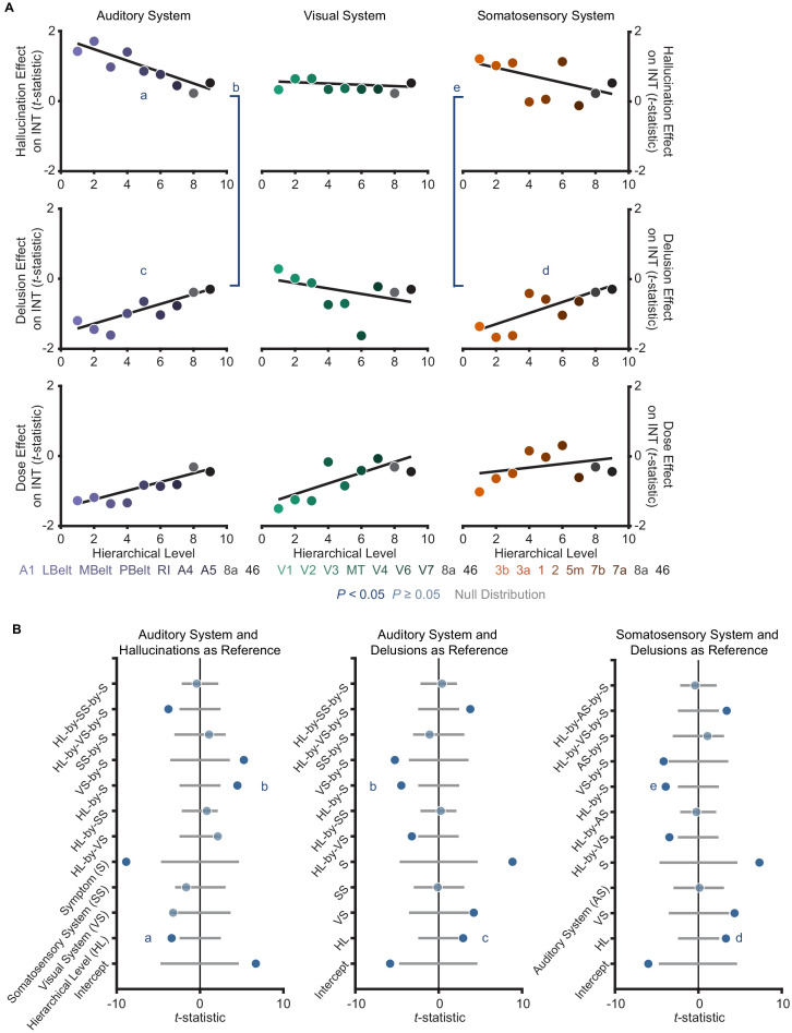
The model (M2; Materials and methods) that we used as a primary test of main effects and interactions of symptoms on hierarchical INT gradients—which also included interaction terms for each sensory system to account for between-system differences—was significant (omnibus F11,41 = 5.52, p<10−4). Critically, within this model we found hierarchical-gradient effects that differed significantly between hallucinations and delusions in the expected directions for 2/3 systems (auditory system, symptom-by-hierarchical-level interaction: t42 = 4.59 [95% bootstrap confidence interval: 3.39, 9.08], Cohen’s f2 = 1.00, Ppermutation = 0.001; visual: t42 = −2.06 [–6.19, 0.16], f2 = 0.11, Ppermutation = 0.083; and somatosensory: t42 = 3.50 [2.19, 7.35], f2 = 0.41, Ppermutation = 0.011; Figure 3). In the auditory system, this interaction was driven by significant hierarchical-gradient effects in opposite directions for hallucinations (hierarchical-level effect: t42 = −3.50 [–8.42,–2.24], f2 = 0.41, Ppermutation = 0.010) and delusions (hierarchical-level effect: t42 = 2.99 [1.18, 6.37], f2 = 0.27, Ppermutation = 0.025). In the somatosensory system, this effect was driven by a trend-level negative hierarchical-gradient effect for hallucinations (hierarchical-level effect: t42 = −2.35 [–5.91,–1.00], f2 = 0.15, Ppermutation = 0.056) and a significant positive hierarchical-gradient effect for delusions (hierarchical-level effect: t42 = 2.60 [1.43, 5.57], f2 = 0.19, Ppermutation = 0.042; Figure 3). In the visual system, hierarchical-gradient effects were not significant for either symptom (hallucination hierarchical-level effect: t42 = 0.90 [–1.06, 3.62], f2 = 0.00, Ppermutation = 0.466; delusion hierarchical-level effect: t42 = −2.01 [–6.01, 0.53], f2 = 0.11, Ppermutation = 0.087). We also found significant interactions with sensory system, indicating differences in the symptom interactions between the visual and the other systems (see statistics in Figure 3B), but these were not a priori tests (see also Discussion for issues of interpretability). Examining the significant symptom effects further, in the auditory system we observed that patients with high-severity hallucinations exhibited a numeric increase in INT at lower levels of the hierarchy relative to those with low-severity hallucinations, leading to a compression of the INT hierarchical gradient (Figure 3C); in contrast, in both the auditory and somatosensory systems, patients with high-severity delusions exhibited a numeric increase in INT at higher levels of the hierarchy relative to those with low-severity delusions, leading to a more pronounced INT hierarchical gradient.

Figure 3. A priori analyses show that hallucinations and delusions exhibit distinct hierarchical-gradient effects on intrinsic neural timescales in the auditory and somatosensory systems.

(A) Scatterplots showing t-statistic values from a regression model (M1primary; Materials and methods) including all seven symptoms and controlling for age, gender, mean framewise displacement, and sample-acquisition site for hallucination-severity (top) and delusion-severity (bottom) effects from parcels within the auditory (left), visual (middle), or somatosensory (right) systems plotted as a function of hierarchical level (using PFC-extended winning hierarchies; Figure 1). (B) Summary of results from a model (M2; Materials and methods) including symptom-severity effect (hallucinations or delusions), hierarchical level, sensory system (auditory, visual, or somatosensory), and their interactions. These results demonstrate: (1) in the auditory system, a significant difference in the relationship between hallucination severity and hierarchical level versus that for delusion severity and hierarchical level (b); (2) in the auditory system, significant hierarchical-gradient effects of hallucination severity (a) and delusion severity (c); (3) in the somatosensory system, a significant difference in the relationship between hallucination severity and hierarchical level versus that for delusion severity and hierarchical level (e); and (4) in the somatosensory system, a significant hierarchical-gradient effect of delusion severity (d). Note that different symptoms and systems were used as references (implicit variable) across three plots to show each of the relevant effects which were tested within a single model (M2). Null distributions were generated by randomly permuting symptom-severity scores across patients in M1primary (2.5th – 97.5th percentiles shown). (C) To illustrate the effects, the group-averaged autocorrelation functions were estimated from subjects with fMRI data acquired with the same repetition time (N = 10 for each group). High-severity patients were the 10 subjects with the highest residual symptom scores after regressing out all other symptoms; low-severity patients were the 10 subjects with the lowest residual symptom scores. The group-averaged autocorrelation functions are shown for high-severity (solid lines) and low-severity (dashed lines) hallucination patients from low and high levels of the auditory hierarchy (A1 and area 46, top). The group-averaged autocorrelation functions are also shown for high-severity and low-severity delusion patients from low and high levels of the auditory hierarchy (middle). Finally, the group-averaged autocorrelation functions are shown for high-severity and low-severity delusion patients from low and high levels of the somatosensory hierarchy (area 3b and area 46, bottom). These plots depict a compression of the auditory hierarchical gradient in high-severity hallucination patients and, instead, an expansion of both the auditory and somatosensory hierarchical gradients in high-severity delusion patients. LBelt, lateral belt; MBelt, medial belt; PBelt, parabelt; RI, retroinsular cortex; MT, middle temproal area.

Figure 3—source data 1. Data and code to reproduce Figure 3.

Figure 3.

Figure 3—figure supplement 1. Controlling for antipsychotic dose does not change the distinct hierarchical-gradient effects of hallucination and delusion severity in the auditory and somatosensory systems.

Figure 3—figure supplement 1.

(A) Scatterplots showing t-statistic values from a regression model including all seven symptoms and and controlling for age, gender, mean framewise displacement, and sample-acquisition site for hallucination-severity (top), delusion-severity (middle), and dose (bottom) effects from parcels within the auditory (left), visual (middle), or somatosensory (right) hierarchies plotted as a function of hierarchical level. (B) Summary of results from a model including symptom-severity effect (hallucination or delusion), hierarchical level, sensory system (auditory, visual, or somatosensory), and their interactions. We repeated the main analysis comparing the effects of hallucination and delusion severity when including antipsychotic dose as a covariate. Only 109 of the 127 patients had available data for chlorpromazine equivalents. All results were similar to those reported in the Main Text where the effect of medication dose was not controlled for. The model explaining symptom effects and their differences by hierarchical-level and their interaction by symptoms and sensory system was significant (omnibus F11,41 = 22.4, p<10−13). Critically, we found significant hierarchical-gradient effects that differed between hallucinations and delusions (auditory system symptom-by-hierarchical-level interaction: t42 = 4.48, Ppermutation = 0.002; visual: t42 = −0.88, Ppermutation = 0.560; and somatosensory: t42 = 3.92, Ppermutation = 0.001). In the auditory system, this effect was driven by significant hierarchical-gradient effects in opposite directions for hallucinations (hierarchical-level effect: t42 = −3.40, Ppermutation = 0.013) and delusions (hierarchical-level effect: t42 = 2.94, Ppermutation = 0.028). In the somatosensory system, this effect was driven by a trend-level negative hierarchical-gradient effect for hallucinations (hierarchical-level effect: t42 = −2.25, Ppermutation = 0.064) and a significant positive hierarchical-gradient effect for delusions (hierarchical-level effect: t42 = 3.30, Ppermutation = 0.017).
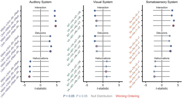
Figure 3—figure supplement 2. Distinct hierarchical-gradient effects of hallucinations and delusions are robust to the choice of sensory hierarchies.

Figure 3—figure supplement 2.

Summary of results from a model including symptom-severity effect (hallucination or delusion), hierarchical level, sensory system (auditory, visual, or somatosensory), and their interactions for each of the four sensory cortex hierarchies tested for the auditory (left), visual (middle), and somatosensory (right) systems during Selection and Multimodal Validation of Neural Hierarchies. For the auditory system, A1 was always the lowest order region while A4 and A5 were always the 6th and 7th level regions. For the visual system, V1, V2, and V3 were always the three lowest order regions. For the somatosensory system, 5m, 7b, and 7a were always the 5th, 6th, and 7th level regions, respectively. For all three sensory systems, areas 8A and 46 were always the two highest order regions. The winning orderings from the selection process (Figure 1) are outlined in red. Hierarchical-gradient effects of hallucinations (Hallucinations; bottom four), delusions (Delusions; middle four) and their interaction (Interaction; top four) are shown (from M2). For this analysis, one sensory system hierarchical-ordering was tested while the other two sensory system hierarchical-orderings were chosen to be the winning orderings. Null distributions were generated by randomly permuting symptom-severity scores across patients in M1primary (2.5th – 97.5th percentiles shown).
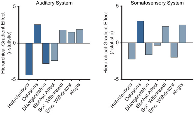
Figure 3—figure supplement 3. Only positive symptoms show hierarchical-gradient effects in the auditory system and only delusions show a hierarchical-gradient effect in the somatosensory system.

Figure 3—figure supplement 3.

Bar graphs showing t-statistic values from a model including symptom-severity effect, heirarchical level, and their interaction (each symptom and sensory system was tested independently) for the hierarchical-gradient effect of each symptom in the auditory and somatosensory systems. In the auditory system, all three positive symptoms (including conceptual disorganization) show significant effects while none of the negative symptoms show significant effects. In the somatosensory system, only delusions show a significant effect, although hallucinations show the strongest negative effect. These results reflect some level of selectivity that should be examined in more detail in future studies. It is important to note that the perceptual-inference model of psychosis does not require these effects to be specific to hallucinations and delusions. Furthermore, an effect of conceptual disorganization—a positive symptom that unlike negative symptoms tends to correlate with hallucinations and delusions—may suggest extensions of the model to account for additional phenomena.
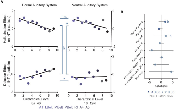
Figure 3—figure supplement 4. Comparison of hierarchical-gradient effects in the dorsal and ventral auditory systems.

Figure 3—figure supplement 4.

(A) Scatterplots showing t-statistic values for hallucination-severity (top) or delusion-severity (bottom) effects from parcels within the dorsal auditory (left; areas 8a and 46) and ventral auditory (right; areas 10 and 12vl) hierarchies plotted as a function of hierarchical level. (B) Summary of results from a model including symptom-severity effect (hallucination or delusion), hierarchical level, sensory system (dorsal or ventral auditory system), and their interactions. These results demonstrate: (1) there is no significant difference in the hierarchical-gradient effect of hallucination severity between the dorsal and ventral auditory systems; (2) there is no significant difference in the relationship between hallucination severity and hierarchical level versus that for delusion severity and hierarchical level in the ventral auditory system (a); (3) there is a significant difference in the hierarchical-gradient effect of delusion severity between the dorsal and ventral auditory systems. These findings add to previous evidence suggesting that the dorsolateral prefrontal cortex (area 46; included in the dorsal but not the ventral auditory system) may play a key role in delusions.
Figure 3—figure supplement 5. Distinct hierarchical-gradient effects of hallucination and delusion severity in the auditory system are observed when using an anatomically agnostic definition of the hierarchy.

Figure 3—figure supplement 5.

Scatterplots showing t-statistic values from a regressino model (M1primary; Materials and methods) including all seven symptoms and controlling for age, gender, mean framewise displacement, and sample-acquisition site for hallucination-severity (left) or delusion-severity (right) effects from bins consisting of the average from 60 voxels within the nine auditory system parcels (600 voxels total) as a function of hierarchical level. Voxels were first ranked using their corresponding intrinsic neural timescale (INT) value in the HCP dataset group-averaged INT map and then averaged within 10 equally spaced bins with increasing hierarchical level (i.e. the lowest level has the shortest average INT in the HCP dataset while the higheset hierarchical level has the longest average INT). Similar to our main analysis results, we observe a significant symptom-by-hierarchical-level interaction, a significant negative hierarchical-gradient effect for hallucinations, and a significant positive hierarchical-gradient effect for delusions.

To correct for multiple comparisons, we carried out a family-wise permutation test determining the probability of spuriously obtaining the set of significant effects we observed in support of our hypothesis. Based on the chance level of jointly observing negative hierarchical-gradient effects of hallucination severity in at least 1/3 systems, and positive hierarchical-gradient effects of delusion severity in at least 2/3 systems, and interaction effects of hierarchy-by-symptom in the expected direction in at least 2/3 systems, this analysis suggested that the observed set of results was statistically above chance (set-level Ppermutation = 0.014). Furthermore, based on the chance level of observing a significant negative hierarchical-gradient effect for hallucinations, and a significant positive hierarchical-gradient effect for delusions, and a significant symptom-by-hierarchical-level interaction (i.e. all three effects in one system), the observed set of results in the auditory system was also statistically above chance (set-level Ppermutation = 0.043).

To rule out an effect of our approach for selecting hierarchical orderings on these results, we tested these symptom effects for each of the four different sensory cortex hierarchical orderings considered a priori candidates for each sensory system. Results were generally consistent across the different hierarchical orderings (Figure 3—figure supplement 2), particularly in the auditory system. A family-wise permutation test similar to the one above, but including all four orderings per system (12 total orderings), showed that the observed set of results was statistically above chance for all systems (set-level Ppermutation = 0.002) and for the auditory system alone (set-level Ppermutation = 0.001).

Post-hoc analysis of the specificity of INT hierarchical-gradient effects

In a post-hoc analysis, we then investigated the specificity of these hierarchical-gradient effects to the positive psychotic symptoms under investigation. To this end, we determined hierarchical-gradient effects individually for each symptom in the auditory and somatosensory systems using a model including symptom-severity effect (only one symptom), hierarchical level, sensory system (auditory, visual, and somatosensory), and their interactions. In the auditory system, conceptual disorganization was the only symptom—other than hallucinations and delusions—that showed a significant effect (hierarchical-level effect: t21 = −2.80 [–5.31,–1.10], f2 = 0.60, Ppermutation = 0.036; Figure 3—figure supplement 3). But this effect was weaker than that for hallucinations (hierarchical-level effect: t21 = −4.38 [–10.31,–3.28], f2 = 10.57, Ppermutation = 0.005; Figure 3—figure supplement 3). These results thus suggest some specificity to positive symptoms, which conceptual disorganization is classically defined as (Association AP, 2013; VandenBos, 2007) (but see van der Gaag et al., 2006), consistent with a stronger correlation of conceptual disorganization with positive symptoms (average rs = 0.48) versus negative symptoms (average rs = 0.23) in our sample. Indeed, a permutation test comparing the average strength of hierarchical-gradient effects (i.e. mean absolute-value of t-statistics) for positive versus negative symptoms (i.e. blunted affect, social withdrawal, emotional withdrawal, and alogia) showed the effects of positive symptoms to be significantly larger than the effects of negative symptoms (Ppermutation = 0.043) in the auditory system. In the somatosensory system, no symptoms other than delusions showed a significant hierarchical-gradient effect. Hallucinations, however, showed the strongest negative effect (hierarchical-level effect: t21 = −2.23 [–6.79,–1.75], f2 = 0.31, Ppermutation = 0.079; Figure 3—figure supplement 3).

Thus, although the hierarchical-gradient effects were not unique to the two symptoms under investigation—which is not required under perceptual-inference models of psychosis and which could suggest model extensions to account for additional phenomena—these effects were strongest for, and relatively specific to, positive symptoms.

Altered E/I ratio as a potential biological mechanism

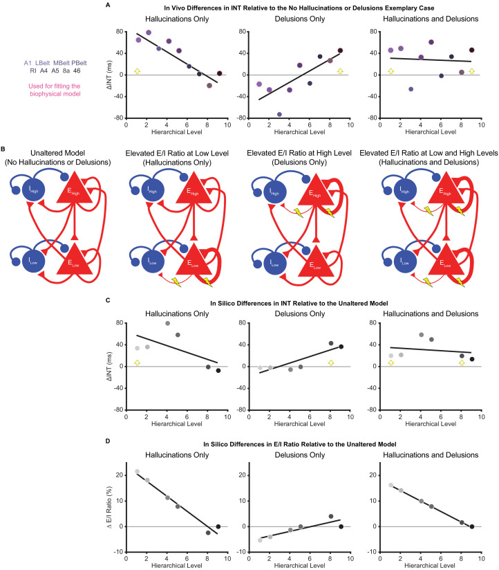
To explore candidate biological mechanisms for the effects we observed in vivo, we leveraged a large-scale biophysical model previously shown to capture intrinsic timescale hierarchies (Chaudhuri et al., 2015). This model depicts the macaque cortex using 29 recurrently connected nodes, with connection strengths based on macaque tract-tracing studies (Figure 4B). Given growing evidence for E/I imbalance in schizophrenia (Foss-Feig et al., 2017; Jardri et al., 2016) and the hypothesized local increases of INT, we fit the biophysical model to our data to explore whether our results could be driven by local increases in E/I ratio. These E/I ratio changes were modeled as a triangle function where a local maximum exhibited a peak E/I ratio increase and other nodes had E/I ratio changes that decreased linearly as a function of absolute distance in hierarchical levels from the peak. This function was described by three free parameters: (i) the hierarchical level of the peak E/I ratio increase, (ii) the magnitude of the E/I ratio increase at the peak, and (iii) the magnitude of the E/I ratio change at the minimum (i.e. at the hierarchical level furthest from the peak).

Figure 4. Hallucination and delusion effects on intrinsic neural timescales are recapitulated by elevated excitation-inhibition ratios at different hierarchical levels.

Figure 4.

(A) Scatterplots showing the difference in estimated intrinsic nerual timescale (INT) values between the three exemplary cases capturing extreme symptom profiles (‘hallucinations only’, ‘delusions only’, and ‘hallucinations and delusions’) with respect to the ‘no hallucinations or delusions’ exemplary case (in vivo ΔINT; fitted parcel-wise data from M1primary). The parcel data used for fitting the biophysical model are outlined in pink. Yellow arrowheads denote the hypothesized hierarchical level of the maximum excitation-inhibition (E/I) ratio increase. (B) Simplified schematic of a large-scale biophysical model of the macaque cortex and its variants (Materials and methods). The model consists of 29 nodes with local excitatory (red triangles) and inhibitory (blue circles) pools of neurons; only two nodes—high (top) and low (bottom) hierarchical levels—are shown for illustrative purposes. These nodes have both local (recurrent) and long-range (across-node) connections. Lightning bolts mark theoretical perturbations to the model. Thicker or thinner lines with respect to the reference scenario reflect increased or decreased connection strengths, respectively. Note that E/I ratio can be increased either by increasing local excitatory-to-excitatory connection strength or by decreasing local excitatory-to-inhibitory connection strength, but these scenarios were modeled individually. (C) Scatterplots showing the difference in simulated INT values between each of the three pathological biophysical models (‘elevated E/I ratio at low level’, ‘elevated E/I ratio at high level’, and ‘elevated E/I ratio at low and high levels’) with respect to the reference biophysical model (‘unaltered model’) using the best-fitting E/I ratio parameters (in silico ΔINT). By allowing the E/I ratios to vary, the biophysical model can recapitulate the in vivo INT changes with a negative (compressed) hierarchical-gradient effect for hallucinations, a positive (expanded) hierarchical-gradient effect for delusions, and an overall INT increase (without a manifest hierarchical-gradient effect) for the combined case of hallucinations and delusions. The visual hierarchy (V1, V2, V4, MT, 8l, and 46d) was used for these simulations given the lack of tract-tracing data for the auditory cortex—other levels of the visual cortex are omitted for the same reason—but the qualitative pattern of results applies to hierarchical-gradient effects in any given sensory system. Yellow arrowheads denote the hierarchical level of the maximum E/I ratio increase. (D) Scatterplots showing the fitted changes to E/I ratios for the pathological biophysical models: the in vivo INT changes associated with hallucinations can be recapitulated by elevated E/I ratio at the lowest hierarchical level and those associated with delusions by elevated E/I ratio (of smaller magnitude) at the highest hierarchical level, with the addition of these two alterations capturing the changes in patients with both hallucinations and delusions. Note that E/I ratio in level 9 of the hierarchy was fixed to its value in the unaltered model to prevent model instability (Materials and methods). LBelt, lateral belt; MBelt, medial belt; PBelt, parabelt; RI, retroinsular cortex.

Figure 4—source data 1. Data and code to reproduce Figure 4.

To fit the biophysical model, we first estimated in vivo data for ‘exemplary cases’ using regression fits from M1primary (Materials and methods) in the auditory system—the system that showed the strongest effects in vivo. The regression fits allowed us to estimate INT values at each level of the hierarchy for exemplary cases representative of extreme symptom profiles (while controlling for variability in other factors). INT values for the auditory hierarchy were estimated for four exemplary cases: (1) no hallucinations or delusions (fitted INT values from M1primary with minimum scores of 0 for both symptoms); (2) hallucinations only (maximum score of 5 for hallucinations and score of 0 for delusions); (3) delusions only (scores of 0 for hallucinations and 5 for delusions); (4) hallucinations and delusions (scores of 5 for both symptoms). For all exemplary cases, the severity of other symptoms and the values of covariates were set to the average values from all patients. Changes of INT for exemplary cases 2–4 were determined as the difference in INT relative to the ‘no hallucinations or delusions’ case (in vivo ΔINT; Figure 4A). We modeled the in vivo ΔINT in the auditory system using the macaque visual system as a model hierarchy with realistic biological constraints due to the lack of tract-tracing data for the auditory system; note that sensory system and species differences limit our ability to derive precise quantitative conclusions from the modeling results but still afford qualitative insights. We specifically used the six biophysical-model nodes that directly corresponded to levels of our visual hierarchy and for which tract-tracing data were available: V1 (level 1), V2 (level 2), V4 (level 4), MT (level 5), 8l (level 8), and 46d (level 9). Model-derived in silico ΔINT (Figure 4C) were calculated for each node as the difference in INT from the biophysical model with no perturbations (‘unaltered model’) and the INT from the best-fitting biophysical model for which the values of the parameters controlling the E/I ratio provided the closest approximation to the in vivo ΔINT across exemplary cases (in the six corresponding parcels of our auditory hierarchy: A1 [level 1], LBelt [level 2], PBelt [level 4], RI [level 5], 8a [level 8], and 46 [level 9]). Specifically, two sets of the 3 E/I ratio parameters were jointly fitted to exemplary cases 2–4, one for hallucinations and one for delusions, with the combined effect of hallucinations and delusions resulting from the sum of the E/I ratio changes for the two individual symptoms.

In silico results using the best-fitting parameters were able to recapitulate the INT effects of hallucinations and delusions (compression versus expansion of the INT hierarchical gradient, respectively) via local increases in E/I ratio at low or high levels of the hierarchy, respectively. Specifically, the best-fitting levels of the peak increase in local E/I ratio were levels 1 and 8 for hallucinations and delusions, respectively (Figure 4D). Interestingly, given the relatively greater strength of both recurrent and long-range connections at higher levels that is built into the biophysical model, the required peak E/I ratio increase to achieve the observed changes of INT was considerably smaller for the delusion-related alteration at level 8 (ΔE/I = 4.02%) compared to the hallucination-related alteration at level 1 (ΔE/I = 21.61%). Also, the in silico ΔINT based on the summed E/I ratio alterations for individual symptoms closely approximated the combined case of hallucinations and delusions (exemplary case 4), which consisted of a general increase in INT with no clear change in the hierarchical gradient. This suggests that additivity of the local symptom-specific alterations could explain symptom co-occurrence.

In a follow-up analysis, we further explored our in vivo data for evidence of the additive effect of hallucinations and delusions, focusing on the auditory system. We first compared the average INT across all auditory system parcels between patients with both high hallucination and delusion scores (i.e. raw average data from subjects with a score of 5 for both symptoms; N = 11) and patients with neither hallucinations nor delusions (i.e. subjects with a score of 0 for both symptoms; N = 18). Here, we observed significantly higher average INT in patients with high-severity hallucinations and delusions (t27 = 1.84, p=0.038; one-tailed two-sample t-test). Second, we fit a linear model predicting auditory parcel INT as a function of hierarchical level, allowing separate intercepts for each of the two groups, and an interaction between hierarchical level and group. Here, we found that the intercept was indeed higher for patients with high-severity hallucinations and delusions compared to patients with neither symptom (t257 = 2.04, p=0.043). Furthermore, we found no difference in the hierarchical gradients between these groups, with a non-significant hierarchical-level-by-group interaction (t257 = 0.65, p=0.519). Although preliminary, these results supply some support for the notion of additive hierarchical alterations in psychosis.

Control analyses examining alternative definitions of auditory hierarchies

Given that the hierarchical-gradient effects supporting our initial hypotheses were clearest in the auditory system—a system thought to comprise dual processing streams—we considered the impact of alternative definitions of the auditory hierarchy on our results.

Diverging auditory streams with downstream projections to dorsal (areas 8a and 46) versus ventral (areas 10 and 12vl) PFC have been described (Kaas and Hackett, 2000). We thus conducted an ordering-selection analysis for the ventral stream, like that presented above for the dorsal stream (Selection and Multimodal Validation of Neural Hierarchies). Within the ventral stream, area 12vl was better predicted as the highest hierarchical level, and the winning ordering explained more variance than chance based on a null distribution of random orderings (ventral auditory system: Ppermutation < 10−4; Figure 1—figure supplement 6A). Furthermore, similar to the dorsal stream, hierarchical level in the ventral stream correlated with INT (in-sample: rs = 0.87, p=0.005, Ppermutation = 0.003; out-of-sample: rs = 0.80, p=0.014; Figure 1—figure supplement 6B) and expression of granular layer IV genes (rs = −0.91, p=0.001). Given these dual auditory streams and their corresponding validated hierarchies, we explored potential differences in hierarchical-gradient effects of hallucination and delusion severity for the dorsal versus ventral streams. The model explaining symptom effects and their differences by hierarchical level and their interaction by symptoms and auditory stream was significant (omnibus F7,27 = 8.60, p<10−4). We further found a significant difference in the symptom-by-hierarchical-level effects between the dorsal and ventral auditory streams (symptom-by-hierarchical-level-by-processing-stream interaction: t28 = 1.75 [1.02, 3.90], f2 = 0.12, Ppermutation = 0.005; Figure 3—figure supplement 4). In the ventral stream, we did not find a significant difference in the hierarchical-gradient effects between hallucinations and delusions (symptom-by-hierarchical-level interaction: t28 = 2.01 [–0.37, 6.43], f2 = 0.17, Ppermutation = 0.159; Figure 3—figure supplement 4); we found a trend-level hierarchical-gradient effect of hallucination severity (hierarchical-level effect: t28 = −2.56 [–7.04,–0.73], f2 = 0.31, Ppermutation = 0.098; Figure 3—figure supplement 4) and no effect of delusion severity (hierarchical-level effect: t28 = 0.28 [–2.42, 3.36], f2 = 0.00, Ppermutation = 0.445; Figure 3—figure supplement 4). Interestingly, comparing the dorsal and ventral auditory streams, we observed a significant difference in the hierarchical-gradient effect of delusion severity (hierarchical-level-by-processing-stream interaction: t28 = 1.89 [1.39,3.49], f2 = 0.15, Ppermutation = 0.003; Figure 3—figure supplement 4). These results thus support the involvement of dorsolateral PFC in delusions, consistent with prior work (Corlett et al., 2007).

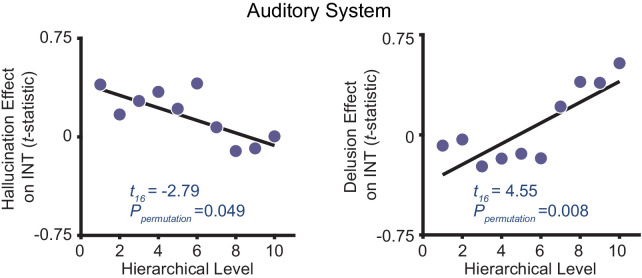
As an additional control for the uncertainty in defining the auditory hierarchy, we also adopted an anatomically agnostic, data-driven approach. First, the symptom effects (M1primary) were estimated for each voxel within the nine auditory-system parcels (600 voxels total). Second, each voxel was ranked based on its INT value in the average INT map from the HCP dataset. Third, 10 equally spaced bins along the INT ranking (60 voxels per bin) were created, which comprised the levels of the data-driven hierarchy, and the voxelwise t-statistics (from M1primary) were averaged per bin. Similar to the main analysis (Figure 3), a model that included main effects and interactions of symptoms on the hierarchical INT gradient was significant (omnibus F4,15 = 10.20, p=0.001). Within this model, we found hierarchical-gradient effects that differed significantly between hallucinations and delusions (symptom-by-hierarchical-level interaction: t16 = 5.19 [0.48 23.26], f2 = 10.12, Ppermutation = 0.003; Figure 3—figure supplement 5). This interaction was driven by significant hierarchical-gradient effects in opposite directions for hallucinations (hierarchical-level effect: t16 = −2.79 [–16.72, 2.40], f2 = 0.25, Ppermutation = 0.049; Figure 3—figure supplement 5) and delusions (hierarchical-level effect: t16 = 4.55, [1.04, 19.76], f2 = 4.04, Ppermutation = 0.008; Figure 3—figure supplement 5).

Discussion

Using a recently developed method for measuring neural timescales from resting-state fMRI data, we set out to test the hypothesis that hallucinations and delusions are associated with dysfunctions at different levels of neural hierarchies. Using established structural indices of hierarchy (myelin and cortical thickness) and INT (a functional index of hierarchy) in independent samples, we first validated extended sensory hierarchies for the auditory, visual, and somatosensory systems that captured substantial variability in the hierarchical MRI indices. After further showing excellent reliability of the INT measure, in exploratory analyses, we showed for the first time that patients with schizophrenia have globally reduced INT. Most importantly, our primary analyses comparing INT effects for hallucinations versus delusions in the validated hierarchies demonstrated that these symptoms are associated with distinct changes of the hierarchical gradients in the auditory and somatosensory systems, an effect we failed to observe in the visual system.

Hierarchical models of perceptual inference posit that perceptions are shaped by prior beliefs (Dayan et al., 1995; Friston and Kiebel, 2009; Kiebel, 2009; Lee and Mumford, 2003; Rao and Ballard, 1999) through reciprocal message passing across different levels of sensory hierarchies, an architecture that mirrors the known anatomy of sensory systems (Felleman and Van Essen, 1991; Glasser et al., 2016; Kaas and Hackett, 2000; Markov et al., 2014a; Van Essen et al., 1992; Young, 1993). In this scheme, higher levels of the neural hierarchy are thought to represent increasingly abstract belief states that evolve at slower timescales (Kiebel, 2009). For instance, during speech perception, the hierarchical structure of linguistic units can be parsed such that lower levels of auditory processing encode syllable information at faster timescales while higher levels encode sentence information at slower timescales (Ding et al., 2016). An emerging body of work in psychosis has linked hallucinations to preferential biases toward prior beliefs in low-level inferences during detection or estimation of stimulus features (Cassidy et al., 2018; Davies et al., 2018; Powers et al., 2017; Teufel et al., 2015) and delusions to preferential biases toward prior beliefs in higher-level inferences about more abstract, hidden states (Baker et al., 2019). The observed biases toward prior beliefs in past behavioral work can be framed as primacy biases (Baker et al., 2019), where past information is weighted more heavily during the inferential process, or equivalently, where information is integrated over longer timescales (Glaze et al., 2015). Temporal integration is at the core of the neural implementation of perceptual inference (Mazurek et al., 2003) and is thought to depend crucially on recurrent network activity (Chaudhuri et al., 2015; Mante et al., 2013). Thus, a plausible neuronal implementation of primacy biases at a given level of the hierarchy would be through increases in the strength of recurrent excitation or decreases in the strength of recurrent inhibition (i.e. elevated E/I ratio) leading to relative increases in neural timescales.

Here, we observed changes in neural timescales across levels of neural hierarchies that differed between hallucinations and delusions, an effect that was most evident in the auditory hierarchy. Patients with more severe hallucinations exhibited a less pronounced INT hierarchical gradient, consistent with increased timescales at lower levels compared to those with less severe hallucinations; those with more severe delusions instead exhibited a more pronounced INT hierarchical gradient, consistent with increased timescales at higher levels compared to those with less severe delusions (Figure 3C). We further recapitulated these findings by respectively elevating E/I ratios at low or high hierarchical levels of a large-scale biophysical model (Chaudhuri et al., 2015). These E/I ratio elevations could, in principle, result from alterations in NMDA or dopamine activation at these levels and are thus plausible under widely supported glutamatergic and dopaminergic theories of psychosis (Brunel and Wang, 2001; Corlett et al., 2009; Corlett et al., 2011; Durstewitz and Seamans, 2002; Jardri et al., 2016; Javitt et al., 2012; Weinstein et al., 2017). These results thus demonstrate distinct hierarchical alterations for hallucinations and delusions that are generally consistent with our hypothesized hierarchical framework, where distinct hierarchical alterations provide symptom-specific pathways that together may explain symptom co-occurrence, thus providing a candidate biological mechanism for the psychotic syndrome.

Such hierarchical alterations may also fit well with the phenomenological timescale of these symptoms. Clinical observation indicates that hallucinations—like rapidly changing sensory events—change transiently and intermittently over seconds or minutes, while delusions—like slowly changing ‘conceptual’ beliefs—evolve more slowly over days or months, but their average severities over a given period typically evolve in parallel. These clinical features are consistent with a hierarchical structure of nested timescales (Kiebel et al., 2008). While our findings generally support this notion, computational work explicitly laying out the proposed model in the context of inferential alterations in psychosis and empirical confirmations are warranted. One outstanding question is how the delusion-related alterations in neural timescales we observed—which may predominate in high levels of the hierarchy but manifest as changes on the order of seconds—might drive delusions evolving over much longer timescales. One possible explanation is that, while delusion maintenance may involve long-term memory processes, the underlying mechanism initiating delusions transpires more rapidly and disrupts inferences at timescales on the order of seconds, consistent with prior work (Baker et al., 2019). Since encoded memories likely reflect inferences summarizing information at a given timepoint (Shadlen and Shohamy, 2016), high-level inferential biases at shorter timescales may be sufficient to shape long-term conceptual memories in a way that further propagates biases over long time-periods, particularly under primacy biases that decrease the relative influence of newer information. Although less critical, it is also worth noting that INT reflects differences in resting circuit dynamics, the timescale of which is likely to be substantially magnified when these circuits are engaged (Chaudhuri et al., 2015; Hasson et al., 2008).

Our opposing findings for diagnosis (globally reduced INT) and symptom severity (focally increased INT) may be reconciled within pathophysiological models of psychosis which posit a key role for compensatory processes in schizophrenia. Hallucinations and delusions have been proposed to represent a temporary state of the illness that results from a failed attempt to compensate for a trait-like, baseline deficit (Adams et al., 2013; Moutoussis et al., 2011). Relatedly, long-standing circuit-level theories have suggested that psychosis-related increases in striatal dopamine transmission are secondary to a primary cortical deficit (Weinberger, 1987). In particular, previous frameworks suggest that psychotic states are associated with excessive prior biases in inferential processes arising as an overcompensation for a baseline trait consisting of the opposite bias (Adams et al., 2013; Horga and Abi-Dargham, 2019). From a biophysical-modeling standpoint, the trait-like baseline deficit in schizophrenia could consist of globally reduced E/I ratio (for instance, arising from NMDA-receptor hypofunction of excitatory neurons Cavanagh et al., 2019), which behaviorally would translate into general recency biases. In contrast, a failed compensatory mechanism could result in local increases in E/I ratio at different levels leading to distinct primacy biases and psychotic symptoms (Lam et al., 2017). While speculative, the compensatory changes could arise from dopaminergic alterations that effectively increase E/I ratio by preferentially boosting NMDA-receptor function of excitatory neurons (or other changes dampening NMDA-receptor function of inhibitory neurons) (Brunel and Wang, 2001).

Our finding of preferential involvement of the auditory system for hallucinations is not surprising, given that in schizophrenia this symptom tends to predominate in the auditory modality despite also presenting in other modalities (Lim et al., 2016; Waters and Fernyhough, 2017); auditory-cortex abnormalities in schizophrenia are also well established (Javitt and Sweet, 2015). Our finding of somatosensory system involvement for delusions is also consistent with previous work on delusions of passivity (Brüne et al., 2008; Spence et al., 1997) and deficits in sensory attenuation via motor predictions in schizophrenia (Shergill et al., 2005; Shergill et al., 2014). However, despite our failure to detect differential alterations in the visual system, substantial evidence also suggests visual-cortex abnormalities in schizophrenia (Butler et al., 2008; Cavuş et al., 2012; Dorph-Petersen et al., 2007). And evidence from subclinical populations suggests symptom-specific hierarchical alterations in visual tasks (Davies et al., 2018). Furthermore, the general differences in INT values between sensory systems (Figure 1), while potentially relevant to psychosis in and of themselves, could imply differential sensitivity in our analyses across sensory domains. Our null findings in the visual system are also qualified by the poorer correspondence between levels of the visual hierarchy and hierarchical MRI indices (not only for INT but also surprisingly for the structural indices) compared to the other systems (Figure 1). This suggests the need for further investigation into the sensitivity of available MRI measures of hierarchy to uncover the underlying gradients within the visual cortex.

Previous empirical work using structural (Bassett et al., 2008) and functional measures (Dondé et al., 2019; Leitman et al., 2010; Yang et al., 2016), suggests hierarchical alterations in schizophrenia. This work, however, did not evaluate hierarchical differences between symptoms and used measures that differ fundamentally from INT. In exploratory analyses testing diagnostic effects, we found global INT reductions in schizophrenia but no clear shifts in the hierarchical INT gradients (see Figure 2—figure supplement 2 for initial evidence of an exponential effect). We used the same approach as a previous study measuring INT in individuals with autism, which reported decreased INT in the visual cortex (and increased INT in the caudate) (Watanabe et al., 2019). Consistent with our interpretation, this INT phenotype was linked to other data in autism supporting excessive weighting of sensory evidence (Gollo, 2019; Lawson et al., 2017)—akin to a decreased primacy bias (i.e. a recency bias).

Some limitations are worth discussing. Because 93% of the patients (with available medication data) were taking antipsychotics, we cannot definitively rule out medication confounds, particularly on diagnosis effects. However, we observed similar effects when controlling for dose in our main analysis, no correlations between dose and symptoms, and did not expect differential neural effects on hallucinations versus delusions (Figure 3—figure supplement 1); future studies should elucidate medication effects on INT. Additionally, our study was limited to investigating the effects of global severity of hallucinations and delusions and could not resolve effects of symptom subtype or content, since detailed assessments were only available in a small subset of our patients. Larger studies with more detailed assessments are needed to tease out these potential effects.

In conclusion, we have presented evidence for distinct hierarchical alterations in neural timescales as a function of hallucination and delusion severity, lending initial neural support for hierarchical views of psychosis. Additionally, our work suggests that INT (Watanabe et al., 2019) provides a reliable and interpretable measure of neural function with the potential to elucidate hierarchical alterations and dysfunctions in circuit dynamics in schizophrenia and other neuropsychiatric disorders.

Materials and methods

Human Connectome Project dataset

T1w/T2w and cortical thickenss maps, and resting-state fMRI data were obtained for a subset of 100 unrelated young and healthy subjects from the Human Connectome Project (HCP) WU-Minn Consortium (Van Essen et al., 2013). The first fMRI run (single-shot EPI with left-to-right phase encoding direction) from the first fMRI session was obtained for each subject during eyes-open-on-fixation with the following scanning parameters: repetition time (TR) = 720 ms; spatial resolution = 2 × 2×2 mm; timepoints = 1200. High-resolution (0.7 mm isotropic voxels) T1w and T2w anatomical images were also acquired. Details regarding subject recruitment and MRI data acquisition have been previously reported (Smith et al., 2013; Van Essen et al., 2012). Preprocessing of the HCP data was performed using the HCP minimal preprocessing pipeline (Glasser et al., 2013). The preprocessed fMRI data were then used for the estimation of INT maps in 32k Conte69 mesh surface space and MNI152_ICBM2009a_nlin volume space with native spatial resolution. The T1w/T2w (myelin) maps (Glasser and Van Essen, 2011) in 32k Conte69 mesh surface space were used to compare functional (INT) and structural (T1w/T2w) measures of hierarchy.

Schizophrenia combined dataset

T1w images and resting-state fMRI data were obtained for 331 healthy control subjects and 254 patients diagnosed with either schizophrenia (N = 241) or schizoaffective disorder (N = 13) from four publicly available datasets. Three of these datasets were from the SchizConnect repository [BrainGluSchi (Bustillo et al., 2016), COBRE (Aine et al., 2017; Çetin et al., 2014), and NMorphCH (Alpert et al., 2016)] and one was from the OpenfMRI repository (UCLA; Poldrack et al., 2016). Data that survived a quality-control check (~95%) and motion-censoring check (~64%) included 140 patients and 225 controls. The quality control check consisted of visual inspection of the spatially normalized images. The motion-censoring check consisted of determining if there were sufficient degrees of freedom after motion censoring to perform nuisance variable regression. A subset of 158 controls was then selected that matched patients on gender and age. To minimize scanner- and site-related differences, we excluded subjects if the signal-to-noise ratio (SNR) was less than 100 for any of the standard regions-of-interest (Power et al., 2011). The final sample after quality-control checks consisted of 127 patients and 152 age- and gender-matched controls (Table 1).

The fMRI data were collected for each subject during eyes-open-on-fixation with the following scanning parameters: TR = 2000 ms (except for NMorphCH, where TR = 2200 ms); timepoints (BrainGluSchi/COBRE/NMorphCH/UCLA) = 165/150/323/152; spatial resolution (mm) = 3.5×3.5×3.5/3.5×3.5×3.5/4×4×4/3×3×3. Data were preprocessed using the AFNI afni_proc.py function (Cox, 1996). The following steps were performed: (1) removal of the first five volumes with the 3dTcat function; (2) slice-timing correction; (3) motion correction; (4) 12-parameter affine registration of the fMRI images to the T1w image; (5) spatial normalization of fMRI images to MNI152_ICBM2009a_nlin volume space using nonlinear warping via the T1w image; (6) single-interpolation resampling of fMRI images combining motion correction and spatial normalization.

Symptom severity in patients was assessed with the Positive and Negative Syndrome Scale (PANSS) (Kay et al., 1987) in the COBRE and BrainGluSchi samples, and with the Scale for the Assessment of Positive Symptoms (SAPS) (Andreasen, 1984) and the Scale for the Assessment of Negative Symptoms (SANS) (Andreasen, 1983) in the UCLA and NMorphCH samples. To appropriately combine the scores across all four samples, we chose the subset of seven items that constituted unequivocal matches between the PANSS and SAPS/SANS (in parentheses): delusions (global rating of delusions), conceptual disorganization (global rating of positive formal thought disorder), hallucinatory behavior (global rating of hallucinations), blunted affect (global rating of affective flattening), emotional withdrawal (global rating of anhedonia/asociality), passive/apathetic social withdrawal (global rating of avolition/apathy), lack of spontaneity and flow of conversation (global rating of alogia). PANSS scores were decreased by one point for all levels of severity and the severe and moderately severe levels were combined into a single level so that scoring conformed to the SAPS/SANS scale (from 0 to 5 with increasing severity).

Intrinsic neural timescale maps

Before estimating the voxelwise or vertexwise INT values, preprocessed fMRI data were further processed with the following steps: (1) regression of white-matter signal, cerebrospinal-fluid signal, global-brain signal, and the six motion parameters along with their first derivatives; (2) bandpass filtering in the 0.01–0.1 Hz range; (3) motion censoring of volumes with framewise displacement (FD) (Power et al., 2012) greater than 0.3 mm along with the volumes directly preceding and following that volume; (4) spatial smoothing with a 4 mm full-width-at-half-maximum Gaussian kernel. INT maps were estimated following the procedure of Watanabe et al., 2019. At a single-participant level, the processed resting-state fMRI data were used to estimate the INT value in each voxel or vertex (for HCP dataset only). First, the autocorrelation function was estimated according to:

ACFk=t=k+1T(yty¯)(ytky¯)t=1T(yty¯)2 (1)

where k is the time lag, T is the total number of timepoints, and y is the fMRI signal. INT was then estimated as the area under the curve of the ACF during the intial positive period:

INT=TRk=1NACFk (2)

where TR is the repetition time of the fMRI signal and N is the lag directly preceeding the first negative ACF value.

After INT estimation, the INT maps for subjects from the BrainGluSchi, COBRE, and NMorphCH samples were resampled to a spatial resolution of 3×3×3 mm to match the UCLA sample.

HCP dataset analyses

Based on previous work showing that lower T1w/T2w map values co-localize with higher hierarchical levels (Burt et al., 2018), as do longer neural timescales (Chaudhuri et al., 2015; Murray et al., 2014), we examined the spatial relationship between T1w/T2w, cortical thickness, and INT values. We restricted this examination to the HCP dataset since its high-resolution and high-quality structural MRI data allows for precise estimation of myelin maps. Group-averaged T1w/T2w, cortical thickness, and INT maps in surface space were parcellated using the HCP-multimodal parcellation (HCP-MMP1.0) (Glasser et al., 2016). The parcels were separated into either the six parcel groups that the 22 sections described by Glasser et al. are divided into: (1) visual (sections 1–5); (2) sensorimotor (sections 6–9); (3) auditory (sections 10–12); (4) remaining temporal cortex (sections 13–14); (5) remaining posterior cortex (sections 15–18); (6) remaining anterior cortex (sections 19–22) (Glasser et al., 2016); or 12 networks (Ji et al., 2019). We tested the parcel-wise spatial relationship between T1w/T2w or cortical thickness and INT values using linear regression.

Findings from the parcel-wise analysis did not support a brain-wide, system- or network-independent alignment of structural and functional hierarchies. This motivated a search for anatomically informed hierarchies within the sensory systems (auditory, visual, and somatosensory). Linear mixed-effects models were used to determine the best-fitting hierarchical ordering for each system. Models predicted hierarchical level from fixed- and random-effects (per subject) of T1w/T2w and cortical thickness values for parcels ordered accordingly. Hierarchical orderings were first determined for the sensory cortices. The four most likely orderings for each sensory system were determined based on the primate anatomy literature (Felleman and Van Essen, 1991; Galaburda and Pandya, 1983; Hyvärinen and Poranen, 1978; Kaas and Hackett, 2000; Morel et al., 1993). The auditory cortex regions were A1, lateral belt (LBelt), medial belt (MBelt), parabelt (PBelt), retroinsular cortex (RI), A4, and A5. The positions of LBelt and MBelt were allowed to take either level 2 or 3 of the hierarchy; PBelt and RI were allowed to take either level 4 or 5; A1 was level 1, A4 was level 6, and A5 was level 7 in all cases. The visual regions were V1, V2, V3, V4, middle temporal area (MT), V6, and V7. The positions of V4 and MT were allowed to take either level 4 or 5 of the hierarchy; V6 and V7 were allowed to take either level 6 or 7; V1 was level 1, V2 was level 2, and V3 was level 3 in all cases. The somatosensory cortex regions were areas 3b, 3a, 1, 2, 5m, 7b, and 7a. The positions of areas 3b and 3a were allowed to take either level 1 or 2 of the hierarchy; areas 1 and 2 were allowed to take either level 3 or 4; area 5m was level 5, area 7b was level 6, and area 7a was level 7 in all cases. Since all compared models had the same number of variables, the winning models for each system were simply determined based on the orderings that explained the most variance (R2). After selection of the hierarchies in the sensory cortices, two downstream prefrontal cortex regions (areas 8a and 46) were added as either level 8 or 9 of the hierarchy based on a second model comparison. The PFC-extended winning hierarchies were then validated by determining the relationship of hierarchical levels with INT values using non-parametric Spearman correlations (rs) both in the HCP (in-sample) dataset and in the control group from the schizophrenia combined (out-of-sample) dataset. Following prior work (Burt et al., 2018), the winning hierarchies were additionally validated against human postmortem gene-expression data from the Allen Human Brain Atlas (Hawrylycz et al., 2012).

HCP robustness analyses

Because the schizophrenia combined dataset was analyzed in volume space, HCP dataset single-subject and group-averaged INT maps were also estimated in volume space and parcellated into 180 cortical parcels using HCP-MMP1.0 in volume space (https://identifiers.org/neurovault.collection:1549) and 8 FreeSurfer (Fischl, 2012) subcortical parcels (thalamus, caudate, putamen, pallidum, hippocampus, amygdala, nucleus accumbens, and ventral diencephalon). The reliability of INT maps was assessed at the voxel level in volume space using the two-way random, single score intraclass correlation coefficient [ICC(2,1)] (Shrout and Fleiss, 1979). INT maps for each of the 100 subjects estimated using the first 5 min of data acquisition (similar to the amount of data available for the schizophrenia datasets) were compared to those estimated using the last 5 min of data acquisition of a single 14-min run. To evaluate potential confounds of the INT values, we examined their relationship with age, gender, and head motion—based on mean framewise displacement (Power et al., 2012) (FD)—using linear regression.

Schizophrenia combined dataset analyses

INT maps were parcellated into 180 cortical parcels using HCP-MMP1.0 in volumetric space (https://identifiers.org/neurovault.collection:1549) and 8 FreeSurfer (Fischl, 2012) subcortical parcels (thalamus, caudate, putamen, pallidum, hippocampus, amygdala, nucleus accumbens, and ventral diencephalon). In an exploratory analysis, differences in INT map values between patients with schizophrenia and healthy controls were investigated using a linear-regression model (M1exploratory) predicting parcel-wise INT as a function of diagnosis while controlling for age, gender, mean FD, and sample-acquisition site (BrainGluSchi, COBRE, NMorphCH, and UCLA). To test our hypothesis of hallucination- and delusion-specific alterations of INT, we evaluated the relationships between symptom severity and INT values using a linear-regression model (M1primary) predicting parcel-wise INT with each of the seven symptoms (hallucinations, delusions, conceptual disorganization, emotional withdrawal, social withdrawal, blunted affect, and alogia) as regressors while controlling for age, gender, mean FD, and sample-acquisition site. We did not use voxelwise statistical parametric mapping approaches because our main focus was on effects along hierarchical gradients not necessarily dependent on anatomical proximity. Our main test focused on differences between hallucinations and delusions in INT gradient effects within anatomically informed hierarchies of the auditory, visual, and somatosensory systems—reflecting symptom-specific INT alterations at different hierarchical levels. We specifically tested our primary hypothesis using a linear-regression model (M2) predicting auditory, visual, and somataosensroy system t-statistics for hallucination and delusion severity from M1primary as a function of symptom, hierarchical level, and sensory system. The interactions of symptom-by-hierarchical-level were used to directly test our hypothesis. This model included full interactions for all varaibles (symptoms, heirarchical level, and sensory systems). We included sensory-system interactions to allow for differences between sensory systems. A post-hoc power analysis for M2 showed our analyses had between 88% and 99% power to detect effect sizes (Cohen’s f2) between 0.19 and 0.36 (α = 0.05).

Permutation testing

To assess statistical significance while controlling for multiple comparisons, we used permutation tests, which provide adequate protection against false positives in fMRI analyses (Eklund et al., 2016). Permutation tests compared observed effects (t-statistics of individual regression coefficients from M2 [or M1exploratory]) to those in a null distribution obtained from 10,000 surrogate datasets in which the values of the predictor variables of interest in M1primary (or M1exploratory) were randomly shuffled. Corrected p-values at 0.05 (‘Ppermutation’), two-sided, are reported. Permutation tests were also used to determine null distributions of the hierarchy model-comparison for determining the hierarchical orderings. There, null distributions were obtained from 10,000 surrogate datasets in which the hierarchical level of each region was randomly assigned. Corrected p-values at 0.05, one-sided, are reported for the model-comparison step while corrected p-values at 0.05, two-sided, are reported for the in-sample INT correlation.

Bootstrap confidence intervals

Bootstrap confidence intervals were determined for the results from M2 using the accelerated bias-corrected (BCa) percentile method (Efron, 1987). 10,000 bootstraps were performed at the level of M1primary and two-sided 95% confidence intervals were determined.

Large-scale biophysical model of cortical neural timescales

We implemented the model of Chaudhuri et al., 2015, a large-scale biophysical model of hierarchical dynamic processing in the primate cortex. We chose this model because it was constructed using gold-standard tract-tracing experiments to determine the directed- and weighted-connectivity strengths between nodes (unlike similar models of the human cortex). Additionally, this model captures the observed hierarchy of intrinsic neural timescales Murray et al., 2014. The model contains 29 nodes, each consisting of an excitatory and inhibitory population. The populations are described by:

τEddtvE=vE+βE[IE]+τIddtvI=vI+βI[II]+ (3)

vE is the firing rate of the excitatory population, with intrinsic time constant τE and input current IE, and for which the f-I curve has the slope βE. IE+= max(IE, 0). The inhibitory population has corresponding parameters vI, τI, II and βI. Values for τE, τI, βE, and βI are given below and taken from prior work (Binzegger et al., 2009).

At each node, the input currents have a component originating within the area (i.e. local input) and another originating from other areas (i.e. long-range input):

IEi=(1+ηhi)(wEEvEi+Ilr,Ei)wEIvIi+Iext,EiIIi=(1+ηhi)(wIEvEi+Ilr,Ii)wIIvIi+Iext,Ii (4)

The super- and sub-script, i, denotes the node (1 – 29), wEE and wEI are couplings to the excitatory population from the local excitatory and inhibitory population respectively, Ilr,Ei is the long-range input to the excitatory population, and Iext,Ei is external input (both stimulus input and any noise added to the system). wIE, wII, Ilr,Ii, and Iext,Ei are the corresponding parameters for the inhibitory population.

The excitatory inputs to an area, both local and long-range, are scaled by its position in the hierarchy, hi (see below for details). hi is normalized between 0 and 1, and η is a scaling parameter that controls the effect of hierarchy. By setting η = 0, the intrinsic differences between areas are removed. Note that both local and long-range projections were scaled by hierarchy, rather than just local projections, following prior observations (Markov et al., 2011).

Long-range input is modeled as excitatory current to both excitatory and inhibitory cells:

Ilr,Ei=μEEj=129FLNijvEjIlr,Ii=μIEj=129FLNijvEj (5)

Here, j ranges over all areas. Ilr,Ei and Ilr,Ii are the long-range inputs to the excitatory and inhibitory populations, vEj is the firing rate of the excitatory population in area j and FLNij is the fraction of labeled neurons (FLN; see below for details) projecting from area j to area i. μEE and μIE are scaling parameters that control the strengths of long-range input to the excitatory and inhibitory populations, respectively, and do not vary between connections; all the specificity comes from the FLN. Long-range connectivity is thus determined by three parameters: μEE and μIE control the connection strengths of long-range projections, and η maps the hierarchy into excitatory connection strengths. The excitatory-to-inhibitory ratio of input current, γ=Iinp,E/Iinp,I, was chosen such that the steady-state firing rate of the excitatory population does not change when the current is present. Given an input of Iinp,E to the excitatory population, an input of γIinp,E to the inhibitory population increases the inhibitory firing rate sufficiently to cancel out the additional input to the excitatory population. μEE and μIE were chosen with a ratio slightly above this value so that projections are weakly excitatory.

Parameter values were: τE = 20 ms, τI = 10 ms, βE = 0.066 Hz/pA, βI = 0.351 Hz/pA, wEE = 24.3 pA/Hz, wIE = 12.2 pA/Hz, wEI = 19.7 pA/Hz, wII = 12.5 pA/Hz, μEE = 33.7 pA/Hz, μIE = 25.3 pA/Hz and η = 0.68. Background input for each area was chosen so that the excitatory and inhibitory populations had rates of 10 and 35 Hz, respectively. As in Chaudhuri et al., we added an external input of white-noise to all areas with a mean of 0 Hz and a standard deviation of 10−5 Hz to simulate the resting-state condition.

Connectivity data are from an ongoing project that is quantitatively measuring all connections between cortical areas in the macaque cortex (Markov et al., 2013; Markov et al., 2014a). The connection strengths between areas are measured by counting the number of neurons labeled by retrograde tracer injections. To control for injection size, these counts are normalized by the total number of neurons labeled in the injection, giving a fraction of labeled neurons (FLN):

FLNji=number of neurons projecting to area i from area j total number of neurons projecting to area i from all areas (6)

These data were also used to estimate the fraction of neurons in a projection originating in the supragranular layers (SLN):

SLNji=number of supragranular neurons projecting to area i from area j number of neurons projecting to area i from area j  (7)

The hierarchy was constructed following a similar framework to Markov et al., 2014b, using a generalized linear model. Hierarchical values were assigned to each area such that the difference in values predicts SLN (Barone et al., 2000):

SLNjig-1hi-hj (8)

where g-1 is a logistic function (logistic regression) and hi is the hierarchy value of area i. In the fit, the contribution of each projection is weighted by the log of its FLN to preferentially match stronger and less noisy projections (Chaudhuri et al., 2015). All connectivity data can be downloaded from www.core-nets.org.

The simulated neuronal activity was converted to blood-oxygen-level-dependent (BOLD) fMRI signal using the Balloon-Windkessel hemodynamic model (Stephan et al., 2007), a dynamical model that describes the transduction of neuronal activity (vE) to changes in a vasodilatory signal (s) that is subject to autoregulatory feedback. This vasodilatory signal is coupled to changes in cerebral blood flow (f) that result in changes to the normalized total deoxyhemoglobin content (q) and normalized venous blood volume (v). For each area (i), these biophysical variables are defined by the following equations:

dsidt=vEiκsiγ(fi1) (9)
dfidt=si (10)
τMTTdvidt=fi-vi1α (11)
τMTTdqidt=fi1-1-ρ1fiρ-vi1αqivi (12)

where τMTT is the mean transit time of blood, ρ is the resting oxygen extraction fraction, and α represents the resistance of the veins (i.e. stiffness). For each area (i), the BOLD signal (B), is a static nonlinear function of deoxyhemoglobin content (q) and venous blood volume (v), that comprises a volume-weighted sum of extravascular and intravascular signals:

Bi=V0[k1(1qi)+k2(1qivi)+k3(1vi)]k1=4.3ϑ0ρTEk2=εr0ρTEk3=1ε (13)

where V0 is the resting venous blood volume fraction, ϑ0 is the frequency offset at the outer surface of the magnetized vessel for fully deoxygenated blood, ε is the ratio of intra- and extra-vascular signals, r0 is the slope of the relation between the intravascular relaxation rate R2I* and oxygen saturation, and TE is the echo time of the fMRI acquisition. Parameters for the Balloon-Windkessel model matched those used previously for 3T fMRI experiments (Stephan et al., 2007). Simulated BOLD signals were downsampled to a temporal resolution of 2 s (i.e. TR = 2 s) to match the in vivo data and INT was estimated as for the in vivo data.

Biophysical model analysis

To simulate the observed effects of hallucination and delusion severity on INT, we perturbed the strength of the couplings to the excitatory population from the local excitatory population (wEE) or to the inhibitory population from the local excitatory population (wIE) for specific nodes. Note that the original definition of the model assigned the same values of wEE and wIE to all nodes, but here we manipulated these values differentially across nodes. We investigated alterations in excitation-inhibition (E/I) ratios by allowing the strength of recurrent connections to vary in five of the six nodes that correspond to levels of our hierarchy (V1, V2, V4, MT, 8l, and 46d, with the latter being fixed) to recapitulate our in vivo observations. Recurrent connection strength was fixed for 46d to avoid model instability upon small parameter changes (E/I ratio changes of ~1%) due to the strong connectivity at this level. The E/I ratio changes were modeled as a triangle function where a local maximum exhibited a peak E/I ratio increase and other nodes had E/I ratio changes that decreased linearly as a function of absolute distance in hierarchical levels from the peak. This function was described by three free parameters. (i) The hierarchical level of the peak E/I ratio increase, which was allowed to take any integer between 1 and 8. Given their stationary nature, these parameters were held constant such that fitting was performed for each combination of peak E/I ratio increase (1–8 for hallucinations and 1–8 for delusions) using a grid search. (ii) The magnitude of the E/I ratio increase at the peak (expressed as percent change to the local recurrent connection strength), which was allowed to vary between 0% and 40%. (iii) The magnitude of the E/I ratio change at the minimum (i.e. at the hierarchical level furthest from the peak), which was allowed to vary between -30% and 40%.

To facilitate fitting the biophysical model, we used regression fits from M1primary in the auditory system to estimate INT values at each level of the hierarchy for 4 'exemplary cases': (1) no hallucinations or delusions (fitted INT values from M1primary with minimum scores of 0 for both symptoms); (2) hallucinations only (maximum score of 5 for hallucinations and score of 0 for delusions); (3) delusions only (scores of 0 for hallucinations and 5 for delusions); (4) hallucinations and delusions (scores of 5 for both symptoms). For all exemplary cases, the severity of other symptoms and the values of covariates were set to the average values from all patients. Changes of INT for exemplary cases 2–4 were determined as the difference in INT relative to the ‘no hallucinations or delusions’ case (in vivo ΔINT). Model-derived in silico ΔINT were calculated for each node as the difference in INT from the unaltered biophysical model (i.e. wIE = 12.2 pA/Hz for all nodes). The parameters describing the E/I ratio changes were fit by minimizing the sum of squared errors between the in silico ΔINT (nodes: V1 [level 1], V2 [level 2], V4 [level 4], MT [level 5], 8l [level 8], and 46d [level 9]) and the in vivo ΔINT (parcels: A1 [level 1], LBelt [level 2], PBelt [level 4], RI [level 5], 8a [level 8], and 46 [level 9]). We simultaneously fit the three free parameters for each symptom (three parameters for hallucinations and three parameters for delusions) using in vivo ΔINT for exemplary cases 2–4 (18 data points) with the combined effect of hallucinations and delusions fit by the sum of E/I ratio changes for hallucinations and the E/I ratio changes for delusions. This was done by calculating the error between the biophysical model with E/I ratio changes for the hallucination parameters and exemplary case 2; the error between the biophysical model with E/I ratio changes for the delusion parameters and exemplary case 3; the error between the biophysical model with E/I ratio changes determined by the sum of the E/I ratio changes for the hallucination parameters and the E/I ratio changes for the delusion parameters, and exemplary case 4; and minimizing the sum of squared errors. Results are shown for reductions to wIE, but similar effects were observed when increasing wEE since both effectively increase the E/I ratio.

Acknowledgements

We thank Drs. Rishidev Chaudhuri and Xiao-Jing Wang for their guidance in implementing the large-scale biophysical model. We also thank Mr. Joshua Burt and Dr. John Murray for sharing their compilation of the Allen Human Brain Atlas data. This work was supported by the National Institute of Mental Health under awards R01MH117323 and R01MH114965. BrainGluSchi: data were downloaded from the COllaborative Informatics and Neuroimaging Suite Data Exchange tool (COINS;http://coins.mrn.org/dx) and data collection was funded by NIMH R01MH084898-01A1. COBRE: Data was downloaded from the COllaborative Informatics and Neuroimaging Suite Data Exchange tool (COINS; http://coins.mrn.org/dx), data collection was performed at the Mind Research Network, and funded by a Center of Biomedical Research Excellence (COBRE) grant 5P20RR021938/P20GM103472 from the NIH to Dr. Vince Calhoun. NMorphCH: data were obtained from the Neuromorphometry by Computer Algorithm Chicago (NMorphCH) dataset (http://nunda.northwestern.edu/nunda/data/projects/NMorphCH); the investigators within NMorphCH contributed to the design and implementation of NMorphCH and/or provided data but did not participate in analysis or writing of this report; data collection and sharing for this project was funded by NIMH grant R01MH056584. UCLA: data was obtained from the OpenfMRI database (its accession number is ds000030) and data collection was funded by the Consortium for Neuropsychiatric Phenomics (NIH Roadmap for Medical Research grants UL1-DE019580, RL1MH083268, RL1MH083269, RL1DA024853, RL1MH083270, RL1LM009833, PL1MH083271, and PL1NS062410). HCP: Data were provided by the Human Connectome Project, WU-Minn Consortium (Principal Investigators: David Van Essen and Kamil Ugurbil; 1U54MH091657) funded by the 16 NIH Institutes and Centers that support the NIH Blueprint for Neuroscience Research; and by the McDonnell Center for Systems Neuroscience at Washington University.

Funding Statement

The funders had no role in study design, data collection and interpretation, or the decision to submit the work for publication.

Contributor Information

Kenneth Wengler, Email: kenneth.wengler@nyspi.columbia.edu.

Guillermo Horga, Email: HorgaG@nyspi.columbia.edu.

Michael J Frank, Brown University, United States.

Michael J Frank, Brown University, United States.

Funding Information

This paper was supported by the following grants:

  • National Institute of Mental Health R01MH117323 to Guillermo Horga.

  • National Institute of Mental Health R01MH114965 to Guillermo Horga.

Additional information

Competing interests

No competing interests declared.

Author contributions

Conceptualization, Data curation, Software, Formal analysis, Supervision, Validation, Investigation, Visualization, Methodology, Writing - original draft, Writing - review and editing.

Data curation, Software, Formal analysis, Investigation.

Data curation, Software, Investigation.

Conceptualization, Resources, Supervision, Funding acquisition, Methodology, Writing - review and editing.

Additional files

Transparent reporting form

Data availability

All data analysed during this study are publicly available.

The following previously published datasets were used:

Poldrack RA, Congdon E, Triplett W, Gorgolewski KJ, Karlsgodt KH, Mumford JA, Sabb FW, Freimer NB, London ED, Cannon TD, Bilder RM. 2016. UCLA. OpenNeuro. 10.12688/f1000research.11964.2

References

  1. Adams RA, Stephan KE, Brown HR, Frith CD, Friston KJ. The computational anatomy of psychosis. Frontiers in Psychiatry. 2013;4:47. doi: 10.3389/fpsyt.2013.00047. [DOI] [PMC free article] [PubMed] [Google Scholar]
  2. Adams RA, Napier G, Roiser JP, Mathys C, Gilleen J. Attractor-like dynamics in belief updating in schizophrenia. The Journal of Neuroscience. 2018;38:9471–9485. doi: 10.1523/JNEUROSCI.3163-17.2018. [DOI] [PMC free article] [PubMed] [Google Scholar]
  3. Aine CJ, Bockholt HJ, Bustillo JR, Cañive JM, Caprihan A, Gasparovic C, Hanlon FM, Houck JM, Jung RE, Lauriello J, Liu J, Mayer AR, Perrone-Bizzozero NI, Posse S, Stephen JM, Turner JA, Clark VP, Calhoun VD. Multimodal neuroimaging in schizophrenia: description and dissemination. Neuroinformatics. 2017;15:343–364. doi: 10.1007/s12021-017-9338-9. [DOI] [PMC free article] [PubMed] [Google Scholar]
  4. Alpert K, Kogan A, Parrish T, Marcus D, Wang L. The northwestern university neuroimaging data archive (NUNDA) NeuroImage. 2016;124:1131–1136. doi: 10.1016/j.neuroimage.2015.05.060. [DOI] [PMC free article] [PubMed] [Google Scholar]
  5. Andreasen N. The Scale for the Assessment of Negative Symptoms (SANS) University of Iowa; 1983. [PubMed] [Google Scholar]
  6. Andreasen NC. Scale for the Assessment of Positive Symptons:(SAPS) University of Iowa; 1984. [Google Scholar]
  7. Association AP . Diagnostic and Statistical Manual of Mental Disorders (DSM-5) American Psychiatric Pub; 2013. [Google Scholar]
  8. Baker SC, Konova AB, Daw ND, Horga G. A distinct inferential mechanism for delusions in schizophrenia. Brain. 2019;142:1797–1812. doi: 10.1093/brain/awz051. [DOI] [PMC free article] [PubMed] [Google Scholar]
  9. Barone P, Batardiere A, Knoblauch K, Kennedy H. Laminar distribution of neurons in extrastriate Areas projecting to visual Areas V1 and V4 correlates with the hierarchical rank and indicates the operation of a distance rule. The Journal of Neuroscience. 2000;20:3263–3281. doi: 10.1523/JNEUROSCI.20-09-03263.2000. [DOI] [PMC free article] [PubMed] [Google Scholar]
  10. Bassett DS, Bullmore E, Verchinski BA, Mattay VS, Weinberger DR, Meyer-Lindenberg A. Hierarchical organization of human cortical networks in health and schizophrenia. Journal of Neuroscience. 2008;28:9239–9248. doi: 10.1523/JNEUROSCI.1929-08.2008. [DOI] [PMC free article] [PubMed] [Google Scholar]
  11. Becker A, Peters B, Schroeder H, Mann T, Huether G, Grecksch G. Ketamine-induced changes in rat behaviour: a possible animal model of schizophrenia. Progress in Neuro-Psychopharmacology and Biological Psychiatry. 2003;27:687–700. doi: 10.1016/S0278-5846(03)00080-0. [DOI] [PubMed] [Google Scholar]
  12. Binzegger T, Douglas RJ, Martin KA. Topology and dynamics of the canonical circuit of cat V1. Neural Networks. 2009;22:1071–1078. doi: 10.1016/j.neunet.2009.07.011. [DOI] [PubMed] [Google Scholar]
  13. Brodmann K. Vergleichende Lokalisationslehre Der Grosshirnrinde in Ihren Prinzipien Dargestellt Auf Grund Des Zellenbaues. JA Barth; 1909. [Google Scholar]
  14. Brüne M, Lissek S, Fuchs N, Witthaus H, Peters S, Nicolas V, Juckel G, Tegenthoff M. An fMRI study of theory of mind in schizophrenic patients with "passivity" symptoms. Neuropsychologia. 2008;46:1992–2001. doi: 10.1016/j.neuropsychologia.2008.01.023. [DOI] [PubMed] [Google Scholar]
  15. Brunel N, Wang XJ. Effects of neuromodulation in a cortical network model of object working memory dominated by recurrent inhibition. Journal of Computational Neuroscience. 2001;11:63–85. doi: 10.1023/a:1011204814320. [DOI] [PubMed] [Google Scholar]
  16. Burt JB, Demirtaş M, Eckner WJ, Navejar NM, Ji JL, Martin WJ, Bernacchia A, Anticevic A, Murray JD. Hierarchy of transcriptomic specialization across human cortex captured by structural neuroimaging topography. Nature Neuroscience. 2018;21:1251–1259. doi: 10.1038/s41593-018-0195-0. [DOI] [PMC free article] [PubMed] [Google Scholar]
  17. Bustillo JR, Jones T, Chen H, Lemke N, Abbott C, Qualls C, Stromberg S, Canive J, Gasparovic C. Glutamatergic and neuronal dysfunction in gray and white matter: a spectroscopic imaging study in a large schizophrenia sample. Schizophrenia Bulletin. 2016;212:611–619. doi: 10.1093/schbul/sbw122. [DOI] [PMC free article] [PubMed] [Google Scholar]
  18. Butler PD, Silverstein SM, Dakin SC. Visual perception and its impairment in schizophrenia. Biological Psychiatry. 2008;64:40–47. doi: 10.1016/j.biopsych.2008.03.023. [DOI] [PMC free article] [PubMed] [Google Scholar]
  19. Cassidy CM, Balsam PD, Weinstein JJ, Rosengard RJ, Slifstein M, Daw ND, Abi-Dargham A, Horga G. A perceptual inference mechanism for hallucinations linked to striatal dopamine. Current Biology. 2018;28:503–514. doi: 10.1016/j.cub.2017.12.059. [DOI] [PMC free article] [PubMed] [Google Scholar]
  20. Cavanagh SE, Lam NH, Murray JD, Hunt LT, Kennerley SW. A circuit mechanism for irrationalities in decision-making and NMDA receptor hypofunction: behaviour, computational modelling, and pharmacology. bioRxiv. 2019 doi: 10.1101/826214. [DOI]
  21. Cavuş I, Reinhart RM, Roach BJ, Gueorguieva R, Teyler TJ, Clapp WC, Ford JM, Krystal JH, Mathalon DH. Impaired visual cortical plasticity in schizophrenia. Biological Psychiatry. 2012;71:512–520. doi: 10.1016/j.biopsych.2012.01.013. [DOI] [PMC free article] [PubMed] [Google Scholar]
  22. Çetin MS, Christensen F, Abbott CC, Stephen JM, Mayer AR, Cañive JM, Bustillo JR, Pearlson GD, Calhoun VD. Thalamus and posterior temporal lobe show greater inter-network connectivity at rest and across sensory paradigms in schizophrenia. NeuroImage. 2014;97:117–126. doi: 10.1016/j.neuroimage.2014.04.009. [DOI] [PMC free article] [PubMed] [Google Scholar]
  23. Chaudhuri R, Knoblauch K, Gariel MA, Kennedy H, Wang XJ. A Large-Scale circuit mechanism for hierarchical dynamical processing in the primate cortex. Neuron. 2015;88:419–431. doi: 10.1016/j.neuron.2015.09.008. [DOI] [PMC free article] [PubMed] [Google Scholar]
  24. Corlett PR, Murray GK, Honey GD, Aitken MR, Shanks DR, Robbins TW, Bullmore ET, Dickinson A, Fletcher PC. Disrupted prediction-error signal in psychosis: evidence for an associative account of delusions. Brain. 2007;130:2387–2400. doi: 10.1093/brain/awm173. [DOI] [PMC free article] [PubMed] [Google Scholar]
  25. Corlett PR, Frith CD, Fletcher PC. From drugs to deprivation: a bayesian framework for understanding models of psychosis. Psychopharmacology. 2009;206:515–530. doi: 10.1007/s00213-009-1561-0. [DOI] [PMC free article] [PubMed] [Google Scholar]
  26. Corlett PR, Honey GD, Krystal JH, Fletcher PC. Glutamatergic model psychoses: prediction error, learning, and inference. Neuropsychopharmacology. 2011;36:294–315. doi: 10.1038/npp.2010.163. [DOI] [PMC free article] [PubMed] [Google Scholar]
  27. Corlett PR, Horga G, Fletcher PC, Alderson-Day B, Schmack K, Powers AR. Hallucinations and strong priors. Trends in Cognitive Sciences. 2019;23:114–127. doi: 10.1016/j.tics.2018.12.001. [DOI] [PMC free article] [PubMed] [Google Scholar]
  28. Cox RW. AFNI: software for analysis and visualization of functional magnetic resonance neuroimages. Computers and Biomedical Research. 1996;29:162–173. doi: 10.1006/cbmr.1996.0014. [DOI] [PubMed] [Google Scholar]
  29. Davies DJ, Teufel C, Fletcher PC. Anomalous perceptions and beliefs are associated with shifts toward different types of prior knowledge in perceptual inference. Schizophrenia Bulletin. 2018;44:1245–1253. doi: 10.1093/schbul/sbx177. [DOI] [PMC free article] [PubMed] [Google Scholar]
  30. Dayan P, Hinton GE, Neal RM, Zemel RS. The Helmholtz machine. Neural Computation. 1995;7:889–904. doi: 10.1162/neco.1995.7.5.889. [DOI] [PubMed] [Google Scholar]
  31. Ding N, Melloni L, Zhang H, Tian X, Poeppel D. Cortical tracking of hierarchical linguistic structures in connected speech. Nature Neuroscience. 2016;19:158–164. doi: 10.1038/nn.4186. [DOI] [PMC free article] [PubMed] [Google Scholar]
  32. Dondé C, Silipo G, Dias EC, Javitt DC. Hierarchical deficits in auditory information processing in schizophrenia. Schizophrenia Research. 2019;206:135–141. doi: 10.1016/j.schres.2018.12.001. [DOI] [PMC free article] [PubMed] [Google Scholar]
  33. Dorph-Petersen KA, Pierri JN, Wu Q, Sampson AR, Lewis DA. Primary visual cortex volume and total neuron number are reduced in schizophrenia. The Journal of Comparative Neurology. 2007;501:290–301. doi: 10.1002/cne.21243. [DOI] [PubMed] [Google Scholar]
  34. Durstewitz D, Seamans JK. The computational role of dopamine D1 receptors in working memory. Neural Networks. 2002;15:561–572. doi: 10.1016/S0893-6080(02)00049-7. [DOI] [PubMed] [Google Scholar]
  35. Efron B. Better bootstrap confidence intervals. Journal of the American Statistical Association. 1987;82:171–185. doi: 10.1080/01621459.1987.10478410. [DOI] [Google Scholar]
  36. Eklund A, Nichols TE, Knutsson H. Cluster failure: why fMRI inferences for spatial extent have inflated false-positive rates. PNAS. 2016;113:7900–7905. doi: 10.1073/pnas.1602413113. [DOI] [PMC free article] [PubMed] [Google Scholar]
  37. Felleman DJ, Van Essen DC. Distributed hierarchical processing in the primate cerebral cortex. Cerebral Cortex. 1991;1:1–47. doi: 10.1093/cercor/1.1.1. [DOI] [PubMed] [Google Scholar]
  38. Fischl B, Rajendran N, Busa E, Augustinack J, Hinds O, Yeo BTT, Mohlberg H, Amunts K, Zilles K. Cortical folding patterns and predicting cytoarchitecture. Cerebral Cortex. 2008;18:1973–1980. doi: 10.1093/cercor/bhm225. [DOI] [PMC free article] [PubMed] [Google Scholar]
  39. Fischl B. FreeSurfer. NeuroImage. 2012;62:774–781. doi: 10.1016/j.neuroimage.2012.01.021. [DOI] [PMC free article] [PubMed] [Google Scholar]
  40. Fletcher PC, Frith CD. Perceiving is believing: a bayesian approach to explaining the positive symptoms of schizophrenia. Nature Reviews Neuroscience. 2009;10:48–58. doi: 10.1038/nrn2536. [DOI] [PubMed] [Google Scholar]
  41. Foss-Feig JH, Adkinson BD, Ji JL, Yang G, Srihari VH, McPartland JC, Krystal JH, Murray JD, Anticevic A. Searching for Cross-Diagnostic convergence: neural mechanisms governing excitation and inhibition balance in schizophrenia and autism spectrum disorders. Biological Psychiatry. 2017;81:848–861. doi: 10.1016/j.biopsych.2017.03.005. [DOI] [PMC free article] [PubMed] [Google Scholar]
  42. Friston K, Kiebel S. Predictive coding under the free-energy principle. Philosophical Transactions of the Royal Society B: Biological Sciences. 2009;364:1211–1221. doi: 10.1098/rstb.2008.0300. [DOI] [PMC free article] [PubMed] [Google Scholar]
  43. Frohlich J, Van Horn JD. Reviewing the ketamine model for schizophrenia. Journal of Psychopharmacology. 2014;28:287–302. doi: 10.1177/0269881113512909. [DOI] [PMC free article] [PubMed] [Google Scholar]
  44. Galaburda AM, Pandya DN. The intrinsic architectonic and connectional organization of the superior temporal region of the rhesus monkey. The Journal of Comparative Neurology. 1983;221:169–184. doi: 10.1002/cne.902210206. [DOI] [PubMed] [Google Scholar]
  45. Glasser MF, Sotiropoulos SN, Wilson JA, Coalson TS, Fischl B, Andersson JL, Xu J, Jbabdi S, Webster M, Polimeni JR, Van Essen DC, Jenkinson M, WU-Minn HCP Consortium The minimal preprocessing pipelines for the human connectome project. NeuroImage. 2013;80:105–124. doi: 10.1016/j.neuroimage.2013.04.127. [DOI] [PMC free article] [PubMed] [Google Scholar]
  46. Glasser MF, Coalson TS, Robinson EC, Hacker CD, Harwell J, Yacoub E, Ugurbil K, Andersson J, Beckmann CF, Jenkinson M, Smith SM, Van Essen DC. A multi-modal parcellation of human cerebral cortex. Nature. 2016;536:171–178. doi: 10.1038/nature18933. [DOI] [PMC free article] [PubMed] [Google Scholar]
  47. Glasser MF, Van Essen DC. Mapping human cortical Areas in vivo based on myelin content as revealed by T1- and T2-weighted MRI. Journal of Neuroscience. 2011;31:11597–11616. doi: 10.1523/JNEUROSCI.2180-11.2011. [DOI] [PMC free article] [PubMed] [Google Scholar]
  48. Glaze CM, Kable JW, Gold JI. Normative evidence accumulation in unpredictable environments. eLife. 2015;4:e08825. doi: 10.7554/eLife.08825. [DOI] [PMC free article] [PubMed] [Google Scholar]
  49. Gollo LL. Computational psychiatry: exploring atypical timescales in the brain. eLife. 2019;8:e45089. doi: 10.7554/eLife.45089. [DOI] [PMC free article] [PubMed] [Google Scholar]
  50. Hasson U, Yang E, Vallines I, Heeger DJ, Rubin N. A hierarchy of temporal receptive windows in human cortex. Journal of Neuroscience. 2008;28:2539–2550. doi: 10.1523/JNEUROSCI.5487-07.2008. [DOI] [PMC free article] [PubMed] [Google Scholar]
  51. Hasson U, Chen J, Honey CJ. Hierarchical process memory: memory as an integral component of information processing. Trends in Cognitive Sciences. 2015;19:304–313. doi: 10.1016/j.tics.2015.04.006. [DOI] [PMC free article] [PubMed] [Google Scholar]
  52. Hawrylycz MJ, Lein ES, Guillozet-Bongaarts AL, Shen EH, Ng L, Miller JA, van de Lagemaat LN, Smith KA, Ebbert A, Riley ZL, Abajian C, Beckmann CF, Bernard A, Bertagnolli D, Boe AF, Cartagena PM, Chakravarty MM, Chapin M, Chong J, Dalley RA, David Daly B, Dang C, Datta S, Dee N, Dolbeare TA, Faber V, Feng D, Fowler DR, Goldy J, Gregor BW, Haradon Z, Haynor DR, Hohmann JG, Horvath S, Howard RE, Jeromin A, Jochim JM, Kinnunen M, Lau C, Lazarz ET, Lee C, Lemon TA, Li L, Li Y, Morris JA, Overly CC, Parker PD, Parry SE, Reding M, Royall JJ, Schulkin J, Sequeira PA, Slaughterbeck CR, Smith SC, Sodt AJ, Sunkin SM, Swanson BE, Vawter MP, Williams D, Wohnoutka P, Zielke HR, Geschwind DH, Hof PR, Smith SM, Koch C, Grant SGN, Jones AR. An anatomically comprehensive atlas of the adult human brain transcriptome. Nature. 2012;489:391–399. doi: 10.1038/nature11405. [DOI] [PMC free article] [PubMed] [Google Scholar]
  53. Hilgetag C-C, Van Essen DC, O'Neill MA, Young MP, Felleman DJ. Indeterminate organization of the visual systemon hierarchies: response to Hilgetag, et al rejoinder: further commentary: determinate or indeterminate organization. Science. 1996;271:776. doi: 10.1126/science.271.5250.776. [DOI] [PubMed] [Google Scholar]
  54. Honey CJ, Thesen T, Donner TH, Silbert LJ, Carlson CE, Devinsky O, Doyle WK, Rubin N, Heeger DJ, Hasson U. Slow cortical dynamics and the accumulation of information over long timescales. Neuron. 2012;76:423–434. doi: 10.1016/j.neuron.2012.08.011. [DOI] [PMC free article] [PubMed] [Google Scholar]
  55. Horga G, Abi-Dargham A. An integrative framework for perceptual disturbances in psychosis. Nature Reviews Neuroscience. 2019;20:763–778. doi: 10.1038/s41583-019-0234-1. [DOI] [PubMed] [Google Scholar]
  56. Hyvärinen J, Poranen A. Receptive field integration and submodality convergence in the hand area of the post-central gyrus of the alert monkey. The Journal of Physiology. 1978;283:539–556. doi: 10.1113/jphysiol.1978.sp012518. [DOI] [PMC free article] [PubMed] [Google Scholar]
  57. Jardri R, Hugdahl K, Hughes M, Brunelin J, Waters F, Alderson-Day B, Smailes D, Sterzer P, Corlett PR, Leptourgos P, Debbané M, Cachia A, Denève S. Are hallucinations due to an imbalance between excitatory and inhibitory influences on the brain? Schizophrenia Bulletin. 2016;42:1124–1134. doi: 10.1093/schbul/sbw075. [DOI] [PMC free article] [PubMed] [Google Scholar]
  58. Javitt DC, Zukin SR, Heresco-Levy U, Umbricht D. Has an angel shown the way? etiological and therapeutic implications of the PCP/NMDA model of schizophrenia. Schizophrenia Bulletin. 2012;38:958–966. doi: 10.1093/schbul/sbs069. [DOI] [PMC free article] [PubMed] [Google Scholar]
  59. Javitt DC, Sweet RA. Auditory dysfunction in schizophrenia: integrating clinical and basic features. Nature Reviews Neuroscience. 2015;16:535–550. doi: 10.1038/nrn4002. [DOI] [PMC free article] [PubMed] [Google Scholar]
  60. Ji JL, Spronk M, Kulkarni K, Repovš G, Anticevic A, Cole MW. Mapping the human brain's cortical-subcortical functional network organization. NeuroImage. 2019;185:35–57. doi: 10.1016/j.neuroimage.2018.10.006. [DOI] [PMC free article] [PubMed] [Google Scholar]
  61. Kaas JH, Hackett TA. Subdivisions of auditory cortex and processing streams in primates. PNAS. 2000;97:11793–11799. doi: 10.1073/pnas.97.22.11793. [DOI] [PMC free article] [PubMed] [Google Scholar]
  62. Kay SR, Fiszbein A, Opler LA. The positive and negative syndrome scale (PANSS) for schizophrenia. Schizophrenia Bulletin. 1987;13:261–276. doi: 10.1093/schbul/13.2.261. [DOI] [PubMed] [Google Scholar]
  63. Kehrer C, Maziashvili N, Dugladze T, Gloveli T. Altered Excitatory-Inhibitory balance in the NMDA-Hypofunction model of schizophrenia. Frontiers in Molecular Neuroscience. 2008;1:6. doi: 10.3389/neuro.02.006.2008. [DOI] [PMC free article] [PubMed] [Google Scholar]
  64. Kiebel SJ, Daunizeau J, Friston KJ. A hierarchy of time-scales and the brain. PLOS Computational Biology. 2008;4:e1000209. doi: 10.1371/journal.pcbi.1000209. [DOI] [PMC free article] [PubMed] [Google Scholar]
  65. Kiebel SJ. Perception and hierarchical dynamics. Frontiers in Neuroinformatics. 2009;3:20. doi: 10.3389/neuro.11.020.2009. [DOI] [PMC free article] [PubMed] [Google Scholar]
  66. Krystal JH, Karper LP, Seibyl JP, Freeman GK, Delaney R, Bremner JD, Heninger GR, Bowers MB, Charney DS. Subanesthetic effects of the noncompetitive NMDA antagonist, ketamine, in humans Psychotomimetic, perceptual, cognitive, and neuroendocrine responses. Archives of General Psychiatry. 1994;51:199–214. doi: 10.1001/archpsyc.1994.03950030035004. [DOI] [PubMed] [Google Scholar]
  67. Krystal JH, D'Souza DC, Mathalon D, Perry E, Belger A, Hoffman R. NMDA receptor antagonist effects, cortical glutamatergic function, and schizophrenia: toward a paradigm shift in medication development. Psychopharmacology. 2003;169:215–233. doi: 10.1007/s00213-003-1582-z. [DOI] [PubMed] [Google Scholar]
  68. Lam NH, Borduqui T, Hallak J, Roque AC, Anticevic A, Krystal JH, Wang X-J, Murray JD. Effects of altered Excitation-Inhibition balance on decision making in a cortical circuit model. bioRxiv. 2017 doi: 10.1101/100347. [DOI] [PMC free article] [PubMed]
  69. Lawson RP, Mathys C, Rees G. Adults with autism overestimate the volatility of the sensory environment. Nature Neuroscience. 2017;20:1293–1299. doi: 10.1038/nn.4615. [DOI] [PMC free article] [PubMed] [Google Scholar]
  70. Lee TS, Mumford D. Hierarchical bayesian inference in the visual cortex. Journal of the Optical Society of America A. 2003;20:1434–1448. doi: 10.1364/JOSAA.20.001434. [DOI] [PubMed] [Google Scholar]
  71. Leitman DI, Sehatpour P, Higgins BA, Foxe JJ, Silipo G, Javitt DC. Sensory deficits and distributed hierarchical dysfunction in schizophrenia. American Journal of Psychiatry. 2010;167:818–827. doi: 10.1176/appi.ajp.2010.09030338. [DOI] [PubMed] [Google Scholar]
  72. Lerner Y, Honey CJ, Silbert LJ, Hasson U. Topographic mapping of a hierarchy of temporal receptive windows using a narrated story. Journal of Neuroscience. 2011;31:2906–2915. doi: 10.1523/JNEUROSCI.3684-10.2011. [DOI] [PMC free article] [PubMed] [Google Scholar]
  73. Lewandowski KE, DePaola J, Camsari GB, Cohen BM, Ongür D. Tactile, olfactory, and gustatory hallucinations in psychotic disorders: a descriptive study. Annals of the Academy of Medicine, Singapore. 2009;38:383–385. [PubMed] [Google Scholar]
  74. Lim A, Hoek HW, Deen ML, Blom JD, GROUP Investigators Prevalence and classification of hallucinations in multiple sensory modalities in schizophrenia spectrum disorders. Schizophrenia Research. 2016;176:493–499. doi: 10.1016/j.schres.2016.06.010. [DOI] [PubMed] [Google Scholar]
  75. Lisman JE, Coyle JT, Green RW, Javitt DC, Benes FM, Heckers S, Grace AA. Circuit-based framework for understanding neurotransmitter and risk gene interactions in schizophrenia. Trends in Neurosciences. 2008;31:234–242. doi: 10.1016/j.tins.2008.02.005. [DOI] [PMC free article] [PubMed] [Google Scholar]
  76. Major G, Tank D. Persistent neural activity: prevalence and mechanisms. Current Opinion in Neurobiology. 2004;14:675–684. doi: 10.1016/j.conb.2004.10.017. [DOI] [PubMed] [Google Scholar]
  77. Mante V, Sussillo D, Shenoy KV, Newsome WT. Context-dependent computation by recurrent dynamics in prefrontal cortex. Nature. 2013;503:78–84. doi: 10.1038/nature12742. [DOI] [PMC free article] [PubMed] [Google Scholar]
  78. Markov NT, Misery P, Falchier A, Lamy C, Vezoli J, Quilodran R, Gariel MA, Giroud P, Ercsey-Ravasz M, Pilaz LJ, Huissoud C, Barone P, Dehay C, Toroczkai Z, Van Essen DC, Kennedy H, Knoblauch K. Weight consistency specifies regularities of macaque cortical networks. Cerebral Cortex. 2011;21:1254–1272. doi: 10.1093/cercor/bhq201. [DOI] [PMC free article] [PubMed] [Google Scholar]
  79. Markov NT, Ercsey-Ravasz M, Lamy C, Ribeiro Gomes AR, Magrou L, Misery P, Giroud P, Barone P, Dehay C, Toroczkai Z, Knoblauch K, Van Essen DC, Kennedy H. The role of long-range connections on the specificity of the macaque interareal cortical network. PNAS. 2013;110:5187–5192. doi: 10.1073/pnas.1218972110. [DOI] [PMC free article] [PubMed] [Google Scholar]
  80. Markov NT, Ercsey-Ravasz MM, Ribeiro Gomes AR, Lamy C, Magrou L, Vezoli J, Misery P, Falchier A, Quilodran R, Gariel MA, Sallet J, Gamanut R, Huissoud C, Clavagnier S, Giroud P, Sappey-Marinier D, Barone P, Dehay C, Toroczkai Z, Knoblauch K, Van Essen DC, Kennedy H. A weighted and directed interareal connectivity matrix for macaque cerebral cortex. Cerebral Cortex. 2014a;24:17–36. doi: 10.1093/cercor/bhs270. [DOI] [PMC free article] [PubMed] [Google Scholar]
  81. Markov NT, Vezoli J, Chameau P, Falchier A, Quilodran R, Huissoud C, Lamy C, Misery P, Giroud P, Ullman S, Barone P, Dehay C, Knoblauch K, Kennedy H. Anatomy of hierarchy: feedforward and feedback pathways in macaque visual cortex. Journal of Comparative Neurology. 2014b;522:225–259. doi: 10.1002/cne.23458. [DOI] [PMC free article] [PubMed] [Google Scholar]
  82. Mazurek ME, Roitman JD, Ditterich J, Shadlen MN. A role for neural integrators in perceptual decision making. Cerebral Cortex. 2003;13:1257–1269. doi: 10.1093/cercor/bhg097. [DOI] [PubMed] [Google Scholar]
  83. Morel A, Garraghty PE, Kaas JH. Tonotopic organization, architectonic fields, and connections of auditory cortex in macaque monkeys. The Journal of Comparative Neurology. 1993;335:437–459. doi: 10.1002/cne.903350312. [DOI] [PubMed] [Google Scholar]
  84. Moutoussis M, Bentall RP, El-Deredy W, Dayan P. Bayesian modelling of Jumping-to-Conclusions Bias in delusional patients. Cognitive Neuropsychiatry. 2011;16:422–447. doi: 10.1080/13546805.2010.548678. [DOI] [PubMed] [Google Scholar]
  85. Murray JD, Bernacchia A, Freedman DJ, Romo R, Wallis JD, Cai X, Padoa-Schioppa C, Pasternak T, Seo H, Lee D, Wang XJ. A hierarchy of intrinsic timescales across primate cortex. Nature Neuroscience. 2014;17:1661–1663. doi: 10.1038/nn.3862. [DOI] [PMC free article] [PubMed] [Google Scholar]
  86. Olney JW, Farber NB. Glutamate receptor dysfunction and schizophrenia. Archives of General Psychiatry. 1995;52:998–1007. doi: 10.1001/archpsyc.1995.03950240016004. [DOI] [PubMed] [Google Scholar]
  87. Poldrack RA, Congdon E, Triplett W, Gorgolewski KJ, Karlsgodt KH, Mumford JA, Sabb FW, Freimer NB, London ED, Cannon TD, Bilder RM. A phenome-wide examination of neural and cognitive function. Scientific Data. 2016;3:160110. doi: 10.1038/sdata.2016.110. [DOI] [PMC free article] [PubMed] [Google Scholar]
  88. Postmes L, Sno HN, Goedhart S, van der Stel J, Heering HD, de Haan L. Schizophrenia as a self-disorder due to perceptual incoherence. Schizophrenia Research. 2014;152:41–50. doi: 10.1016/j.schres.2013.07.027. [DOI] [PubMed] [Google Scholar]
  89. Power JD, Cohen AL, Nelson SM, Wig GS, Barnes KA, Church JA, Vogel AC, Laumann TO, Miezin FM, Schlaggar BL, Petersen SE. Functional network organization of the human brain. Neuron. 2011;72:665–678. doi: 10.1016/j.neuron.2011.09.006. [DOI] [PMC free article] [PubMed] [Google Scholar]
  90. Power JD, Barnes KA, Snyder AZ, Schlaggar BL, Petersen SE. Spurious but systematic correlations in functional connectivity MRI networks arise from subject motion. NeuroImage. 2012;59:2142–2154. doi: 10.1016/j.neuroimage.2011.10.018. [DOI] [PMC free article] [PubMed] [Google Scholar]
  91. Powers AR, Mathys C, Corlett PR. Pavlovian conditioning-induced hallucinations result from overweighting of perceptual priors. Science. 2017;357:596–600. doi: 10.1126/science.aan3458. [DOI] [PMC free article] [PubMed] [Google Scholar]
  92. Rao RP, Ballard DH. Predictive coding in the visual cortex: a functional interpretation of some extra-classical receptive-field effects. Nature Neuroscience. 1999;2:79–87. doi: 10.1038/4580. [DOI] [PubMed] [Google Scholar]
  93. Sarkissov S, Filimonoff I, Kononowa E, Preobraschenskaja I, Kukuew L. Atlas of the Cytoarchitectonics of the Human Cerebral Cortex. Moscow Medgiz; 1955. [Google Scholar]
  94. Shadlen MN, Shohamy D. Decision making and sequential sampling from memory. Neuron. 2016;90:927–939. doi: 10.1016/j.neuron.2016.04.036. [DOI] [PMC free article] [PubMed] [Google Scholar]
  95. Shergill SS, Samson G, Bays PM, Frith CD, Wolpert DM. Evidence for sensory prediction deficits in schizophrenia. American Journal of Psychiatry. 2005;162:2384–2386. doi: 10.1176/appi.ajp.162.12.2384. [DOI] [PubMed] [Google Scholar]
  96. Shergill SS, White TP, Joyce DW, Bays PM, Wolpert DM, Frith CD. Functional magnetic resonance imaging of impaired sensory prediction in schizophrenia. JAMA Psychiatry. 2014;71:28–35. doi: 10.1001/jamapsychiatry.2013.2974. [DOI] [PubMed] [Google Scholar]
  97. Shrout PE, Fleiss JL. Intraclass correlations: uses in assessing rater reliability. Psychological Bulletin. 1979;86:420–428. doi: 10.1037/0033-2909.86.2.420. [DOI] [PubMed] [Google Scholar]
  98. Smith SM, Beckmann CF, Andersson J, Auerbach EJ, Bijsterbosch J, Douaud G, Duff E, Feinberg DA, Griffanti L, Harms MP, Kelly M, Laumann T, Miller KL, Moeller S, Petersen S, Power J, Salimi-Khorshidi G, Snyder AZ, Vu AT, Woolrich MW, Xu J, Yacoub E, Uğurbil K, Van Essen DC, Glasser MF, WU-Minn HCP Consortium Resting-state fMRI in the human connectome project. NeuroImage. 2013;80:144–168. doi: 10.1016/j.neuroimage.2013.05.039. [DOI] [PMC free article] [PubMed] [Google Scholar]
  99. Spence SA, Brooks DJ, Hirsch SR, Liddle PF, Meehan J, Grasby PM. A PET study of voluntary movement in schizophrenic patients experiencing passivity phenomena (delusions of alien control) Brain. 1997;120:1997–2011. doi: 10.1093/brain/120.11.1997. [DOI] [PubMed] [Google Scholar]
  100. Stephan KE, Weiskopf N, Drysdale PM, Robinson PA, Friston KJ. Comparing hemodynamic models with DCM. NeuroImage. 2007;38:387–401. doi: 10.1016/j.neuroimage.2007.07.040. [DOI] [PMC free article] [PubMed] [Google Scholar]
  101. Stephens GJ, Honey CJ, Hasson U. A place for time: the spatiotemporal structure of neural dynamics during natural audition. Journal of Neurophysiology. 2013;110:2019–2026. doi: 10.1152/jn.00268.2013. [DOI] [PMC free article] [PubMed] [Google Scholar]
  102. Sterzer P, Adams RA, Fletcher P, Frith C, Lawrie SM, Muckli L, Petrovic P, Uhlhaas P, Voss M, Corlett PR. The predictive coding account of psychosis. Biological Psychiatry. 2018;84:634–643. doi: 10.1016/j.biopsych.2018.05.015. [DOI] [PMC free article] [PubMed] [Google Scholar]
  103. Teufel C, Subramaniam N, Dobler V, Perez J, Finnemann J, Mehta PR, Goodyer IM, Fletcher PC. Shift toward prior knowledge confers a perceptual advantage in early psychosis and psychosis-prone healthy individuals. PNAS. 2015;112:13401–13406. doi: 10.1073/pnas.1503916112. [DOI] [PMC free article] [PubMed] [Google Scholar]
  104. Transtrum MK, Machta BB, Sethna JP. Why are nonlinear fits to data so challenging? Physical Review Letters. 2010;104:060201. doi: 10.1103/PhysRevLett.104.060201. [DOI] [PubMed] [Google Scholar]
  105. van der Gaag M, Hoffman T, Remijsen M, Hijman R, de Haan L, van Meijel B, van Harten PN, Valmaggia L, de Hert M, Cuijpers A, Wiersma D. The five-factor model of the positive and negative syndrome scale II: a ten-fold cross-validation of a revised model. Schizophrenia Research. 2006;85:280–287. doi: 10.1016/j.schres.2006.03.021. [DOI] [PubMed] [Google Scholar]
  106. Van Essen DC, Anderson CH, Felleman DJ. Information processing in the primate visual system: an integrated systems perspective. Science. 1992;255:419–423. doi: 10.1126/science.1734518. [DOI] [PubMed] [Google Scholar]
  107. Van Essen DC, Ugurbil K, Auerbach E, Barch D, Behrens TE, Bucholz R, Chang A, Chen L, Corbetta M, Curtiss SW, Della Penna S, Feinberg D, Glasser MF, Harel N, Heath AC, Larson-Prior L, Marcus D, Michalareas G, Moeller S, Oostenveld R, Petersen SE, Prior F, Schlaggar BL, Smith SM, Snyder AZ, Xu J, Yacoub E, WU-Minn HCP Consortium The human connectome project: a data acquisition perspective. NeuroImage. 2012;62:2222–2231. doi: 10.1016/j.neuroimage.2012.02.018. [DOI] [PMC free article] [PubMed] [Google Scholar]
  108. Van Essen DC, Smith SM, Barch DM, Behrens TE, Yacoub E, Ugurbil K, WU-Minn HCP Consortium The WU-Minn human connectome project: an overview. NeuroImage. 2013;80:62–79. doi: 10.1016/j.neuroimage.2013.05.041. [DOI] [PMC free article] [PubMed] [Google Scholar]
  109. VandenBos GR. APA Dictionary of Psychology. American Psychological Association; 2007. [Google Scholar]
  110. Vázquez-Rodríguez B, Suárez LE, Markello RD, Shafiei G, Paquola C, Hagmann P, van den Heuvel MP, Bernhardt BC, Spreng RN, Misic B. Gradients of structure-function tethering across neocortex. PNAS. 2019;116:21219–21227. doi: 10.1073/pnas.1903403116. [DOI] [PMC free article] [PubMed] [Google Scholar]
  111. Vogt O. Die myeloarchitektonik des isocortex parietalis. J Psychology Neurology. 1911;18:107–118. [Google Scholar]
  112. Von Economo C. The Cytoarchitectonics of the Human Cerebral Cortex. Milford Oxford University Press; 1929. [Google Scholar]
  113. Wagstyl K, Ronan L, Goodyer IM, Fletcher PC. Cortical thickness gradients in structural hierarchies. NeuroImage. 2015;111:241–250. doi: 10.1016/j.neuroimage.2015.02.036. [DOI] [PMC free article] [PubMed] [Google Scholar]
  114. Watanabe T, Rees G, Masuda N. Atypical intrinsic neural timescale in autism. eLife. 2019;8:e42256. doi: 10.7554/eLife.42256. [DOI] [PMC free article] [PubMed] [Google Scholar]
  115. Waters F, Fernyhough C. Hallucinations: a systematic review of points of similarity and difference across diagnostic classes. Schizophrenia Bulletin. 2017;43:32–43. doi: 10.1093/schbul/sbw132. [DOI] [PMC free article] [PubMed] [Google Scholar]
  116. Weinberger DR. Implications of normal brain development for the pathogenesis of schizophrenia. Archives of General Psychiatry. 1987;44:660–669. doi: 10.1001/archpsyc.1987.01800190080012. [DOI] [PubMed] [Google Scholar]
  117. Weinstein JJ, Chohan MO, Slifstein M, Kegeles LS, Moore H, Abi-Dargham A. Pathway-Specific dopamine abnormalities in schizophrenia. Biological Psychiatry. 2017;81:31–42. doi: 10.1016/j.biopsych.2016.03.2104. [DOI] [PMC free article] [PubMed] [Google Scholar]
  118. Yang GJ, Murray JD, Wang XJ, Glahn DC, Pearlson GD, Repovs G, Krystal JH, Anticevic A. Functional hierarchy underlies preferential connectivity disturbances in schizophrenia. PNAS. 2016;113:E219–E228. doi: 10.1073/pnas.1508436113. [DOI] [PMC free article] [PubMed] [Google Scholar]
  119. Young MP. The organization of neural systems in the primate cerebral cortex. Proceedings of the Royal Society of London. Series B, Biological Sciences. 1993;252:13–18. doi: 10.1098/rspb.1993.0040. [DOI] [PubMed] [Google Scholar]

Decision letter

Editor: Michael J Frank1
Reviewed by: Claire M Gillan2, Philip R Corlett3

In the interests of transparency, eLife publishes the most substantive revision requests and the accompanying author responses.

Acceptance summary:

This works provides rigorous and novel insight into the brain mechanisms underlying psychosis, with distinct processes relating to hallucinations and delusions. The authors provide evidence for a hierarchical process in which alterations in the dynamics of somatosensory brain systems are related to delusions, whereas alterations in dynamics of auditory perceptual brain systems are related to hallucinations. Simulations from a computer model recapitulate these findings by altering the balance between excitation and inhibition in distinct hierarchical layers of a simulated circuit.

Decision letter after peer review:

[Editors’ note: the authors submitted for reconsideration following the decision after peer review. What follows is the decision letter after the first round of review.]

Thank you for submitting your work entitled "Distinct hierarchical alterations of intrinsic neural timescales account for different manifestations of psychosis" for consideration by eLife. Your article has been reviewed by three peer reviewers, including Claire M Gillan as the Reviewing Editor and Reviewer #1, and the evaluation has been overseen by a Senior Editor. The following individual involved in review of your submission has agreed to reveal their identity: Philip R Corlett (Reviewer #2).

Our decision has been reached after consultation between the reviewers. Based on these discussions and the individual reviews below, we regret to inform you that your work will not be considered further for publication in eLife.

As will be clear from the reviews, the reviewers agreed that the study posed an interesting, important and timely question, given much discussion in the field around abnormal hierarchical processing dysfunctions in schizophrenia. The premise was compelling and the use of relatively large pre-existing datasets and a new analysis methodology were all strengths. We each enjoyed reading it, but had similar reservations that led us to agree that this study would be better suited to a more specialist psychiatry journal. We think this work lays an important foundation for future research, which we suspect may require even larger samples to arrive at definitive conclusions.

In terms of the key factors that contributed to our decision the main result regarding the relationship between INT and auditory hierarchies was particularly striking, but all of the reviewers ultimately questioned the statistical robustness of the conclusion. This was due to a combination of factors that include (i) the somewhat arbitrary decision with respect to the ordering of regions within the auditory hierarchy, (ii) the fact that the result was marginal and would not survive some reasonable tests of alternative orderings within the hierarchy, (iii) that exploratory analyses appear to indicate effects of a similar magnitude for other symptoms of schizophrenia. The interpretation of the results with respect to delusions were perhaps less well-received, with multiple reviewers noting that results which failed to reach significance were, in parts, over-stated and over-discussed. The authors may like to take these opinions into account in a future submission to a more specialist journal, reducing the emphasis on the delusion result and presenting more fully the exploratory analyses so that future work can build on this excellent study in a more systematic way.

Reviewer #1:

This is a well-motivated and interesting paper that applies recently developed fMRI methods to study intrinsic neural timescales (INT) in a relatively large sample of schizophrenia patients. The findings are novel and I read it with great interest, the key result being that hallucinations are associated with an increase in INT at lower levels of the hierarchy of auditory cortex. Delusions did not show this pattern and trended towards the opposite, an increase in INT at higher levels of the hierarchy. The authors frame this in the context of recent theories of schizophrenia, where delusions are thought to arise from alterations in higher-order processing of information (concepts, beliefs, etc), while hallucinations are posited to stem from alternations in low-level stimulus processing. Again, I thought this was well-presented and I enjoyed reading it.

My key concern is that the results themselves are not 100% compelling. There is no statement about statistical power. All of the key effects are quite small and the significance levels are all just under p<.05. The key result is from auditory cortex, but there is clearly multiple testing (e.g. analyses at whole brain, and also in multiple sub-regions), but no correction has been applied beyond the permutation testing (which as I understand does not control for this), nor are stronger interaction tests (by brain region) carried out. Were a more strict criterion applied, results would not achieve significance. The exploratory analyses, which were not the focus of the study, indicate several other symptoms of schizophrenia that are associated with alterations in the gradient. Hallucinations was significant at p=.04, conceptual disorganisation p=.06 and blunted affect at p=.08 (the authors don't indicate direction). This casts some doubt over the specificity of these results, although I appreciate these are exploratory analyses that were in part predicated on seeing an opposing pattern or delusions. It would of course be more compelling to observe significant differences between hallucinations and all of the other symptoms.

That said, this is a new area and this paper serves as a nice foundational set of analyses for others to probe in the future. Should the authors be penalised for results that are more equivocal than they would have liked (or simply just a bit weaker)? Probably not. I suspect people will read with interest, we just need to ensure that the results are not over-stated.

Reviewer #2:

I read and enjoyed Wengler and colleagues' report of intrinsic resting state functional connectivity within sensory hierarchies and its relationship to hallucinations and delusions in patients with schizophrenia. They claim that hallucinations are related to perturbations lower in the hierarchy whereas delusions are related to higher hierarchical problems, in the auditory (and sensorimotor) but not visual hierarchies.

This is an important finding that may help to contextualize behavioural and computational findings that appear to show delusions relate to aberrant prediction errors (and apparently weak priors) and hallucinations to strong priors.

Whilst I am positively predisposed to this work, I have some concerns that I think should be addressed before publication.

1) Statistics. The claims the authors are making demand a significant omnibus f-test for the interaction between symptom (delusions vs hallucinations), system (visual, auditory, sensorimotor), vs level (high vs low). They report various components of this analysis as post-hoc t-tests, but no overall f-value rather t scores for some comparisons but not others, and the absence of significant effects for some comparisons, rather than testing the full interaction. If the overall comparison is significant, the unpacking will be appropriate and the result will be more believable.

2) Symptom contents. This may prove enlightening. There are some delusions that are more hallucination like, like delusions of parasitosis: the belief that one is infested with insects, which may be associated with tactile and visual hallucinations, where do people with these delusions fall on the hierarchical perturbations

3) Supplementary analyses. It is my understanding that eLife does not permit supplements. Why are supplements mentioned throughout? And why were some things relegated to the supplement? The sensorimotor analysis which is consistent with the auditory result is relevant to delusions of passivity too (and the apparent failures of corollary discharge/forward modeling that may under write them, this should be explored too if possible, are passivity delusions particularly related to changes in the sensorimotor hierarchy?

Furthermore, why did the authors exclude and then re-include DLPFC? It would seem very relevant to delusions from the lesion studies and some fMRI work.

Reviewer #3:

In this study, the authors use an approach first published by Watanabe et al., 2019, to estimate intrinsic neural timescales, INT (i.e. the rate of decay of the autocorrelation function) from resting state fMRI data in subjects with schizophrenia. They do this first in 100 healthy subjects from the HCP dataset, and find that INT can be reliably estimated from rsfMRI data, and that INT increases as one ascends the auditory and visual (and somatosensory) hierarchies. It doesn't have a clear relationship to other brain hierarchies (assessed using their T1w/T2w myelin content) however. They then analyse INT in some open schizophrenia datasets, and find INT is reduced globally in schizophrenia. They look at relations of INT gradient with hallucinations and delusions in the auditory and visual systems, and find that subjects with hallucinations have a positive relationship between INT and hallucinations in lower parts of the auditory hierarchy, despite their lower INT overall. There is a less convincing positive relationship between delusions and INT in the upper part of the auditory hierarchy. Neither is the case in the visual hierarchy. The authors go on to simulate these INT differences using a biophysical model, by increasing the self-connectivity in pyramidal cell populations more at the lower or higher ends of the hierarchy respectively.

This is an interesting paper and an important analysis to perform, given the widespread hypotheses about abnormal hierarchical message passing and pyramidal cell dysfunction in schizophrenia. The relationship between INT and auditory and visual hierarchies is striking. I do have some major reservations about some aspects of the paper, however:

1) My biggest reservation is that (unless I have misunderstood the statistics) the post-hoc test of the relative increase in INT at higher hierarchical levels in the auditory hierarchy in those with worse delusions is not significant (effect of hierarchy p=0.11). The actual p value for the delusion effect shown in Figure 3A seems to be given 32 pages later in the supplement (p=0.21). Yet the whole paper is framed around the hallucination and delusion effects. Really, all mention of any delusion effect should be removed from the paper, such an effect has not been found (unless I misunderstand, if so, many apologies). In addition, I find the motivation for the delusion effect far less persuasive than that for the hallucination effect (see below).

2) I am also not clear on to what extent the significance of the results depends on the strict order of areas given here. For example, what is the evidence that the auditory hierarchy is a linear progression from A1-LBelt-MBelt-PBelt-RI-A4-A5? To my (imperfect) knowledge the auditory hierarchy is complex and not well understood, it may contain two parallel hierarchies (e.g. Hackett, 2011, Hearing Research) and numerous regions are on the same “level” and thus could be listed in any order (e.g. Figure 1, Kaas and Hackett, 2000). Do the key results stand up to different reasonable permutations of the “hierarchical level” order in Figure 3A? Given the closeness of the p values to 0.05 I am concerned they would not…

3) I applaud the use of the simulations but I wonder how much they really add to the paper. In a sense it is a trivial result to show that increasing self-connection strength increases autocorrelation and hence INT: how could it not do so? Perhaps the simulations could be used to more closely match the size of the empirical effects, and thus estimate the rough order of magnitude of the possible changes in parameters that underlie them?

Some other points follow:

Introduction: the authors hypothesize that "INT at these respective levels would increase with more severe symptoms, reflecting increased neural integration of prior information". To me this prediction does not make sense with respect to delusions. From a neurophysiological point of view, I would expect intrinsic neural timescales as measured by these studies to reflect the ability to sustain neural activity, e.g. due to NMDAR function in pyramidal cells, or pyramidal interactions with interneurons, or network attractor dynamics: all of these processes are of the order of up to a few seconds. Delusions seem a different process entirely, likely encoded by long term synaptic plasticity? I don't see why they would have anything to do with INT? (Ongoing hallucinations on the other hand do fit this hypothesis).

Results and Figure 1: I don't understand why Figure 1E shows a mixture of best fit lines from a) 3 networks and b) 3 groups which have no network relationships i.e. “anterior”, “posterior” and “temporal”. What is the logic behind these latter groupings? Why not use other network groupings?

Subsection “Hierarchical Differences in Intrinsic Neural Timescales Between Hallucinations and Delusions”: I don't think the authors can interpret an effect at p=0.11 with any confidence, I would suggest removing the sentence about delusions being associated with an increase in higher level INT from the manuscript. The authors also refer to this "expanded hierarchical gradient related to delusions" elsewhere in the manuscript, e.g. in Figure 3B, 4, Discussion etc. I don't think this can be accepted as a finding, if they wish to include all the simulations then that is fine, but they should not be described as if referring to an empirical result.

In the Discussion the authors state "patients with more severe hallucinations exhibited a less pronounced hierarchical gradient, consistent with increased timescales at lower levels". Could this be rephrased to emphasise the timescales at lower levels are not increased relative to controls, but just that their gradient is more shallow?

In the Discussion the authors say "distinct hierarchical alterations provide symptom-specific pathways that together may explain symptom co-occurrence", but given that apparently opposite relationships exist between INT gradients and delusions vs hallucinations, how does this explain symptom co-occurrence? Would one not expect these symptoms to correlate negatively if these opposite relationships were correct and causal? Also in the next paragraph, the authors claim these findings "fit well with the timescale of symptoms", but this is not the case for delusions, for which it is hard to motivate a relationship with INT (as discussed above).

Apologies if I missed it but does post-hoc testing show a significant effect of hierarchy for hallucinations in the somatosensory system? Or is it just the interactions that are significant?

Materials and methods: Motion is clearly a concern given the authors have shown it is associated with reduced INT in the HCP sample. I see that motion scrubbing was performed, as well as a motion quality check, but was the motion in the schizophrenia group significantly higher than the control group even after these procedures? And if motion is used as a nuisance regressor, is the schizophrenia group still significantly associated with lower INT?

In the simulations, were wEE in V1, V2 and V4 and also V2, V4 and MT increased by 10%, 5% and 2.5% respectively in both cases? Unless I misunderstand something there must be a misprint here, do you mean 2.5%, 5% and 10% for the latter set of areas?

In any case, are the effects in Figure 4 of the same order of magnitude as the effects observed in the fMRI data? The upper and lower hierarchy effects are also quite different to each other. Why not simulate effects of similar orders of magnitude to the detected effects, this would convey what magnitude of changes to these parameters might be needed to cause these pathologies… Also, I would have thought the most realistic simulations would in fact be ones in which wEEdecreased throughout the hierarchy but in differing amounts depending on hierarchical level in the hallucination vs delusion cases. The simulation as it is has INT increasing above “normal” in the pathological cases, which is not what was observed in any area in the schizophrenia group, unless I'm mistaken?

[Editors’ note: further revisions were suggested prior to acceptance, as described below.]

Thank you for resubmitting your article "Distinct hierarchical alterations of intrinsic neural timescales account for different manifestations of psychosis" for consideration by eLife. Your revised article has been reviewed by two peer reviewers, and the evaluation has been overseen by Michael Frank as the Senior Editor and Reviewing Editor. The reviewers have opted to remain anonymous.

The reviewers have discussed the reviews with one another and the Reviewing Editor has drafted this decision to help you prepare a revised submission.

Overall the reviewers and I were impressed by your revision. As you will see however, there was variability in how convinced the reviewers were by the new results given that they depend on a new hierarchy. In the consultation session among reviewers, one reviewer noted that while the paper is innovating and exciting, they are troubled because they felt that links between brain and behavior should be tethered/grounded at both ends. They would like to be more sure that the new way that you have chosen to define the brain hierarchy isn't the one that happens to correlate with delusions, noting "would like to be convinced that there is independent validation of the hierarchy and that is indeed the one that we find in the brain, rather than the one that happens to work best for the authors' purpose".

The other reviewer was more convinced and thought your way of establishing the hierarchy via T1w/T2w and thickness was as good as you might get in humans, and that establishing this hierarchy would be a paper in itself, and that all 4 auditory rankings you obtained from the literature (without checking the primary sources) showed significant AVH effects and 3/4 significant delusion effects. So maybe the fine details of the middle-order ranking don't matter so much? In any case, looking at the Glasser et al., 2016, supplement, the myelin content fits the hierarchy you came up with. So all in all, this reviewer was fairly confident this auditory hierarchy is reasonable and not just picked for its data-fitting qualities.

They then followed this up noting that your winning visual hierarchy falls within the null distribution of model fits for myelin/thickness (Figure 1B). So it seems visual hierarchy is not very reliably measured at all. But the intrinsic timescale results weren't significant in the visual system either so that doesn't really matter. They noted "If anything I think they should stop treating all the sensory hierarchies similarly and point out the visual one seems quite different but this is a side issue".

Given these divergent opinions but with overall positive inclinations, I would like you to consider some more moderate way you could address this, e.g. by reporting more fully the AVH and delusion affects for all 4 auditory rankings and discussing the implications of the revised approach which then leads to relation to delusions.

Reviewer #2:

This revision and appeal is much improved.

It is challenging since we should not moderate our enthusiasm for a piece based on the specific results, however, the fact that the gradients now relate significantly and oppositely to hallucinations and delusions is encouraging.

Here is my remaining concern. The authors can't have it both ways. They reclassified the hierarchy and got this interesting and compelling pattern of findings. The pattern is even significant compared to a random ordering of regions. However, I would like to be reassured further that:

1) This is the most appropriate construction of hierarchy, i.e. the choice of hierarchy construction reflects biological reality (leveraging for example postmortem data on which there are also MRI data).

2) What impact the choice of hierarchy construction has on the symptom associations, that is, compared to some control other than random, how robust are the associations, given that they made some different choices and got a less robust set of effects.

To summarize, I would like to be more convinced that these effects are not being driven by the authors new choices about anatomy and hierarchy, and would like to be reassured that these are the best choices given what we know about the brain

Reviewer #3:

I think the authors have done a great job in responding to the comments and the paper is definitely stronger as a result. I have only a couple of comments.

I have some trouble understanding the new modelling part, the description is not clear in the text and neither in the figure legend. The different panels in Figure 4 are also not explicitly referenced in the text (at least not in the rebuttal letter). There is also not much labelling in Figure 4 itself. Could this all please be clarified? Some specific issues too:

The authors state "the best-fitting levels of the peak increase in local E/I ratio were levels 1 and 8" but this is a six node hierarchy? Should this be levels 1 and 6?

The in silico plots in Figure 4B look identical all along the row. Is that meant to be the case? I'm also not clear why in vivo auditory results are being compared with in silico visual ones?

The legend descriptions "insets for A" and "Insets for B" should be B and C respectively, I think?

Also in the phrase "Insets for A show predicted INT values" does “predicted” mean estimated from in vivo data? “Predicted” sounds like a model has been involved but I assume that is not the case? I don't understand the difference between the Insets for A and B?

eLife. 2020 Oct 27;9:e56151. doi: 10.7554/eLife.56151.sa2

Author response


[Editors’ note: The authors appealed the original decision. What follows is the authors’ response to the first round of review.]

In terms of the key factors that contributed to our decision, the main result regarding the relationship between INT and auditory hierarchies was particularly striking, but all of the reviewers ultimately questioned the statistical robustness of the conclusion. This was due to a combination of factors that include (i) the somewhat arbitrary decision with respect to the ordering of regions within the auditory hierarchy,

We agree with the reviewers that our justification for the ordering of regions within the auditory hierarchy was insufficient. While the ordering we chose was based on anatomy studies, it is true that there is no widely accepted ordering of the auditory hierarchy. To address this concern, we have performed an anatomically constrained model comparison of plausible models to determine the best ordering of regions in the auditory, visual, and somatosensory hierarchies. This was performed using myelin (T1w/T2w ratio) maps and cortical thickness maps – both widely used and validated structural measures of hierarchy – from high-quality data in 100 healthy HCP subjects. Furthermore, to also address a comment regarding the DLPFC (by the second reviewer) and to increase the range of the hierarchy (thereby improving statistical power), after determining the best order for the sensory cortex regions in the HCP dataset, we added two known downstream projections of the auditory, visual, and somatosensory cortices in the prefrontal cortex: area 8a and area 46. A second model comparison step was performed to determine the order of areas 8a and 46. Finally, we validated this hierarchy (determined via structural MRI measures of hierarchy) by showing that it significantly explains a substantial amount of variance in INT values (a functional MRI measure of hierarchy) in both the HCP subjects and, separately, on an external dataset (the 158 healthy control subjects in the combined schizophrenia dataset; both R2>0.63 for auditory system). Furthermore, we show that the explained variance of the chosen orders explains significantly more variance than random orderings. Thus, we now present a principled method for choosing the hierarchical ordering that we believe minimizes the need for arbitrary choices, since it is strongly rooted in primate anatomy and empirically validated via structural and functional human MRI measures. As an additional control analysis, we used an anatomically-agnostic definition of the hierarchy by performing a voxelwise analysis and determining the hierarchy by binning voxels based on INT values in the HCP data (with the lowest INT bin reflecting the lowest hierarchical level and the highest INT bin reflecting the highest hierarchical level). Results from this analysis are convergent with the main results (with the hierarchical gradients being significantly compressed for hallucinations and significantly expanded for delusions).

The following section has been added to the revised manuscript:

“Selection and Multimodal Validation of Neural Hierarchies

Our hypothesis of symptom-specific INT differences in hierarchical gradients was agnostic with respect to the specific neural hierarchies involved in psychosis, but involvement of most sensory modalities has been reported (Lewandowski et al., 2009; Postmes et al., 2014). […] Thus, we empirically validated extended sensory hierarchies that captured variability in structural and functional hierarchical indices across two independent samples, although this was surprisingly less evident for the visual system.”

See Figure 3—figure supplement 5.

(ii) the fact that the result was marginal and would not survive some reasonable tests of alternative orderings within the hierarchy,

We believe that our new method for empirical validation of the hierarchies circumvents the need for testing of alternative orderings, since we show that the selected auditory hierarchy captures a substantial amount of variability in structural measures of hierarchy (explaining over 80% of the variance) and in functional measures of hierarchy in two samples (explaining over 63% of the variance in each). Indeed, while the auditory hierarchy is less well established than the visual hierarchy, which is not generally seen as controversial, our winning hierarchical ordering for the auditory hierarchy explains considerably more variance in structural and functional MRI measures than that for the visual hierarchy. Therefore, we believe that the hierarchical ordering that we selected, even if imperfect, is a reasonable approximation to the true underlying hierarchy and thus allows us to test our hypothesis. That being said, all four of the auditory cortex orderings compared within the model comparison analysis (i.e., the orderings we considered to be most plausible given prior anatomical studies) showed significant negative hierarchical-gradient effects of hallucinations (all Ppermutation < 0.044) and all four showed significant (3 out of 4) or trend-level (1 out of 4) positive hierarchical-gradient effects of delusions (all Ppermutation < 0.064). Furthermore, an additional analysis shows that our results are significant when compared to a null distribution of randomly ordered auditory hierarchies (all Ppermutation < 0.001 for positive effect of delusions, negative effect of hallucinations, and their interaction), instead of compared to a null distribution of randomly permuted symptom scores.

(iii) that exploratory analyses appear to indicate effects of a similar magnitude for other symptoms of schizophrenia.

Using our newly defined auditory hierarchy including prefrontal cortex projections, we observe the strongest effects for positive symptoms, with hallucinations (t = -5.51 , Ppermutation = 0.005) being the strongest negative effect and delusions (t = 3.12, Ppermutation = 0.031) being the strongest positive effect; conceptual disorganization (t = -3.19, Ppermutation = 0.026) is the only other significant effect observed. All other symptoms are associated with non-significant effects Ppermutation > 0.11. Thus, all three positive symptoms (including disorganization) and none of the negative symptoms show significant effects, which we take to reflect some level of selectivity that we now examine in more detail. It is important to note that the perceptual-inference model of psychosis we set out to test does not require these effects to be specific to hallucinations and delusions. Also, we don’t believe that an effect of disorganization (a positive symptom that unlike negative symptoms tends to correlate with hallucinations and delusions) provides a challenge for the hypothesized model – indeed it may suggest extensions of the model to account for additional phenomena. Finally, we chose to include the analyses of other symptoms for completeness since this is the first study to investigate INT in schizophrenia, hoping the exploratory analysis results would provide interesting future directions. But our hypothesis was exclusively focused on hallucinations and delusions. This is partly because we reasoned that finding distinct correlates of symptoms that tend to correlate would provide a stringent test, the results of which would be most informative for the hypothesized model of psychosis.

To elaborate on this point, the following has been added to the revised manuscript:

“Post-Hoc Analysis on the Specificity of INT Hierarchical-Gradient Effects

In a post-hoc analysis, we then investigated the specificity of these hierarchical-gradient effects to the positive psychotic symptoms under investigation. […] Thus, although the hierarchical-gradient effects were not unique to the two symptoms under investigation – which is not to be required under perceptual-inference models of psychosis and which could suggest model extensions to account for additional phenomena – these effects were strongest for, and relatively specific to, positive symptoms.”

The interpretation of the results with respect to delusions were perhaps less well-received, with multiple reviewers noting that results which failed to reach significance were, in parts, over-stated and over-discussed.

We agree that our results for delusions were over-stated and over-discussed. Our intention was to emphasize that based on the perceptual-inference model of psychosis, the critical test was the interaction between hallucinations and delusions. And since we showed a significant effect for hallucination severity that was significantly moderated by delusion severity, we took this to support the model, which we wanted to elaborate upon in the discussion (which, partly due to its complexity, could not be fully described in the Introduction). Furthermore, the results for delusions were significant in some of the control analyses and in all analyses using a standard test not based on permutations, so we thought of it as a trend. But we realize the rationale was unclear. That said, this should be less problematic now, since the analysis using the newly defined hierarchy yields significant main effects for both delusions and hallucinations as well as a significant interaction effect (all via a stringent permutation test). This is also true in the control analysis using INT-based bins instead of an anatomically informed ordering (see Figure 3—figure supplement 5). Furthermore, we also observe a significant hierarchical-gradient effect for delusions in the somatosensory cortex.

Reviewer #1:

[…]

My key concern is that the results themselves are not 100% compelling. There is no statement about statistical power. All of the key effects are quite small and the significance levels are all just under p<.05.

We thank the reviewer for raising this concern. To address this, we conducted an anatomically constrained data-driven model selection of neural hierarchies. By including PFC downstream regions of sensory cortex, again informed by known anatomical projections and supported by empirical proxy measures of structural hierarchy (myelin content and cortical thickness; see above), we aimed to augment the hierarchical range under study and thereby increase sensitivity and statistical power for our a priori hypothesis testing. Following this approach, we specifically added two additional levels to the hierarchies that are downstream projections for both the auditory and visual cortices, areas 8a and 46 (the latter being particularly relevant to delusions and psychosis based on Corlett et al., 2007). Although the P-values for some of the key effects are still not very small (Ppermutation = 0.006, 0.029, and 0.045 for hierarchical interaction effect between hallucinations and delusions, hierarchical effect for hallucinations, and hierarchical effect for delusions, respectively, for the auditory hierarchy), the effect sizes fall within the large range (Cohen’s f2 = 1.00, 0.41, and 0.27, respectively). Particularly, the interaction effect, which we believe is key to testing our hypothesis (as it provides statistical support that gradient effects differ between hallucinations and delusions), is consistently significant across all analyses and well below the significance threshold. A post-hoc power analysis of the effect sizes suggests >96% power for these observed effects (a = 0.05). In addition, the tests we use here are based on a non-parametric permutation test that is substantially stricter than a standard parametric test (i.e., all the relevant p values are substantially smaller for standard parametric significance tests). Furthermore, we hope that the reviewer finds our main results more compelling in light of the evidence against vibration of effects (i.e., different control models and analysis showing similar results) and our converging findings using a purely data-driven approach to determine auditory hierarchies (based on INTbased bins). Additionally, and as recommended in previous work dealing with issues of statistical power and confidence in observed results (Button et al., Nature Reviews Neuroscience 2013), we now report bootstrapping-based confidence intervals for significant effects. A final consideration is that we show that the INT measure has excellent test-retest reliability, substantially higher than for many other widely used fMRI-based measures (Plichta et al., NeuroImage, 2012; Birn et al., NeuroImage, 2013; Noble et al., Cerebral Cortex, 2017; Choe et al., NeuroImage, 2017; Zhang et al., NeuroImage, 2018), a factor that contributes to increasing the statistical power (and decrease the required sample size) of tests involving this measure (Zuo, Xu and Milham, Nature Human Behavior, 2019).

In response to this comment, we added measures of effect size for all main analyses and the following statement about statistical power:

“A post-hoc power analysis for M2 showed our analyses had between 88% and 99% power to detect effect sizes (Cohen’s f2) between 0.19 and 0.36 (a = 0.05).”

The key result is from auditory cortex, but there is clearly multiple testing (e.g. analyses at whole brain, and also in multiple sub-regions), but no correction has been applied beyond the permutation testing (which as I understand does not control for this), nor are stronger interaction tests (by brain region) carried out. Were a more strict criterion applied, results would not achieve significance.

We thank the reviewer for the opportunity to clarify our analysis plan. We did run a number of analyses (e.g. analyses for validation of hierarchies, which we take as a selection step, or exploratory analyses of diagnostic differences and of potential confounds) but we would like to clarify that our main, a priori analysis is that testing for differences in hierarchical INT effects for hallucinations and delusions (within the second-level model testing hierarchy-by-symptom-by-sensory-system effects). Within this model, we do test for hierarchical gradient symptom effects and differences by symptom in 3 sensory systems. We chose to use a single model that included all effects of interest (hierarchy-by-symptom-by-sensory-system). Within this model, individual parametric tests of regression coefficients account for multiple comparisons by adjusting the degrees of freedom (with more complex models effectively increasing the threshold for significance), thus guarding against false positives. However, this is only true for the parametric t-tests, which despite this correction we found to be too lenient (note that all positive effects reported here were significant within the model after this adjustment of degrees of freedom in the t-test for individual regression coefficients). Since we chose to use a permutation test of individual regression coefficients that is more stringent but formally lacks this adjustment, in response to this point we have added a family-wise correction using permutation test to further account for potential false positives. Here, we determined the chance level of observing the set of significant effects we report (i.e., at least 2 interaction effects of hierarchy-by-symptom, 1 negative effects of hierarchy for hallucination severity, and 2 positive effect of hierarchy for delusion severity, all consistent with our hypothesis) in the context of all the tests we run for coefficients within the main model (i.e., one test for each of the 2 symptoms plus one interaction test for each of 3 systems, for a total of 9 tests). This test shows that our results (with 5 out of 9 tests showing significant effects in the expected direction) indeed survive this family-wise-error correction. The following was added to the Results section of the revised manuscript:

“To correct for multiple comparisons, we carried out a family-wise permutation test determining the probability of spuriously obtaining the set of significant a priori effects we observed in support of our original hypothesis. […] Furthermore, based on the chance level of observing a significant negative hierarchical-gradient effect for hallucinations, and a significant positive hierarchical-gradient effect for delusions, and a significant symptom-by-hierarchical-level interaction (i.e., all 3 effects in one system), this analysis suggested that the observed set of results in the auditory system was also statistically above chance (set-level Ppermutation = 0.043).”

The exploratory analyses, which were not the focus of the study, indicate several other symptoms of schizophrenia that are associated with alterations in the gradient. Hallucinations was significant at p=.04, conceptual disorganisation p=.06 and blunted affect at p=.08 (the authors don't indicate direction). This casts some doubt over the specificity of these results, although I appreciate these are exploratory analyses that were in part predicated on seeing an opposing pattern or delusions. It would of course be more compelling to observe significant differences between hallucinations and all of the other symptoms.

Please see our response to the third major concern from the editorial summary.

Reviewer #2:

[…]

1) Statistics. The claims the authors are making demand a significant omnibus f-test for the interaction between symptom (delusions vs hallucinations), system (visual, auditory, sensorimotor), vs level (high vs low). They report various components of this analysis as post-hoc t-tests, but no overall f-value (rather t scores for some comparisons but not others, and the absence of significant effects for some comparisons, rather than testing the full interaction. If the overall comparison is significant, the unpacking will be appropriate and the result will be more believable.

We thank the reviewer for pointing this out; we apologize for omitting this from the original submission. The omnibus f-test in the main model (and other control models) is significant, which thus justifies the main tests of interactions by symptom that we were primarily interested in. We also made an effort to report relevant statistics more systematically and comprehensively throughout the manuscript. (Note also that the graphs in Figure 3B provide complete t statistics for each term in the main regression model, which we show with alternative coding of reference categories to parse out the main effects. If the reviewer thinks it would be clearer, we could also add a supplemental table with complete statistics.) Regarding the omnibus test, this has been added to the Results section of the revised manuscript:

“The model explaining symptom effects and their differences by hierarchical-level and their interaction by symptoms and sensory system was significant (omnibus F11,41 = 5.52, P < 10-4).”

2) Symptom contents. This may prove enlightening. There are some delusions that are more hallucination like, like delusions of parasitosis: the belief that one is infested with insects, which may be associated with tactile and visual hallucinations, where do people with these delusions fall on the hierarchical perturbations

We agree with the reviewer that this is a very interesting question and is something we are looking into exploring in the future. Unfortunately, we do not have the data to answer this question with sufficient confidence. The SAPS scale was only available for roughly half of our subjects (N = 56) and a comprehensive investigation of this question would require a very large dataset. That said, here we present exploratory analyses using the SAPS scale in this smaller subset of subjects, which allowed for a more fine-grained investigation of symptom content. Specifically, we performed an additional analysis where delusion (or hallucination) severity was replaced with the score of one delusion (or hallucination) SAPS subitem in the firstlevel model. The strengths of these effects were then carried to the second-level model to determine their hierarchical effects (as in our main analyses) and these effects were then ranked to qualitatively determine if certain hallucination or delusion subitems could be driving the hierarchical effects we observed for the parent symptom. We observed some qualitative effects that we would have expected a priori: e.g., auditory hallucinations had the strongest negative hierarchical-gradient effect in the auditory system (with this symptom modality thus being likely to drive the observed gradient effect in the corresponding system), somatic hallucinations had the strongest negative hierarchical-gradient effect in the somatosensory system (with the symptom modality again matching the system), and delusions of being controlled had the strongest positive hierarchical gradient effect in the somatosensory system (consistent with corollary discharge and related models). Nonetheless, there are a number of effects that are more counterintuitive and some that showed opposite effects to the observed hierarchical-gradient effects of hallucinations and delusions in general. Thus, we believe that these results are difficult to interpret. We again agree with the reviewer that this type of analysis would be of great interest. But due to the limited sample size with sufficiently fine-grained clinical information, we believe our study is not well suited to address questions of symptom content with sufficient precision. Beyond the sample size, our confidence in this analysis is particularly low given the skewed distribution of symptom scores for the delusion and hallucination SAPS subitems, with most patients having a score of 0 for many individual subitems. Thus, we believe that a meaningful answer to this question would require a very large sample that ensures a wide range of severity in each subitem, which was clearly not the case in the subsample with available SAPS data. In relation to this important point, the following text has been added to the discussion as a future direction:

“Furthermore, our study was limited to investigating the effects of global severity of hallucinations and delusions and could not resolve effects of symptom subtype or content, since detailed assessments were only available in a small subset of our patients. Larger studies with more detailed assessments are needed to tease out these potential effects.”

3) Supplementary analyses. It is my understanding that eLife does not permit supplements. Why are supplements mentioned throughout? And why were some things relegated to the supplement? The sensorimotor analysis, which is consistent with the auditory result is relevant to delusions of passivity too (and the apparent failures of corollary discharge/forward modeling that may under write them, this should be explored too if possible, are passivity delusions particularly related to changes in the sensorimotor hierarchy?

Figure supplements are allowed and encouraged by eLife. While traditional supplemental information is discouraged, we did include 3 supplements: Methods, Exploratory Analyses, and Discussion. We have moved elements of the original supplemental discussion to the main text but would like to keep the supplemental methods as a supplement due to their length (mainly regarding the biophysical model, as we believe is important to give specific details on implementation that do not fit in the main text). That said, we will remove those sections, and perhaps add some more detail in figure captions, if the reviewer and the editors deem it appropriate. As for the Exploratory Analyses, these were relegated to the supplement because they do not directly pertain to our hypothesis regarding hierarchical perceptual-inference models of psychosis and we did not want them to distract readers from the goal of the paper. We have, however, included a figure depicting the hierarchical-gradient effects of all symptoms into the main text to address concerns regarding specificity of effects. Furthermore, given the relevance of the somatosensory system to psychosis, we have included it in all main analyses throughout the manuscript. With regard to the relevance of the somatosensory system to passivity delusions, we found some evidence for this but are not confident about these results for the reasons explained above in response to Comment 2.

Furthermore, why did the authors exclude and then re-include DLPFC? It would seem very relevant to delusions from the lesion studies and some fMRI work.

Thank you for pointing this out. Originally we did not include the DLPFC in our main text because of difficulties in assigning an exact hierarchical level to it (although it is well known from tract tracing studies that DLPFC is a downstream region receiving projections from the highest-level regions within auditory, visual, and somatosensory cortices). In response to this suggestion and the first reviewer’s comment about statistical power, however, we have now chosen to include the DLPFC (area 46) and area 8a – both downstream targets of auditory, visual, and somatosensory cortices (Felleman and Van Essen, 1991; Kaas and Hackett, 2000) – to our hierarchies in order to expand the covered range of these hierarchies, refrain from excluding relevant anatomical regions to delusions, and to increase statistical power (see response for point 1 in the editorial summary for further details).

Reviewer #3:

[…]

1) My biggest reservation is that (unless I have misunderstood the statistics) the post-hoc test of the relative increase in INT at higher hierarchical levels in the auditory hierarchy in those with worse delusions is not significant (effect of hierarchy p=0.11). The actual p value for the delusion effect shown in Figure 3A seems to be given 32 pages later in the supplement (p=0.21). Yet the whole paper is framed around the hallucination and delusion effects. Really, all mention of any delusion effect should be removed from the paper, such an effect has not been found (unless I misunderstand, if so, many apologies). In addition, I find the motivation for the delusion effect far less persuasive than that for the hallucination effect (see below).

Please see our response to the fourth point from the editorial summary.

2) I am also not clear on to what extent the significance of the results depends on the strict order of areas given here. For example, what is the evidence that the auditory hierarchy is a linear progression from A1-LBelt-MBelt-PBelt-RI-A4-A5? To my (imperfect) knowledge the auditory hierarchy is complex and not well understood, it may contain two parallel hierarchies (e.g. Hackett 2011 Hearing Research p138) and numerous regions are on the same “level” and thus could be listed in any order (e.g. Figure 1, Kaas and Hackett, 2000). Do the key results stand up to different reasonable permutations of the “hierarchical level” order in Figure 3A? Given the closeness of the p values to 0.05 I am concerned they would not…

Please see our response to the first and second points from the editorial summary.

3) I applaud the use of the simulations but I wonder how much they really add to the paper. In a sense it is a trivial result to show that increasing self-connection strength increases autocorrelation and hence INT: how could it not do so? Perhaps the simulations could be used to more closely match the size of the empirical effects, and thus estimate the rough order of magnitude of the possible changes in parameters that underlie them?

We thank the reviewer for this insightful comment and suggestion, which encouraged us to more carefully evaluate the model in question and its implications. We chose to include the simulations because we believe they are helpful in suggesting biologically plausible mechanisms that could be more directly examined in the future. While we agree that our simulation showing that increasing self-connection strength increases INT may seem trivial to those with a working knowledge of biophysical models, we do not believe this to be apparent to a general readership including non-modelers. Furthermore, because we use a large-scale network that captures brain-wide hierarchies which features short- and long-range connections of varying strengths between every node (unless no connections exist anatomically), changing the self-connection strength in one node affects all connected nodes. This model is thus more complex than a single-layer model where increasing the self-connection strength will always lead to a proportional local increase in INT, and we thus felt that actually showing the effects of manipulating the large-scale biophysical model was informative. That said, we agree that a more comprehensive examination of the model makes it even more valuable. Therefore, we have followed the reviewer’s suggestion and now directly fit the biophysical model to the estimated changes in INT as a function of symptom severity of hallucinations and delusions (from the first-level GLM; M1primary). We do this for different relevant clinical profiles: hallucinations only, delusions only, and both hallucinations and delusions. Because of the large number of possible free parameters, and current working models implicating changes in E/I ratio, we use 3 parameters for each symptom to describe a gradient of changes to self-connection strengths of 5 nodes in the hierarchy during fitting. Similar to our previous results, we observe a gradient of alteration in E/I ratio for hallucinations (larger increase in E/I ratio at low levels than at high levels) and for delusions (larger increase in E/I ratios at high levels than at low levels). These parameters were determined relative to an unaltered model. (Note that our focus was on modeling changes in INT comparing highly symptomatic versus asymptomatic patients and that using an unaltered model or using an altered model reflecting a schizophrenia phenotype leads to equivalent results; we thus chose the unaltered model as reference for simplicity and clarity). Our results also show that the effect of both delusions and hallucinations could be caused by a combined effect of the individual low-level effects of hallucinations and high-level effects of delusions resulting in increased E/I ratio throughout the hierarchy. The following was added to the revised manuscript:

“Altered E/I Ratio as a Potential Biological Mechanism

To explore candidate biological mechanisms for the effects we observed in vivo, we leveraged a large-scale biophysical model previously shown to capture intrinsic timescale hierarchies (Chaudhuri et al., 2015). […] Although preliminary, these results provide some support for the notion of additive hierarchical alterations underlying hallucinations and delusions.”

“Large-Scale Biophysical Model of Cortical Neural Timescales

We used a computational model of macaque cortex previously shown to capture the hierarchy of neural timescales observed using electrophysiology (Chaudhuri et al., 2015). […] A linear gradient in E/I ratio change was assumed between the peak and the minimum that was symmetrical around the peak.”

Some other points follow:

Introduction: the authors hypothesize that "INT at these respective levels would increase with more severe symptoms, reflecting increased neural integration of prior information". To me this prediction does not make sense with respect to delusions. From a neurophysiological point of view, I would expect intrinsic neural timescales as measured by these studies to reflect the ability to sustain neural activity, e.g. due to NMDAR function in pyramidal cells, or pyramidal interactions with interneurons, or network attractor dynamics: all of these processes are of the order of up to a few seconds. Delusions seem a different process entirely, likely encoded by long term synaptic plasticity? I don't see why they would have anything to do with INT? (Ongoing hallucinations on the other hand do fit this hypothesis).

We thank the reviewer for raising this important point. While we do not necessarily argue against the view that delusions may require an additional involvement of qualitatively different processes like long-term synaptic plasticity changes contributing to their maintenance, our hypothesis of a unifying mechanism for psychosis is motivated by converging lines of theoretical and empirical work. These include (1) theoretical models of psychosis which aim to explain hallucinations and delusions as resulting from altered inference, (2) initial empirical support from our group and others involving altered inference at the timescale of sensory events occurring on the order of a few hundred milliseconds to a few seconds in the pathophysiology of both hallucinations and delusions, (3) multiple clinical studies showing that hallucinations and delusions tend to selectively cluster, across and within subjects (e.g. Breier and Berg, Biological Psychiatry, 1999), suggesting a common syndromal mechanism underlying both symptoms, (4) converging work suggesting excess striatal dopamine transmission underlying hallucinations and delusions (but not necessarily other aspects of the illness) and non-dopaminergic pharmacological manipulations that can induce both symptoms (e.g. ketamine; Corlett, Honey, and Fletcher, 2016; Corlett et al., 2011), and (5) clinical trials showing beneficial effects of antidopaminergic drugs on psychotic symptoms, including hallucinations and delusions (Breier and Berg, Biological Psychiatry, 1999), with a similar time course of response across these symptoms (Gunduz-Bruce et al., American Journal of Psychiatry, 2005). Given this, we believe hierarchical-inference models of psychosis have the advantage that they are more parsimonious than other views where hallucinations and delusions may results from completely different mechanisms (which in principle would not explain their clustering, common underlying neurobiology, or common response to pharmacotherapy) and have the flexibility to accommodate inferential subprocesses which are distinct but interdependent with subprocesses at other levels of the hierarchy. Note that other integrative models of schizophrenia (e.g. Maia and Frank, Biological Psychiatry, 2017), which similarly aim to provide a parsimonious account of the psychotic syndrome, also assume that hallucinations and delusions arise from a shared mechanism (e.g. related to dopamine alterations) rather than from completely different mechanisms. Thus, we believe that assuming a shared mechanism, even one that affecting distinct but interdependent levels of processing, is an important constraint to ensure a parsimonious explanation (in line with Corlett, Frith, and Fletcher, 2009; Adams et al., 2013; Horga and Abi-Dargham, 2019).

We nonetheless agree that it may be difficult relating changes in timescales of the observed magnitude to delusions, which are likely to occur, or at least be maintained, at a much longer timescale. However, there are several considerations that suggest a possible reconciliation. First, it is not unlikely that delusions arise within a relatively short timescale – e.g. in line with phenomenological descriptions of “delusional perception” or the “apophenia” that marks the beginning of the delusional process during the initial stages of psychosis described by Klaus Conrad and others – but persist over much longer timescales, similar to salient events or realizations occurring at a particular point in life that can be remembered for decades. Indeed, our previous work suggests that an alteration in higher-level inferences on hidden states which we measured in the lab within the timescale of seconds (and is thus unlikely to depend primarily on long-term plastic changes) selectively correlates with delusional severity in schizophrenia (Baker et al., 2019). This work indeed suggests a primacy bias in belief updating as a possible neurocognitive candidate for delusions, a bias that leads to excessive weighting of prior information and that could thus exaggerate the influence of older beliefs over time, making them persist over longer time periods. We take this to mean that the relevant timescale for altered inferences in relation to delusions may be the relevant timescale for integrating across events or information samples relevant to a given inferential process (for instance, when trying to infer on someone’s intentions by integrating information from different statements within a single conversation), and which in some cases may be in the order of seconds. Sometimes the inferential process of evidence integration will occur over longer timescales (e.g. days) but it may be already biased by integration of events close in time (e.g. separated by seconds), or may be rehashed after a memory-retrieval step that need not itself be altered. One could readily simulate an inferential process of evidence accumulation taking place within a minutes-long session (integrating several events close in time) and across daily or monthly sessions (integrating the stored inferences from previous sessions with new events in a separate session) to show that within-session biases occurring at a shorter timescale would affect inferences over a longer timescale, assuming that final inferences from one session are retrieved and used as a starting point for evidence integration in the next session (even when assuming some memory decay or noise). Given this theoretical argument and our prior empirical work, we thus believe that the seemingly short timescale we evaluate here is likely relevant to delusions.

Furthermore, it is important to note that the higher-level regions whose “resting” or “intrinsic” timescale (resting-state INT) is in the order of a few seconds, may substantially increase during processes engaging these regions. This is supported by results from simulations using the biophysical model from Chaudhuri et al., 2015, who showed changes in timescales as a function of stimulation, and by fMRI work showing substantially longer timescales measured under stimulation (Hasson et al., 2008). Thus, the small absolute differences in INT across levels of the hierarchy or as a function of symptom severity, may reflect a circuit-level alteration identifiable in spontaneous activity but which translates into substantially larger timescale differences in a system engaged in inferential processes.

Finally, the reviewer is completely correct in assuming that INT depends on sustained activity and the factors that may control it, such as NMDAR function, or pyramidal interactions with interneurons, or network attractor dynamics. But in contrast to the reviewer’s suggestion, these factors have been repeatedly linked to delusions in the literature. In particular, attractor models have been invoked to explain delusions (Chen, Canadian Journal of Psychiatry, 1994; Adams et al., Journal of Neuroscience, 2018), and that these models have been linked to NMDAR function (Loh, Rolls, and Deco, Pharmacopsychiatry, 2007).

For these reasons, we would argue that we have a strong rationale to study hallucinations and delusions within a hierarchical framework assuming alterations at different yet interconnected hierarchical levels. Similar frameworks have been successfully used to explain temporal nesting of information processing during speech in theoretical and empirical work: from the lower level of processing of syllable sounds, which require processing at a fast timescale, to higher levels related to semantic contexts and conceptual beliefs, which like delusions can be argued to persist over very long time periods (and which likely involve longer-term memory as well). We thus believe that, while many aspects will require further examination (e.g. the role of long-term memory processes), this framework provides a promising integrative mechanism to explain various aspects of the psychotic syndrome within a unified framework.

The following was added to the revised manuscript for clarification:

“One outstanding question is how the alterations in neural timescales we observed here in relation to delusions, which may predominate in high levels of the hierarchy yet manifest as changes on the order of seconds, may drive delusions evolving over much longer timescales. […] Although less critical, it is also worth noting that INT reflects differences in resting circuit dynamics, the timescale of which is likely to be substantially magnified when these circuits are engaged (Chaudhuri et al., 2015; Hasson et al., 2008).”

Results and Figure 1: I don't understand why Figure 1E shows a mixture of best fit lines from a) 3 networks and b) 3 groups which have no network relationships, i.e. “anterior”, “posterior” and “temporal”. What is the logic behind these latter groupings? Why not use other network groupings?

These groupings were defined by Glasser et al. in their parcellation paper. The “anterior”, “posterior”, and “temporal” groups are defined in such a way because many of these parcels are involved in multiple networks, making it difficult to separate them into network groupings. To better investigate the T1w/T2w-INT relationship at a network level, we have included the same figure showing parcel groupings according to the Cole-Anticevic Networks. The results are similar to the original groupings, where a model including separate intercepts and slopes for each network better describes the data, again supporting the lack of a universal relationship between T1w/T2w and INT across the whole brain. This is in line with the Chaudhuri et al. biophysical model, which suggests that disproportionate stimulation of a given sensory system (e.g. more auditory than visual stimulation during a resting-state fMRI scan) could exaggerate INT hierarchical gradients in the more stimulated system and change the pattern of INT across different regions. See Figure 1—figure supplement 2.

Subsection “Hierarchical Differences in Intrinsic Neural Timescales Between Hallucinations and Delusions”: I don't think the authors can interpret an effect at p=0.11 with any confidence, I would suggest removing the sentence about delusions being associated with an increase in higher level INT from the manuscript. The authors also refer to this "expanded hierarchical gradient related to delusions" elsewhere in the manuscript, e.g. in Figure 3B, 4, Discussion etc. I don't think this can be accepted as a finding, if they wish to include all the simulations then that is fine, but they should not be described as if referring to an empirical result.

We agree that our results for delusions were over-stated and over-discussed. Our intention was to emphasize that based on the perceptual-inference model of psychosis, the critical test was the interaction between hallucinations and delusions. And since we showed a significant effect for hallucination severity that was significantly moderated by delusion severity, we took this to support the model, which we wanted to elaborate upon in the discussion (which, partly due to its complexity, could not be fully described in the Introduction). Furthermore, the results for delusions were significant in some of the control analyses and in all analyses using a standard test not based on permutations, so we thought of it as a trend. But we realize the rationale was unclear. That said, this should be less problematic now, since the analysis using the newly defined hierarchy yields significant main effects for both delusions and hallucinations as well as a significant interaction effect (all via a stringent permutation test). This is also true in the control analysis using INT-based bins instead of an anatomically informed ordering (see Figure 3—figure supplement 3 above). Furthermore, we also observe a significant hierarchical-gradient effect for delusions in the somatosensory cortex.

In the Discussion the authors state "patients with more severe hallucinations exhibited a less pronounced hierarchical gradient, consistent with increased timescales at lower levels". Could this be rephrased to emphasise the timescales at lower levels are not increased relative to controls, but just that their gradient is more shallow?

Thank you for pointing this out, we agree that the suggested phrasing better reflects the results. For clarification, we have rephrased this in the revised manuscript as:

“Patients with more severe hallucinations exhibited a less pronounced hierarchical gradient, consistent with increased timescales at lower levels compared to those with less severe hallucinations, and those with more severe delusions instead exhibited a more pronounced hierarchical gradient, consistent with increased timescales at higher levels compared to those with less severe delusions (Figure 3c).”

In the Discussion the authors say "distinct hierarchical alterations provide symptom-specific pathways that together may explain symptom co-occurrence", but given that apparently opposite relationships exist between INT gradients and delusions vs hallucinations, how does this explain symptom co-occurrence? Would one not expect these symptoms to correlate negatively if these opposite relationships were correct and causal? Also in the next paragraph, the authors claim these findings "fit well with the timescale of symptoms", but this is not the case for delusions, for which it is hard to motivate a relationship with INT (as discussed above).

We thank the reviewer for allowing us to clarify this point. Our hypothesis was that hallucinations and delusions would predominantly affect different levels of neural hierarchies, which would manifest as distinct changes in the observed hierarchical gradients consistent with our observations. But given that one underlying mechanism for this local increases in E/I in low or high levels for hallucinations or delusions, respectively, these local alterations could combine in an additive manner: i.e., a patient could have increased E/I at both levels and present with both symptoms. If this were the case, as we now show in the extended simulations of the biophysical model, rather than canceling out, the combination of low- and high-level alterations would lead to increased INT across the levels of the hierarchy, with the hierarchical gradient being flatter but its intercept being higher. Thus, this model suggests that patients may exhibit changes in the hierarchical INT gradients if one of the positive symptoms predominates (with hallucinations flattening the gradient and delusions making it steeper) and with overall increases in INT throughout the hierarchy if both symptoms are severe. Our data is consistent with this: (1) the average INT in all 9 auditory parcels was significantly higher for subjects with both symptoms compared to those with neither; and (2) a mixed-effects model estimating the hierarchical effect in the auditory system (INT ~ 1 + hierarchy) showed a significantly larger intercept for subjects with both symptoms compared to those with neither. Give this, we argue that similar mechanisms in partially separable pathways may be responsible for hallucinations and delusions. Since both effects can be additive, and since both could be caused by a common underlying alteration, this framework accommodates clinical presentations featuring one symptom in isolation or their co-occurrence. Future work is of course needed to uncover the common underlying alterations and further elaborate on this model, but we think this is an important first step. In particular, the model we use is not “trained” or “optimized” to perform a certain task via Hebbian plasticity rules (unlike models such as Soltani and Wang, Nature Neuroscience, 2010). We envision that a biophysically realistic model trained to perform relevant tasks with hierarchical structure will likely feature partially related changes in plasticity at different hierarchical levels, thus potentially providing an explanation for why a single alteration in inferential circuits will tend to simultaneously affect low and high hierarchical levels. But this is beyond the scope of the current work. The following has been added to the revised manuscript:

“Importantly, an additive combination of the low-level (hallucinations) and the high-level (delusions) changes closely approximated the combined effect of hallucinations and delusions, which consisted of a general increase of INT with no clear change in the hierarchical gradient. […] Although preliminary, these results provide some support for the notion of additive hierarchical alterations underlying hallucinations and delusions.”

Apologies if I missed it but does post-hoc testing show a significant effect of hierarchy for hallucinations in the somatosensory system? Or is it just the interactions that are significant?

We have included the somatosensory system in all main analyses of the revised manuscript. The results are now reported more clearly, and we find a significant effect of hierarchy for delusions (in the same direction as in the auditory system) as well as a significant interaction of hierarchy between hallucinations and delusions.

Materials and methods: Motion is clearly a concern given the authors have shown it is associated with reduced INT in the HCP sample. I see that motion scrubbing was performed, as well as a motion quality check, but was the motion in the schizophrenia group significantly higher than the control group even after these procedures? And if motion is used as a nuisance regressor, is the schizophrenia group still significantly associated with lower INT?

Our apologies for not making this clear. There was no significant difference in the amount of motion between the schizophrenia group and the control group either before or after scrubbing (excessive motion was an exclusion criteria). The average value before scrubbing was used as a nuisance regressor in all analyses (although results are the same with or without the nuisance regressor). We chose to use the pre-scrubbing average motion because we believe it better represents the quality of the scan and potential impact of motion since motion scrubbing is not a perfect procedure (Power, Schlaggar, and Petersen, Neuroimage, 2015). Also, similar FD effects were observed in the HCP sample if either the pre- or post-scrubbing average FD was used as the regressor.

In the simulations, were wEE in V1, V2 and V4 and also V2, V4 and MT increased by 10%, 5% and 2.5% respectively in both cases? Unless I misunderstand something there must be a misprint here, do you mean 2.5%, 5% and 10% for the latter set of areas?

In any case, are the effects in Figure 4 of the same order of magnitude as the effects observed in the fMRI data? The upper and lower hierarchy effects are also quite different to each other. Why not simulate effects of similar orders of magnitude to the detected effects, this would convey what magnitude of changes to these parameters might be needed to cause these pathologies… Also, I would have thought the most realistic simulations would in fact be ones in which wEE decreased throughout the hierarchy but in differing amounts depending on hierarchical level in the hallucination vs delusion cases. The simulation as it is has INT increasing above “normal” in the pathological cases, which is not what was observed in any area in the schizophrenia group, unless I'm mistaken?

We thank the reviewer for pointing this out. In the revised manuscript we now directly fit the biophysical model to the predicted changes in INT for hallucinations only, delusions only, and both hallucinations and delusions. Critically, we fit the change in INT (ΔINT) compared to the fitted values for patients with no hallucinations or delusions. We chose to take the unaltered model as the baseline for the biophysical modeling because our data and analyses are designed to investigate relative change within patients with schizophrenia, but assuming a starting point of decrease in wEE does not meaningfully change any of the results or conclusions. In any case, the results of the simulation are mostly useful in terms of illustrating plausible mechanisms but the ability to draw quantitative conclusions is limited by several factors, including the fact that this model is based on non-human primate anatomical connections which lacks detail in several relevant connections such as those in the auditory system.

[Editors’ note: what follows is the authors’ response to the second round of review.]

Overall the reviewers and I were impressed by your revision. As you will see however, there was variability in how convinced the reviewers were by the new results given that they depend on a new hierarchy. In the consultation session among reviewers, one reviewer noted that while the paper is innovating and exciting, they are troubled because they felt that links between brain and behavior should be tethered/grounded at both ends. They would like to be more sure that the new way that you have chosen to define the brain hierarchy isn't the one that happens to correlate with delusions, noting " would like to be convinced that there is independent validation of the hierarchy and that is indeed the one that we find in the brain, rather than the one that happens to work best for the authors' purpose".

We thank the reviewer for raising this concern as we fully appreciate the importance of defining hierarchies in an unbiased manner. Based on the reviewers’ comments from the first round of reviews, we indeed aimed to devise an unbiased method to determine hierarchies that could be justified a priori without regard to our aims or circularity. We thought of this as an entirely independent, basic question: how can we determine the hierarchical orderings that best capture hierarchical gradients of specific sensory systems in humans, in general. First, we wanted an approach that could empirically determine the hierarchies and resolve some of the ambiguities that currently exist in the primate anatomy literature. We reviewed the primate literature to determine appropriate measures to define these hierarchies. Although several measures of hierarchy have been previously proposed (e.g. receptive-field size or tissue composition), the most widely accepted measure of hierarchy is calculated using invasive neuronal tract-tracing methods to determine the fraction of supragranular layer neurons – the number of feedforward vs. feedback projections in a given brain region (i.e. is it more feedforward or feedback). Unfortunately, these data are only available for nonhuman primates but not for humans. Despite the substantial overlap in macaque and human neuroanatomy, the lack of a one-to-one correspondence suggested the need for proxy measures for hierarchy in humans. Based on a detailed review of the literature, we determined that T1w/T2w (a ratio used to derive “myelin maps”) and cortical thickness are the two most extensively characterized and validated proxy measures of hierarchy in humans. As mentioned in the paper, the use of these measures is supported by the classic use of myeloarchitecture and cytoarchitecture in anatomical studies of cortical parcellation. But it is the extensive validation of these specific measures, which we omitted in the previous version of the manuscript, that positions them ideally to define hierarchies. In particular, Burt et al., 2018, validated T1w/T2w in macaques against the gold-standard tract-tracing measure of hierarchy, showing excellent agreement between the two (parcel-wise correlation = -0.78). Burt et al. went on to provide a comprehensive and compelling validation in macaques and humans of T1w/T2w and cortical thickness, using several measures including human postmortem gene-expression data from the Allen Human Brain Atlas (in particular granular layer-IV-specific gene expression, a proxy for cytoarchitecture structural type and the gold-standard tract-tracing measure of hierarchy), which established these two MRI measures as well justified candidates for our purpose. Note that Burt et al. already demonstrated that these measures (T1w/T2w and cortical thickness) capture a brain-wide hierarchy, but they did not use them to define hierarchies for separate sensory systems.

Having determined which proxy MRI measures to use for defining these hierarchies in humans, we determined what parcels to include in the sensory hierarchies and how to validate the specific ordering. We decided it was critical to maximize the variance of measures across the hierarchy through model selection and by including downstream prefrontal cortex regions. This second point is critical because adding prefrontal cortex regions downstream from sensory cortex to the hierarchies should a priori maximize our ability to capture hierarchical gradients in general. If present, this should also increase our ability to detect any changes in these hierarchies as a function of symptom severity by increasing the dynamic range, but without biasing results towards detecting symptom effects. Indeed, several previous studies in nonhuman primates have examined hierarchical effects of perceptual decisions and intrinsic timescales by examining these types of extended hierarchies including regions within sensory cortex and in downstream prefrontal cortex (de Lafuente and Romo Proc Natl Acad Sci 2006, Murray et al., 2014, and van Vugt et al., Science 2018 among others). This inclusion of prefrontal downstream regions was overlooked in our initial manuscript and was included in the revised version in response to an excellent suggestion by one of the reviewers, which was partly motivated by the prior literature supporting an involvement of the prefrontal cortex in the pathophysiology of delusions.

We then determined which were the most likely orderings for each hierarchy based on a review of the anatomy literature. The reason for this is that we wanted a robust data-driven validation that was informed by anatomically plausible orderings and did not consider implausible orderings at odds with the extensive neuroanatomical studies in primates (e.g. we can say with high certainty that V1 is not the highest level of the visual hierarchy and so it is not worth considering this option as plausible). We specifically came up with the 4 most likely candidate orderings for each hierarchy, which we took to reflect the main ambiguities with respect to plausible orderings of regions in human sensory hierarchies.

Having determined the appropriate MRI measures and the general method to provide an anatomically informed, data-driven refinement of the candidate hierarchies, we decided to use an independent sample (HCP data) from our schizophrenia subjects to prevent circularity. Also, the HCP dataset allowed us to use the T1w/T2w measure, which was not available in our other dataset. Note that because these structural measures are only used to define the hierarchies and not to test our hypothesis regarding the hierarchical model of psychosis, this is a completely independent test. Given all this, we firmly believe that our method represents the most accurate method we could have used to determine hierarchical orderings based on MRI data, regardless of our aims, and that our approach is well justified and unbiased with respect to our main hypothesis. And unlike much work investigating the hierarchical organization of the brain, that assumes a particular hierarchy we are actually testing and validating the hierarchical orderings.

We indeed provide evidence that the selected hierarchies based on our method capture functional hierarchical gradients in two independent datasets (resting-state-fMRI-based INT from the HCP dataset and, separately, from the controls in the combined schizophrenia dataset), at least in the auditory and somatosensory systems.

To further address this concern, we now provide an additional validation of the selected hierarchies using human postmortem gene-expression data from the Allen Human Brain Atlas. Based on Burt et al., we focused on the average expression of genes preferentially expressed in granular layer IV, a marker of cytoarchitecture structural type that indicates how granular or agranular a region is and which is to our knowledge the closest proxy for the goldstandard tract-tracing measure of hierarchy in humans. Similar to the brain-wide validation provided by Burt et al., we found that genes preferentially expressed in granular layer IV were predictive of the selected hierarchical orderings in all three sensory systems (see Figure 1—figure supplement 3). Again, we thought it was worth confirming this in our hierarchies only because Burt et al. showed the measures to correlate across the whole brain but not separately by sensory system. Therefore, this provides a third independent validation that our orderings capture substantial variance associated with biological gradients reflective of hierarchies. We thus consider this a comprehensive validation of our method that shows that, at the very least, our selection approach provided a reasonable definition to capture variance associated with hierarchical gradients.

Finally, to further convince ourselves of the hallucination and delusion effects on hierarchical gradients, particularly in the auditory system, we also used an anatomically agnostic definition of the auditory hierarchy – voxels within the auditory hierarchy parcels were binned according to their INT values in the HCP subjects. This approach also produced significant hierarchical-gradient effects of both hallucinations and delusions, with the effect for delusions even numerically greater than that of hallucinations. These results, reported in the paper (see Figure 3—figure supplement 5), also indicate that our main findings are robust to the definition of the hierarchies, which we further support below.

The following was added to the revised manuscript to address this comment:

Results:

“In particular, Burt et al., 2018, validated T1w/T2w in macaques by showing strong agreement with a goldstandard tract-tracing measure of hierarchy. […] Consistent with this, expression of granular layer IV genes showed strong, negative correlations with hierarchical level in all three winning hierarchies (auditory: rs = 0.88, P = 0.003; visual: rs = -0.75, P = 0.026; somatosensory: rs = 0.87, P = 0.005; Figure 1—figure supplement 3).”

They then followed this up noting that your winning visual hierarchy falls within the null distribution of model fits for myelin/thickness (Figure 1B). So it seems visual hierarchy is not very reliably measured at all. But the intrinsic timescale results weren't significant in the visual system either so that doesn't really matter. They noted "If anything I think they should stop treating all the sensory hierarchies similarly and point out the visual one seems quite different but this is a side issue".

We appreciate the reviewers noticing this and agree that it is a very interesting and surprising result in and of itself. We have checked that our T1w/T2w maps and cortical thickness maps are similar to others in the literature and have found very strong relationships with others, suggesting that this phenomena of a limited hierarchical gradient of myelin content in the visual cortex has been observed several times (if only indirectly) but not thoroughly investigated. We have revised the manuscript to more clearly point out our uncertainty with regard to the selected visual system hierarchy based on the structural MRI measures used here (or by INT for that matter). Interestingly, while the visual system hierarchy including prefrontal cortex regions was predicted by layer-IV gene expression, this was not the case in the visual cortex (without prefrontal regions). This is an interesting finding that should be studied further; especially given the canonical nature of the visual hierarchy in the literature. Importantly, we do not see this as a problem with our hierarchy selection method but rather as an indication that hierarchical measures (structural, genetic, and functional) do not show very pronounced gradients within visual cortex. Even if we focus on the earliest levels of the hierarchy regardless of our selected orderings, which are uncontroversially arranged from V1 to V2 and V3, we do not see a clear alignment of the visual hierarchy with the gene expression data. For INT, we also see that V4 has lower values than V3 in two datasets, which goes against the predicted hierarchy. Similarly, for T1w/T2w, V6 has higher myelin content than V3 contrary to predictions. These examples illustrate that visual cortex does not demonstrate hierarchical gradients that can be reliably captured with these measures, at least compared to other systems. We now acknowledge this limitation more clearly. But since we suspected from the previous version of the manuscript that effects would be stronger for the non-visual systems (perhaps because we can more reliably define the hierarchies in these systems) we thought removing the visual system for the main hypothesis tests would inflate our results, so we decided to keep the visual hierarchy along with the other systems to provide a more stringent and unbiased test. Because each sensory system is included as a separate regressor and we test for interactions between them in our main model (M2), we make no assumptions that the systems are similar and treat them independently.

The following was added to the revised manuscript to address this comment:

Results:

“Positive but non-significant correlations were observed in the visual system (in-sample: rs = 0.27, P =0.49, Ppermutation = 0.47; out-of-sample: rs = 0.22, P = 0.58; Figure 1C). […] Thus, we empirically validated extended sensory hierarchies that captured variability in hierarchical indices across three independent datasets, although this was generally less clear for the visual system.”

Discussion:

“Our null findings in the visual system are also qualified by the poorer correspondence between levels of the visual hierarchy and hierarchical MRI indices (not only for INT but also surprisingly for the structural indices) compared to the other systems (Figure 1). This suggests the need for further investigation into the sensitivity of available MRI measures of hierarchy to uncover the underlying gradients within the visual cortex.”

Given these divergent opinions but with overall positive inclinations, I would like you to consider some more moderate way you could address this, e.g. by reporting more fully the AVH and delusion affects for all 4 auditory rankings and discussing the implications of the revised approach which then leads to relation to delusions.

We appreciate the opportunity to fully address this concern. We have added a figure supplement for Figure 3 that shows the t-statistics for the hierarchical-gradient effects of hallucination and delusion severities, and their interactions for each of the 4 auditory, visual and somatosensory orderings we considered based on the anatomy literature. As can be seen, the results are similar for the selected and 3 non-selected orderings, indicating the results were robust to our selection approach. Indeed, a family-wise error-corrected test including all 4 orderings by system found that our observation of significant negative hierarchical-gradient effects of hallucinations in 4 out of 12 orderings, significant positive hierarchical-gradient effects of delusions in 6 out of 12 orderings, and significant positive interactions between hierarchical-gradient effects of hallucinations and delusions in 8 out of 12 orderings was statistically above chance (set-level Ppermutation = 0.009).

As for the implications of the revised hierarchy-selection approach, we feel that this approach is better because it is more principled and less circular. We are confident that this approach does not induce a bias towards us finding the hypothesized results. We believe that our approach, and particularly the inclusion of downstream prefrontal regions, increased our sensitivity to detect (true) effects by maximizing the variance related to hierarchical gradients in our main measure of interest, namely INT (even if INT data was not used for selecting the orderings). As mentioned above, inclusion of prefrontal regions was suggested by a reviewer and is common in hierarchical studies to capture full hierarchies (from earliest to highest levels of the hierarchy). In essence, we believe this approach increases the dynamic range in our functional hierarchy measure, which therefore increased our statistical power. In addition, including prefrontal regions can be justified by the previous literature involving prefrontal cortex in the pathophysiology of delusions. But it is notable that our main findings on INT clearly represent continuous changes in the slope of hierarchical gradients rather than effects driven only by the lowest or highest level of the hierarchy (even if the effects can be modeled by localized changes in E/I ratio), suggesting that inclusion of prefrontal regions did not induce an artifact but afforded increased power to detect gradient changes.

The following was added to the revised manuscript to address this comment:

Results:

“To rule out an effect of our approach for selecting hierarchical orderings on these results, we tested these symptom effects for each of the 4 different sensory cortex hierarchical orderings considered a priori candidates for each sensory system. Results were generally consistent across the different hierarchical orderings (Figure 3—figure supplement 2), particularly in the auditory system. A family-wise permutation test similar to the one above, but including all 4 orderings per system (12 total orderings), showed that the observed set of results was statistically above chance for all systems (set-level Ppermutation = 0.002) and for the auditory system alone (set-level Ppermutation = 0.001).”

Reviewer #2:

This revision and appeal is much improved.

It is challenging since we should not moderate our enthusiasm for a piece based on the specific results, however, the fact that the gradients now relate significantly and oppositely to hallucinations and delusions is encouraging.

Here is my remaining concern. The authors can't have it both ways. They reclassified the hierarchy and got this interesting and compelling pattern of findings. The pattern is even significant compared to a random ordering of regions. However, I would like to be reassured further that:

1) This is the most appropriate construction of hierarchy, i.e.the choice of hierarchy construction reflects biological reality (leveraging for example postmortem data on which there are also MRI data).

We thank the reviewer for raising this important concern. Please see our main response above.

2) What impact the choice of hierarchy construction has on the symptom associations, that is, compared to some control other than random, how robust are the associations, given that they made some different choices and got a less robust set of effects.

We appreciate the reviewer raising this concern. We agree that the choice of hierarchy construction has a relevant role in determining the symptom effects that we hypothesized and observed. Here, however, we made an effort to use the most principled and unbiased method to define the hierarchies, as indicated above. We believe that the change that made the biggest difference was adding the prefrontal regions (in response to a comment from this reviewer), which as we mention above, increased the dynamic range of the hierarchy and our statistical power to detect symptom-related changes in hierarchies. We also presented data above showing that results using an anatomically agnostic hierarchical ordering within auditory cortex were consistent with our main results, suggesting their robustness.

To further support the robustness of our results to other reasonable choices of hierarchical orderings, we have conducted an additional analysis looking at the 4 orderings we used for each sensory system based on the anatomy literature. These results (below) show that the main results are robust to this choice. They are now included as a figure supplement for Figure 3 that shows similar t-statistics for the hierarchical-gradient effects of hallucination and delusion severities, and their interactions for each of the 4 auditory, visual and somatosensory orderings. Accounting for all orderings and systems, a family-wise error-corrected test including all 4 orderings by system found that our observation of significant negative hierarchical-gradient effects of hallucinations in 4 out of 12 orderings, significant positive hierarchical-gradient effects of delusions in 6 out of 12 orderings, and significant positive interactions between hierarchical-gradient effects of hallucinations and delusions in 8 out of 12 orderings was statistically above chance (set-level Ppermutation = 0.009).

The following was added to the revised manuscript to address this comment:

“Results:

To rule out an effect of our approach for selecting hierarchical orderings on these results, we tested these symptom effects for each of the 4 different sensory cortex hierarchical orderings considered a priori candidates for each sensory system. Results were generally consistent across the different hierarchical orderings (Figure 3—figure supplement 2), particularly in the auditory system. A family-wise permutation test similar to the one above, but including all 4 orderings per system (12 total orderings), showed that the observed set of results was statistically above chance for all systems (set-level Ppermutation = 0.002) and for the auditory system alone (set-level Ppermutation = 0.001).”

Reviewer #3:

I think the authors have done a great job in responding to the comments and the paper is definitely stronger as a result. I have only a couple of comments.

I have some trouble understanding the new modelling part, the description is not clear in the text and neither in the figure legend. The different panels in Figure 4 are also not explicitly referenced in the text (at least not in the rebuttal letter). There is also not much labelling in Figure 4 itself. Could this all please be clarified?

We apologize for the lack of clarity and thank the reviewer for giving us the opportunity to revise for clarification. We have also modified Figure 4 for improved clarity.

“Results:

Altered E/I Ratio as a Potential Biological Mechanism

To explore candidate biological mechanisms for the effects we observed in vivo, we leveraged a largescale biophysical model previously shown to capture intrinsic timescale hierarchies (Chaudhuri et al., 2015). […] This suggests that additivity of the local symptom-specific alterations could explain symptom co-occurrence.”

Materials and methods:

“The E/I ratio changes were modeled as a triangle function where a local maximum exhibited a peak E/I ratio increase and other nodes had E/I ratio changes that decreased linearly as a function of absolute distance in hierarchical levels from the peak. […] This was done by calculating the error between the biophysical model with E/I ratio changes for the hallucination parameters and exemplary case 2; the error between the biophysical model with E/I ratio changes for the delusion parameters and exemplary case 3; the error between the biophysical model with E/I ratio changes determined by the sum of the E/I ratio changes for the hallucination parameters and the E/I ratio changes for the delusion parameters, and exemplary case 4; and minimizing the sum of squared errors.”

Some specific issues too:

The authors state "the best-fitting levels of the peak increase in local E/I ratio were levels 1 and 8" but this is a six node hierarchy? Should this be levels 1 and 6?

We appreciate the reviewer allowing us to clarify this issue. While we only use 6 nodes for the biophysical-model hierarchy, we assigned the hierarchical levels of the nodes according to their corresponding hierarchical levels in vivo to capture the underlying spacing between levels of the hierarchy even if we need to skip some levels. Because retrograde tracer data are not yet available for all brain regions, our hierarchy is missing levels 3, 6, and 7 (regions V3, V6, and V7 respectively). But we still wanted our hierarchy scale to reflect, for instance, that level 8 is more separate from level 5 than level 2 is from level 1. We have further clarified this in the revised manuscript.

The following was added to the revised manuscript to address this comment:

“Results:

We specifically used the 6 biophysical-model nodes that directly corresponded to levels of our visual hierarchy and for which tract-tracing data were available: V1 (level 1), V2 (level 2), V4 (level 4), MT (level 5), 8l (level 8), and 46d (level 9).”

The in silico plots in Figure 4B look identical all along the row. Is that meant to be the case?

We thank the reviewer for pointing this out. While the in silico plots do look very similar along the row, there are subtle differences throughout; the differences are difficult to display because of the large range of values between the lowest and highest nodes. Given the difficulty in visualizing this difference and to simplify the figure, we have removed Figure 4B.

I'm also not clear why in vivo auditory results are being compared with in silico visual ones?

We thank the reviewer for raising this issue. In vivo auditory results were used because this was the hierarchy where we observed the clearest effects for hallucinations and delusions. In silico visual results were used because retrograde tracer data are not available for the majority of auditory cortex regions. Similarly, retrograde tracer data are not available for the lowest levels of the somatosensory hierarchy (areas 3b and 3a) hindering our ability to sufficiently model the entire hierarchy. While we agree that this is not ideal, the dynamics of the biophysical model should be similar along the hierarchies for each of the sensory systems. We would further like to clarify that the results of the model fitting (i.e., the magnitude of E/I ratio changes) are most helpful to provide qualitative insights but cannot be taken to reflect the biological system in a quantitatively accurate way. This is because, even if auditory nodes were available, the biophysical model uses anatomical macaque data rather than human data. On the contrary, the qualitative patterns and general conclusions (i.e., that there are local E/I ratio changes and a hierarchical gradient of effects) should hold true regardless of which sensory hierarchy in macaques is used and if a human model is used. In other words, we are using the macaque visual hierarchy as a model hierarchy with biophysical and anatomically realistic constraints to reproduce observed effects and gain qualitative insights on candidate underlying processes.

The following was added to the revised manuscript to address this comment:

“Results:

We modeled the in vivo DINT in the auditory system using the macaque visual system as a model hierarchy with realistic biological constraints due to the lack of tract-tracing data for the auditory system; note that sensory system and species differences limit our ability to derive precise quantitative conclusions from the modeling results but still afford qualitative insights. We specifically used the 6 biophysical-model nodes that directly corresponded to levels of our visual hierarchy and for which tracttracing data were available: V1 (level 1), V2 (level 2), V4 (level 4), MT (level 5), 8l (level 8), and 46d (level 9).”

The legend descriptions "insets for A" and "Insets for B" should be B and C respectively, I think?

We thank the reviewer for bringing this to our attention. The revised legend has been corrected.

Also in the phrase "Insets for A show predicted INT values" does “predicted” mean estimated from in vivo data? “Predicted” sounds like a model has been involved but I assume that is not the case?

We appreciate the reviewer raising this question and apologize for the lack of clarity. The in vivo data displayed are from “exemplary cases” based on regression fits of model M1primary. From this model we can estimate INT values per hierarchical level from exemplary cases capturing the extreme symptom profiles (high hallucination only, high delusion only, both high, or both low), which allows us to produce more idealized and extreme patterns that facilitate model fitting. We have revised the manuscript to improve the clarity of this section.

The following was added to the revised manuscript to address this comment:

“Results:

To fit the biophysical model, we first estimated in vivo data for exemplary cases using regression fits from M1primary (Materials and methods) in the auditory system – the system that showed the strongest effects. […] For all exemplary cases, the severity of other symptoms and the values of covariates were set to the average values from all patients. Changes of INT for exemplary cases 2–4 were determined as the difference in INT relative to the ‘no hallucinations or delusions’ case (in vivo DINT; Figure 4A).”

I don't understand the difference between the Insets for A and B?

We apologize again for the lack of clarity. We have modified this figure to no longer include the insets. Figure 4A now shows the difference in estimated INT values (from regression fits of model M1primary) between the 3 pathological exemplary cases (hallucinations, delusions, or both) and the reference exemplary cases (no hallucinations or delusions). Figure 4B now shows the biophysical model. Figure 4C now shows the difference in simulated INT values (from the biophysical model) between the three pathological scenarios and the reference model. Figure 4D now shows the changes in E/I ratio used to produce the results in Figure 4C (determined by fitting the results in Figure 4A).

Associated Data

    This section collects any data citations, data availability statements, or supplementary materials included in this article.

    Data Citations

    1. Poldrack RA, Congdon E, Triplett W, Gorgolewski KJ, Karlsgodt KH, Mumford JA, Sabb FW, Freimer NB, London ED, Cannon TD, Bilder RM. 2016. UCLA. OpenNeuro. 10.12688/f1000research.11964.2 [DOI] [PMC free article] [PubMed]

    Supplementary Materials

    Figure 1—source data 1. Data and code to reproduce Figure 1.
    Figure 2—source data 1. Data and code to reproduce Figure 2.
    Table 1—source data 1. Raw data for each individual subject in Table 1.
    Figure 3—source data 1. Data and code to reproduce Figure 3.
    Figure 4—source data 1. Data and code to reproduce Figure 4.
    Transparent reporting form

    Data Availability Statement

    All data analysed during this study are publicly available.

    The following previously published datasets were used:

    Poldrack RA, Congdon E, Triplett W, Gorgolewski KJ, Karlsgodt KH, Mumford JA, Sabb FW, Freimer NB, London ED, Cannon TD, Bilder RM. 2016. UCLA. OpenNeuro. 10.12688/f1000research.11964.2


    Articles from eLife are provided here courtesy of eLife Sciences Publications, Ltd

    RESOURCES