



Abstract
Despite the thousands of articles discussing appendicitis in the literature, the dilemma of how to best diagnosis and manage pediatric appendicitis remains unsettled. Over the past decade, evidence has been mounting about the use of antibiotics as the sole therapy in uncomplicated appendicitis in the adult population. This debate has even recently bled over into the lay press. While this change in practice pattern is still in its infancy for the pediatric population, radiologists should be aware of this change in therapy and how it can impact the imaging work-up and relevant findings. This article concisely summarizes the imaging findings and various imaging pathways to arrive at the diagnose of appendicitis with an emphasis of how to best be of use to our surgical colleagues in this evolving paradigm. It also highlights venues for further research, namely increasing accuracy of differentiating complicated from uncomplicated appendicitis.
Introduction
For a tiny blind ending tube of hollow viscus, the appendix has garnered quite the reputation. Innumerable articles and debates have centered over this structure whose very function, if it has one, remains shrouded in uncertainty. Most recently, the appendix has even made headlines in the lay press in the USA regarding the utility of appendectomies as a treatment for appendicitis because some believe the medical community may be causing unintended harm by removing this purportedly beneficent organ.1
The appendix has been a source of consternation for diagnosticians for at least the last hundred years. Prior to that time, few in the medical community thought this diminutive structure could wreak such havoc within the abdomen.2 Confoundingly, the initial symptoms of appendicitis overlap substantially with a myriad of other abdominal and pelvic pathologies, making accurate diagnosis difficult. While urgent appendectomy has been considered, the standard treatment of appendicitis for over a hundred years, a burgeoning school of thought suggests that a more measured approach can be taken with antibiotics as an initial therapy in uncomplicated appendicitis.3,4 In fact, there is an active trial underway which pits placebo vs antibiotics.5 Given the evolving landscape of appendicitis, this article will review the role of radiology, particularly pediatric radiology, in aiding clinicians in their decision making.
Epidemiology and clinical evaluation
As the most common intra-abdominal surgical emergency, appendicitis affects 7–10% of the population at some point during their life, with the highest incidence seen in the teenage years.6,7 Common symptoms of appendicitis are well known. In the most straightforward scenario, a patient will present with periumbilical pain that subsequently migrates to the right lower quadrant. Adjunct symptoms include anorexia, vomiting, and fever. In real world practice, however, the diagnosis is frequently anything but straightforward. The differential diagnosis for right lower quadrant pain is extensive and includes ovarian torsion, omental infarction, pyelonephritis, and gastroenteritis.
The physical exam abounds with eponymous findings including McBurney’s point tenderness (focal tenderness in the right lower quadrant), Rosving’s sign (palpation of the left lower quadrant eliciting pain in the right lower quadrant), Cope psoas sign (pain with hyperextension of the right leg) and the Markle heel drop test (pain elicited by dropping from one’s toes to heels with a jarring landing). However, these clinical findings are notoriously unreliable.8 Pediatrics, in particular, has to contend with the difficulties of obtaining a reliable history and physical exam.
Laboratory values, including leukocytosis, elevated polymorphonuclear neutrophils (PMNs) and elevated C-reactive protein (CRP) used in the evaluation of potential appendicitis are all nonspecific markers of inflammation. Therefore their primary utility is ruling out significant pathology when negative.
In order to standardize the approach in the clinical diagnosis of appendicitis, a variety of clinical grading schemes have been described to stratify a patient’s risk of having appendicitis. The Alvarado Scale and Pediatric Appendicitis Scoring (PAS) system are the most frequently used, seen in Tables 1 and 2 respectively. The most commonly described cutoffs for both use a score of 5 or less as not having appendicitis and 6 or 7 and higher as highly associated with appendicitis. In the community setting, with a paucity of dedicated pediatric ER physicians and imagers, these rules are especially valuable to guide the appropriateness of imaging and referral. As a standalone tool, however, they are imperfect. A recent prospective study in the pediatric population (using a cutoff of 7 in both scoring paradigms) demonstrated a sensitivity and specificity of 89 and 59% for the Alvarado score and of 86 and 50% for the PAS system.6
Table 1. .
Alvarado scoring system
| Variable | Value |
| Migration of pain | 1 |
| Anorexia | 1 |
| Nausea/Vomiting | 1 |
| Right lower quadrant tenderness | 2 |
| Rebound pain | 1 |
| Elevation of temperature ≥37.3°C | 1 |
| Leukocytosis ≥10×109/L | 2 |
| Polymorphonuclear neutrophilia ≥75% | 1 |
Table 2. .
Pediatric appendicitis scoring system
| Variable | Value |
| Migration of pain | 1 |
| Anorexia | 1 |
| Nausea/Vomiting | 1 |
| Right lower quadrant tenderness | 2 |
| Rebound pain | 2 |
| Elevation of temperature ≥38°C | 1 |
| Leukocytosis ≥10×109/L | 1 |
| Polymorphonuclear neutrophilia ≥75% | 1 |
Therapeutic interventions
In both the medical and lay press, the increasing use of antibiotic therapy for appendicitis in the adult population is gaining traction.1,3,4 The rationale behind changing course after over a century of accepted surgical practice is that complicated (perforated) and uncomplicated (non-perforated) appendicitis are thought to represent two distinct clinical scenarios. Previously, it was thought that uncomplicated appendicitis invariably progressed to complicated appendicitis in the absence of medical intervention. Yet, many recent studies suggest that uncomplicated appendicitis can be managed more conservatively with antibiotics instead of surgery.9 While this debate is far from settled, several promising large-scale studies in adults have prompted similar attention in the pediatric population. A meta-analysis by Huang et al analyzed 5 studies, totaling 404 patients, which demonstrated that nonoperative management was successful in 152 of 168 patients (90.5%).10 With these promising results, a multicenter randomized control trial is currently underway to further investigate these findings.7
Importantly, the use of antibiotics in these studies is limited to “uncomplicated” appendicitis; this classification was defined by most studies in the meta-analysis as symptoms shorter than 48 h, white blood cell count of less than 180,000 /µL, no evidence of pan peritonitis on physical exam, and no evidence of abscess/phlegmon formation on imaging.10 The presence of an appendicolith was viewed with concern due to the hypothesis that it may lead to obstruction of the appendiceal lumen, a scenario that would not be addressed by antibiotics alone.
In the ongoing multicenter trial, one of the inclusion criteria is “clinical and or/radiological diagnosis of acute non-perforated appendicitis.”7 Thus in this paradigm, not only is the radiologist important in ascertaining the presence of appendicitis but also whether it is perforated. As demonstrated below, the differentiation between these entities on imaging studies remains challenging, warranting continued attention to optimize techniques and refine diagnostic criteria.
Overview of imaging techniques
Ultrasound
Technique
Before the transducer is laid on the patient, the decision should be made in which patient population should ultrasound be utilized. The easy answer is that any hemodynamically stable pediatric patient is a viable candidate regardless of body habitus or age. The literature bears this out with no weight or BMI limit identified at which an ultrasound should not at least be attempted.11,12 No upper (or lower) limits on age exist, with the pediatric literature routinely including patients in their early 20 s.
The basic approach to performing an ultrasound of the appendix has changed little since Puylaert initially described it in the late 1980s.13 After asking the patient to localize the pain, a graded compression technique of the right lower quadrant is performed. To clarify a point that is frequently conflated, the graded compression technique does not refer to the compressibility of the appendix, but instead the incremental increase in pressure applied via the transducer with the goal of eliminating the overlying bowel gas. A linear transducer (ranging from 9 to 18 MHz) is the probe most frequently described in the literature, although in the setting of a large body habitus a curvilinear transducer can be employed. Cundy et al reported great success using a tightly curved array transducer, the thought being the small footprint allows greater targeted compression in the area of interest.14
The patient is initially imaged in a supine position. The iliac vessels are a useful and relatively easy anatomic landmark to identify, over which the appendix is frequently draped (Figure 1). In 80% of patients, the appendix is located in this vicinity, either retro-ileal or subcecal.15 If the appendix is unable to be visualized with the patient supine, other techniques that can be used are posterior manual compression or rolling the patient in a left lateral decubitus position.
Figure 1. .
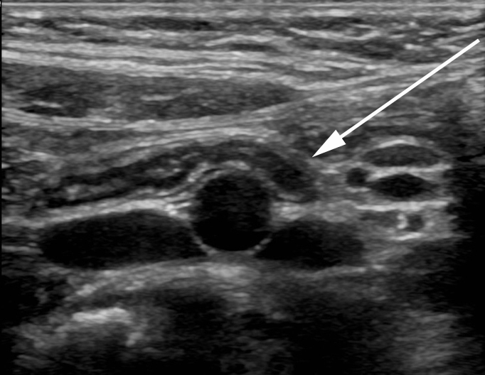
A 8-year-old male with abdominal pain who was diagnosed with gastroenteritis. An image obtained with a 15 MHz linear transducer demonstrates a normal appendix draped over the iliac vessels. The appendix measured 5 mm in diameter (normal), with normal mural architecture and thickness and no increased echogenicity of the periappendiceal fat.
Imaging findings
The diagnostic criteria for appendicitis have evolved in the past three decades. Until recently, any appendix with a diameter of greater than 6 mm was considered highly suspicious for appendicitis. However, recent literature suggests that this is too sensitive of a threshold with 7 mm being a more useful cutoff, as close to 40% of otherwise normal appendices measure greater than 6 mm.16–18 Regardless of the exact diameter used, having a strict criterion for diameter may be a useful tool but should not be used in isolation to make the diagnosis of appendicitis.
Secondary signs of appendicitis have also been in flux, possibly due to improved transducers allowing better visualization of the appendix and surrounding structures. Initially the presence of an appendicolith was considered suspicious. However, appendicoliths are not uncommonly seen in a normal appendix, a fact that was even noted in the original description of appendicitis.2 Thus in the absence of other concerning features, an appendicolith is not a source of alarm with a recent study noting that (adult) patients with incidentally discovered appendicoliths were at no higher risk of developing appendicitis than the general population.19 When there is high suspicion for appendicitis, however, an appendicolith should be reported as its presence has the ability to change management of the patient.10
The degree of compressibility has also fallen out of favor as a number of factors are at play that reduce the consistency of this finding, namely body habitus, the pressure applied by the sonographer, and the location of the appendix.16 The degree of hyperemia is similarly subject to the pitfalls of technique and subjective determination of what constitutes increased blood flow.15,16
Although not initially described, increased echogenicity of the periappendiceal fat has been demonstrated to a useful secondary signs (Figure 2).17,20 The loss of mural stratification has been shown repeatedly to be a helpful adjunct, with apparent discontinuity of the wall increasing the likelihood of perforated appendicitis. Changes to the adjacent bowel (loss of normal peristalsis, thickening) are also useful with the caveat that the primary inflammatory process can actually be the bowel with secondary inflammatory changes of the appendix.
Figure 2. .
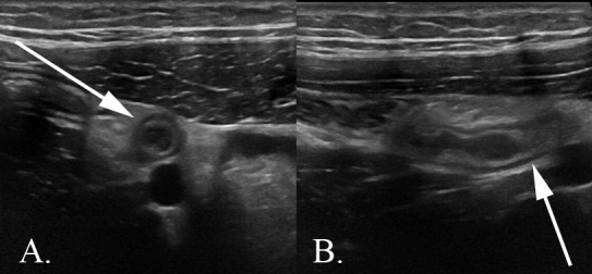
A 14-year-old male with abdominal pain who was diagnosed with uncomplicated appendicitis. Images obtained with a 15 Hz linear transducer. (A) The appendix in short axis and (B) in long axis is enlarged, measuring 8 mm in diameter. The surrounding periappendiceal fat demonstrates increased echogenicity with subtle mass effect on the overlying abdominal musculature.
Small volume, simple free fluid is frequently seen in the setting of a normal appendix.15,17,20 However, the presence of a large amount of fluid, particularly when it is complex, remains helpful as does the presence of a frank abscess. In the setting of other secondary findings, the presence of complex fluid should be reported as highly suspicious for complicated appendicitis (Figure 3).21,22 The presence of free air on ultrasound is also pathognomonic for perforation, although small amounts can be challenging to appreciate. Unfortunately, the sensitivity for the detection of perforation on ultrasound is unreliable, ranging from 29 to 84%.40,41 The sonographic findings of acute appendicitis are summarized in Table 3.
Figure 3. .
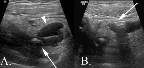
A 14-year-old male with abdominal pain diagnosed with complicated appendicitis. Images obtained with a 9 MHz linear probe. (A) An arrow indicates the shadowing appendicolith contained with a thickened appendix. The arrowhead demonstrates a small pocket of complex fluid. It is important to note in the report whether or not the appendicolith is intraluminal or contained within the abscess as it can serve as a nidus of recurrent infection. (B) The arrow demonstrates free air casting a “dirty shadow” within the pocket of complex fluid. Both images show marked echogenicity of the surrounding periappendiceal fat.
Table 3. .
Major imaging findings for uncomplicated and complicated appendicitisModality
| Uncomplicated | Complicated | |
| Ultrasound | Enlarged appendix (>6–8 mm), mural thickening, increased echogenicity of the periappendiceal fat, complex free fluid17–20 | Abscess formation, frank mural discontinuity, extraluminal appendicolith, free air (although frequently challenging to detect on us)21,22 |
| CT | Enlarged appendix (>6–8 mm), mural thickening, hyperenhancement of the wall, stranding of the surrounding mesenteric fat23–25 25–28 29 30 31 32 | Abscess formation, frank mural discontinuity, extraluminal appendicolith, free air33 34 35 |
| MRI | Enlarged appendix (>7–8 mm), mural thickening, edema of the wall of the appendix and of the surrounding mesenteric fat36 37 38 39 | Abscess formation, frank mural discontinuity, extraluminal appendicolith, free air (although both free air and an extraluminal appendicolith can be challenging to detect on MR) |
With this arsenal of sonographic findings, a positive exam holds great clinical weight as does a negative exam with a fully visualized appendix. Conversely, a radiologist can also readily determine when an ultrasound is indeterminate and recommend advanced imaging for definitive evaluation. The challenge arises when the appendix is not visualized or only partially visualized. The general trend is that non-visualization of the appendix in the absence of secondary signs can be considered negative.42,43 But this is certainly not unanimous with recent studies showing a lack of sensitivity based solely on the secondary signs in the setting of a non-visualized appendix.44,45
Computed tomography
Technique
During the 1990s, abdominopelvic CT became integral to the imaging work-up of pediatric patients with suspected appendicitis.23,46–50 Over the past 30 years, both imaging protocols and CT technology have evolved.51,52 More recently in response to the Image Gently® campaign, efforts to optimize appendix CT have focused on limiting its use to patients with equivocal ultrasound exams,53 and reducing radiation doses while maintaining diagnostic efficacy.54–57
Despite decades of research and routine clinical use, there remains no universally accepted protocol for CT of suspected appendicitis.58 Various authors have shown extremely high sensitivities and specificities with a variety of techniques: using only rectal contrast59; using only i.v. contrast60; and even no oral, rectal, or i.v. contrast.61 With this last technique, the authors acknowledged that several of their false positives and false negatives were young patients with little intra-abdominal fat, highlighting the potential benefits of contrast material for evaluating pediatric appendicitis. In general, the most widely accepted CT protocols include both i.v. and oral contrast material.58
As mentioned above, concerns over the risks of ionizing radiation in children have led national organizations to encourage reductions in CT utilization when alternative imaging modalities can provide the same diagnostic information. Recently, a study of 45 pediatric hospitals in the USA showed there is a transition away from CT use for appendicitis: in 2005, 59.1% of children with appendicitis had a CT while only 25% had an ultrasound; in contrast in 2014, 32.7% had a CT and 61% had an ultrasound.62
When CT is used, dose reduction techniques should be employed to minimize radiation exposure while maintaining diagnostic utility. In 2015, Callahan et al reported that helical CT using a standard reconstruction algorithm could be performed at 50% dose, resulting in similar rates of appendix visualization55 and similar diagnostic yield of CT in clinical practice.54 Didier et al reported that using iterative reconstruction methods reduced CT radiation dose by 45% without compromising diagnostic accuracy.57 Updating CT protocols to reflect up-to-date dose reduction techniques, and adopting a stepwise Ultrasound-CT imaging pathway may represent the greatest current opportunity for imaging centers to implement quality improvement in their practices.63–69
Imaging findings
The most commonly described CT finding of appendicitis is dilation, however, the definition of appendiceal dilation has been inconsistent. Two recent articles provide valuable normative data for the pediatric appendix,24,26 noting that appendiceal diameters are normally distributed, with 95% confidence interval including diameters up to 8.7 mm (Figure 4).26 These data highlight that appendiceal diameter alone is not enough to confirm a diagnosis of appendicitis on CT.
Figure 4. .
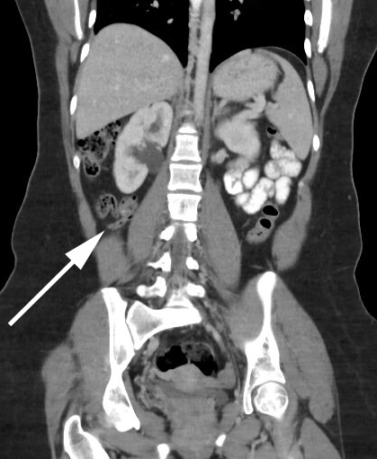
A 16-year-old female with abdominal mass (not pictured). A CT coronal image demonstrates a normal caliber appendix, coiled in the right mid abdomen. There is no evidence of mural thickening. The surrounding periappendiceal fat is pristine, and there is intraluminal gas. Incidentally noted is mild right sided urinary tract dilatation.
Additional suggestive findings of appendicitis include: mural thickening and increased enhancement; presence of an obstructing appendicolith23,25,27,28; evidence of periappendiceal inflammation, including fat stranding, fascial thickening, cecal wall thickening; and presence of free fluid within the right lower quadrant or deep pelvis (Figure 5).23,25,27–32 Importantly in the absence of such findings, CT can be interpreted as negative for appendicitis with a negative predictive value of 98.7%, even if the appendix is not visualized.51
Figure 5. .
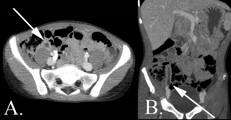
A 13-year-old male with abdominal pain who was diagnosed with uncomplicated appendicitis. (A) Axial CT and (B) coronal CT images demonstrates a dilated fluid filled appendix with a thickened and mildly hyperenhancing appendiceal wall. This appendix has a taut, elongated morphology as opposed to the redundant and coiled appearance of the normal appendix. The paucity of intraabdominal fat in this child makes the assessment of periappendiceal fat stranding challenging.
Specific imaging features that suggest perforation include focal defect in the enhancing appendiceal wall, extraluminal air, extraluminal appendicolith, prominent periappendiceal inflammation or phlegmon, and abscess (Figure 6).33,34 However, these findings are not particularly sensitive for perforation.33,34 In a multireader analysis of 200 patients, reviewers had an overall sensitivity of 62% and specificity of 81% for determining perforation by CT.70 Thus, despite the variability in referenced criteria for differentiating uncomplicated and complicated appendicitis on CT, the overall performance remains excellent with sensitivities in recent studies ranging from 94 to 95% and specificities of 92 to 95%.35,71 Table 3 summarizes the CT findings of acute appendicitis.
Figure 6. .

A 14-year-old male with abdominal pain who was diagnosed with complicated appendicitis (same patient as Figure 3). Axial CT image demonstrates an appendicolith within the lumen of the dilated appendix. There is a surrounding air and fluid containing collection consistent with an abscess.
Magnetic resonance (MR)
Technique
In recent years, MR has emerged as a viable modality to assess for appendicitis in children. This growing utilization has been in part due to increasing awareness of potential long-term effects of radiation associated with CT in childhood.72,73
Review of the literature illustrates the wide variety of MR protocols used in the evaluation for appendicitis. A commonality found in all protocols is the inclusion of T2 weighted single shot fast spin echo sequences, both with and without fat saturation, usually in the axial and coronal planes.74 Some authors have shown success with these sequences alone,75,76 while other studies have shown advantages of including additional sequences. Diffusion-weighted imaging in particular has been shown to be helpful in detecting restricted diffusion in the appendiceal wall77–79 as well as increasing the conspicuity of potential abscesses.80 As is frequently the case with MR, a balance must be struck between the utility of adding sequences and the length of the study.
Use of i.v. gadolinium-based contrast for evaluation of appendicitis has been controversial, particularly given current safety concerns regarding the potential for deposition of free gadolinium in the brain and soft tissues.81 Several studies have shown similar effectiveness between the non-contrast enhanced protocols and the protocols using gadolinium enhanced T1 weighted images (in addition to the T2 weighted imaging).82–84 Given the concern over gadolinium exposure, non-contrast enhanced MR is preferable as it is both a safe and diagnostic option for evaluation of pediatric appendicitis.
In general, MR examinations are lengthy. This creates the potential problem of patient motion, a problem potentiated in the pediatric population. Sedated exams can be performed to combat this challenge. However, sedation is associated with inherent risks, such as aspiration and respiratory depression, as well as increased cost and personnel demands.85,86 Particularly in the younger patient population (<2 years of age), there is increasing reticence to use sedation.87 An additional pragmatic consideration is that sedated exams can tie up the scanner for longer periods of time, a fact which may be untenable in a busy department. However, studies have shown success utilizing non-contrast-enhanced MRI in pediatric patients as young as 4 years old without sedation, especially with emerging faster, shorter protocols.76
Imaging findings
The normal appendiceal diameter on MR has been shown to average 5–7 mm (Figure 7).36 Dieder et al showed that a diameter greater than 7 mm and appendiceal wall thickness greater than 2 mm to have good predictive power for acute appendicitis.37 As with ultrasound and CT, an enlarged appendiceal diameter cannot be used as sole criteria for appendicitis on MR, but rather needs to be used in context with additional findings to make accurate diagnosis.74 Other positive prognostic factors for appendicitis include focal periappendiceal inflammation and fluid, manifested by increased T2 signal in the surrounding fat; an appendicolith, manifested by a focus of diminished signal intensity on all sequences; and abscess formation, manifested by T2 hyperintense collection with a wall (Figure 8). Destruction of the appendiceal wall can be seen in both perforated and nonperforated appendicitis.38 Free air is pathognomonic for perforation but is rarely seen with routine MR sequences (Figure 9).38 Table 3 presents a concise description of the MR findings in acute appendicitis.
Figure 7. .
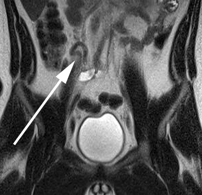
A 17-year-old male with abdominal pain. Coronal T2 weighted MR image without fat saturation demonstrates a normal caliber appendix adjacent to the cecum, draping over the iliac vessels. No fluid or edema is seen surrounding the appendix, and there is no evidence of mural thickening. As on the CT image of the normal appendix, it is coiled upon itself as opposed to the taut morphology seen in an inflamed appendix.
Figure 8. .
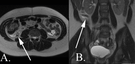
A 14-year-old female with abdominal pain who was diagnosed with acute uncomplicated appendicitis. (A) Axial T 2 weighted MR image without fat saturation demonstrates a dilated appendix with surrounding fluid and edema of the mesenteric fat. Within the appendiceal lumen, there is a round T2 hypointense structure consistent with an appendicolith. (B) Coronal T2 weighted MR image without fat saturation again shows the dilated appendix with surrounding small volume free fluid.
Figure 9. .

A 9-year-old male with abdominal pain who was diagnosed with complicated appendicitis. (A) Axial T2 weighted MR image without fat saturation demonstrates a markedly dilated appendix (arrow) with pronounced inflammatory changes of the periappendiceal fat. There is a complex fluid collection containing a fluid–fluid level and a locule of free air (arrowhead). (B) Coronal T2 weighted MR image without fat saturation again demonstrates a markedly dilated appendix. Small volume free fluid is noted throughout the abdomen with dilated loops of small bowel consistent with developing small bowel obstruction.
As with the prior modalities, the distinction between perforated and non-perforated appendicitis is challenging. While Dillman et al demonstrated a sensitivity of 90% and specificity of 99% for the evaluation of perforation utilizing MR,75 the results of other studies are not nearly as encouraging. In the adult population, Leewenburgh et al described a sensitivity of 57% and specificity of 86%.38 I.v. contrast does not play a specific role in this distinction, although peritoneal enhancement may help with determining perforation.79
Imaging algorithm
For the “appropriate” work-up of appendicitis, there is not a one size fits all approach. The imaging algorithm will vary depending on the resources available at a specific institution. Ultrasound is a reasonable first line imaging modality. However, institutions who do not perform routine pediatric imaging may feel uncomfortable with both performing and interpreting appendicitis ultrasound. There is a relatively steep learning curve in both of these aspects, and, if the volume at a particular facility is not sufficient, it can be challenging to maintain an adequate level of competency. Even in an era of hypervigilance regarding radiation, if it is not feasible to make the correct diagnosis with ultrasound, there should be no hesitancy in proceeding to CT (or MR, as available). Additionally, the work flow of an institution, particularly if patient through put is paramount, may make ultrasound an untenable solution as the exam itself can be lengthy relative to CT. Moreover, while not a requisite of appendicitis ultrasound per se, a full bladder in the pediatric female population is desirable to exclude pelvic pathology mimicking appendicitis, which may further increase the length of time associated with the exam.
MR is the newest imaging modality in the assessment of appendicitis thus many of the “kinks” in perfecting it for this application are still being worked out. Additionally, body MR, and pediatric body MR specifically, is not necessarily widely read by general radiologists. There is also the very real limitation of scanner accessibility. However, if an institution has adequate MR access, the protocols are relatively straightforward to implement. In addition, compared to ultrasound, the learning curve for image acquisition and interpretation for MR is arguably not as steep. A recent study by Covelli et al demonstrated the feasibility of implementing a pediatric appendicitis MR protocol in a general adult hospital with non-pediatric radiologists, emergency physicians, and surgeons.39 While this is a single study, it lays the foundation for pediatric appendicitis MR to move from academic centers into community hospitals. Another study published earlier this year explored the utility of using MR as a first line imaging modality in children with suspected appendicitis.88 Given its lack of ionizing radiation, feasibility without i.v. contrast, and relative operator independence (as well as its exquisite soft tissue contrast to aid in the detection of other intra-abdominal pathology), MRI may very well supplant CT and ultrasound as the first line imaging choice for suspected appendicitis in the future.
Finally, there is CT. This imaging modality has been the lynchpin for the diagnosis of appendicitis for the past three decades, particularly in the adult population. While the paucity of intraabdominal fat in the younger patients can increase the level of difficulty in interpretation, CT has three big advantages: it is readily available, quick to obtain, and highly specific and sensitive for the diagnosis in the hands of both general radiologists and pediatric subspecialists. Thus, even if CT is the only imaging modality available, the diagnosis can be made quickly and correctly, which is ultimately what is most important for patient care.
In a scenario with a full complement of resources available, a reasonable work-up of appendicitis begins with ultrasound. For equivocal or indeterminate cases, this may be followed by a non-contrast enhanced MR CT can be used as a problem-solving tool on an as needed basis.
Standardized reporting schema
Regardless of the imaging modality, it is of utmost importance that the radiology report conveys the relevant findings correctly and concisely. A number of studies have demonstrated that standardizing the reporting format across radiologists not only increased clinician satisfaction but impacts the accuracy of reports.89,90 In the age of report templates, this should be relatively easy to implement and share within a group.
In an ideal world, parsing out the individual imaging findings in a granular format and codifying this across institutions would make a powerful research tool. With the ever expanding world of artificial intelligence and clinical decision support, it is possible that specific imaging features in conjunction with specific clinical findings, could be combined in yet undetermined ways to not only ascertain the presence of appendicitis but also assess the likelihood of perforation.
Conclusion
The primary role of imaging in the work-up of appendicitis now and in the immediate future is to determine the presence of appendicitis. As described above, each of the imaging modalities can readily arrive at the diagnosis and which modality to choose depends on the resources at one’s institution. In an experienced hand and an ideal patient, ultrasound can provide unparalleled resolution of the appendix. Conversely, ultrasound can also fall short of a diagnostic exam. Avoiding the radiation associated with CT or the expense associated with MRI can be admirable goals. However, they should not deter the radiologist or clinician from pursuing a definitive diagnosis. In the end the best test is the one that provides the patient with an answer.
As the debate over the appropriateness of antibiotics as the primary therapy of appendicitis continues, radiologists have an opportunity to play an important role in differentiating complicated from uncomplicated appendicitis. Yet, according to currently available data, no modality is adequate for accurately assessing the presence of appendiceal perforation. However, this distinction may not be a reliable indicator of the true clinical picture. All perforations are not created equal; missing a microperforation found at the appendiceal tip by the pathologists likely does not have the same clinical ramifications as missing frank purulence found at the time of surgery. Arguably, the former could still be considered “uncomplicated appendicitis.” While some studies have relied on the criteria of the presence of phlegmon, abscess, and/or signs of peritonitis for the diagnosis of complicated appendicitis,10 other research, most notably the ongoing multicenter clinical trial, equates perforated appendicitis with complicated appendicitis.7 The distinction between these two entities needs to be formally elucidated with the help of clinical colleagues and then renewed focus can be paid to what imaging features differentiates complicated vs uncomplicated appendicitis.
Over the past century and a half, our understanding of appendicitis has dramatically evolved. From not even recognizing it as a common instigator of abdominopelvic pathology to accepting a 20% false-positive rate at the time of appendectomy to exploring the possibility of a nonsurgical approach to therapy, radiology has been and will continue to be integral in this transformation.
Contributor Information
Cassandra Sams, Email: cassandra.sams@lifespan.org.
Rama S Ayyala, Email: rayyala@gmail.com.
David W. Swenson, Email: swenson.david@gmail.com.
References
- 1. Brody JE. The Case Against Appendectomies. The New York Times [Internet].. 2018. Available from: https://www.nytimes.com/2018/11/19/well/live/the-case-against-appendectomies.html.
- 2. Fitz R. Perforating inflammation of the vermiform appendix: with special reference to its early diagnosis and treatment. Philadelphia, PA; 1886. [Google Scholar]
- 3. Lundholm K, Hansson-Assarsson J, Engström C, Iresjö B-M. Long-Term results following antibiotic treatment of acute appendicitis in adults. World J Surg 2017; 41: 2245–50. doi: 10.1007/s00268-017-3987-6 [DOI] [PMC free article] [PubMed] [Google Scholar]
- 4. Salminen P, Tuominen R, Paajanen H, Rautio T, Nordström P, Aarnio M, et al. Five-Year follow-up of antibiotic therapy for uncomplicated acute appendicitis in the APPAC randomized clinical trial. JAMA 2018; 320: 1259–65. doi: 10.1001/jama.2018.13201 [DOI] [PMC free article] [PubMed] [Google Scholar]
- 5. Sippola S, Grönroos J, Sallinen V, Rautio T, Nordström P, Rantanen T, et al. A randomised placebo-controlled double-blind multicentre trial comparing antibiotic therapy with placebo in the treatment of uncomplicated acute appendicitis: APPAC III trial study protocol. BMJ Open 2018; 8: 117. doi: 10.1136/bmjopen-2018-023623 [DOI] [PMC free article] [PubMed] [Google Scholar]
- 6. Pogorelić Z, Rak S, Mrklić I, Jurić I. Prospective Validation of Alvarado Score and Pediatric Appendicitis Score for the Diagnosis of Acute Appendicitis in Children [Internet. 2015; 31: 164–8. [DOI] [PubMed] [Google Scholar]
- 7. Hall NJ, Eaton S, Abbo O, Arnaud AP, Beaudin M, Brindle M, et al. Appendectomy versus non-operative treatment for acute uncomplicated appendicitis in children: study protocol for a multicentre, open-label, non-inferiority, randomised controlled trial. BMJ Paediatr Open 2017; 1: bmjpo-2017-000028. doi: 10.1136/bmjpo-2017-000028 [DOI] [PMC free article] [PubMed] [Google Scholar]
- 8. Benabbas R, Hanna M, Shah J, Sinert R. Diagnostic accuracy of history, physical examination, laboratory tests, and point-of-care ultrasound for pediatric acute appendicitis in the emergency department: a systematic review and meta-analysis. Acad Emerg Med 2017; 24: 523–51. doi: 10.1111/acem.13181 [DOI] [PubMed] [Google Scholar]
- 9. Mason RJ. Surgery for appendicitis: is it necessary? Surg Infect 2008; 9: 481–8. doi: 10.1089/sur.2007.079 [DOI] [PubMed] [Google Scholar]
- 10. Huang L, Yin Y, Yang L, Wang C, Li Y, Zhou Z. Comparison of antibiotic therapy and appendectomy for acute uncomplicated appendicitis in children: a meta-analysis. JAMA Pediatr 2017; 171: 426–34. doi: 10.1001/jamapediatrics.2017.0057 [DOI] [PMC free article] [PubMed] [Google Scholar]
- 11. Abo A, Shannon M, Taylor G, Bachur R. The influence of body mass index on the accuracy of ultrasound and computed tomography in diagnosing appendicitis in children. Pediatr Emerg Care 2011; 27: 731–6. doi: 10.1097/PEC.0b013e318226c8b0 [DOI] [PubMed] [Google Scholar]
- 12. Butler M, Servaes S, Srinivasan A, Edgar JC, Del Pozo G, Darge K. Us depiction of the appendix: role of abdominal wall thickness and appendiceal location. Emerg Radiol 2011; 18: 525–31. doi: 10.1007/s10140-011-0977-0 [DOI] [PubMed] [Google Scholar]
- 13. Puylaert JB. Acute appendicitis: US evaluation using graded compression. Radiology 1986; 158: 355–60. doi: 10.1148/radiology.158.2.2934762 [DOI] [PubMed] [Google Scholar]
- 14. Cundy TP, Gent R, Frauenfelder C, Lukic L, Linke RJ, Goh DW. Benchmarking the value of ultrasound for acute appendicitis in children. J Pediatr Surg 2016; 51: 1939–43. doi: 10.1016/j.jpedsurg.2016.09.009 [DOI] [PubMed] [Google Scholar]
- 15. Gongidi P, Bellah RD. Ultrasound of the pediatric appendix. Pediatr Radiol 2017; 47: 1091–100. doi: 10.1007/s00247-017-3928-4 [DOI] [PubMed] [Google Scholar]
- 16. Trout AT, Sanchez R, Ladino-Torres MF, Pai DR, Strouse PJ. A critical evaluation of US for the diagnosis of pediatric acute appendicitis in a real-life setting: how can we improve the diagnostic value of sonography? Pediatr Radiol 2012; 42: 813–23. doi: 10.1007/s00247-012-2358-6 [DOI] [PubMed] [Google Scholar]
- 17. Goldin AB, Khanna P, Thapa M, McBroom JA, Garrison MM, Parisi MT. Revised ultrasound criteria for appendicitis in children improve diagnostic accuracy. Pediatr Radiol 2011; 41: 993–9. doi: 10.1007/s00247-011-2018-2 [DOI] [PubMed] [Google Scholar]
- 18. Chicaiza HP, Malia L, Mulvey CH, Smith SR. Revisiting the appendiceal diameter via ultrasound for the diagnosis of acute appendicitis. Pediatr Emerg Care 2018; 34: 757–60. doi: 10.1097/PEC.0000000000001278 [DOI] [PubMed] [Google Scholar]
- 19. Khan MS, Chaudhry MBH, Shahzad N, Tariq M, Memon WA, Alvi AR. Risk of appendicitis in patients with incidentally discovered appendicoliths. J Surg Res 2018; 221: 84–7. doi: 10.1016/j.jss.2017.08.021 [DOI] [PubMed] [Google Scholar]
- 20. Estey A, Poonai N, Lim R. Appendix not seen: the predictive value of secondary inflammatory sonographic signs. Pediatr Emerg Care 2013; 29: 435–9. doi: 10.1097/PEC.0b013e318289e8d5 [DOI] [PubMed] [Google Scholar]
- 21. Carpenter JL, Orth RC, Zhang W, Lopez ME, Mangona KL, Guillerman RP. Diagnostic performance of US for differentiating perforated from Nonperforated pediatric appendicitis: a prospective cohort study. Radiology 2017; 282: 835–41. doi: 10.1148/radiol.2016160175 [DOI] [PubMed] [Google Scholar]
- 22. Tulin-Silver S, Babb J, Pinkney L, Strubel N, Lala S, Milla SS, et al. The challenging ultrasound diagnosis of perforated appendicitis in children: constellations of sonographic findings improve specificity. Pediatr Radiol 2015; 45: 820–30. doi: 10.1007/s00247-014-3232-5 [DOI] [PubMed] [Google Scholar]
- 23. Callahan MJ, Rodriguez DP, Taylor GA. Ct of appendicitis in children. Radiology 2002; 224: 325–32. doi: 10.1148/radiol.2242010998 [DOI] [PubMed] [Google Scholar]
- 24. Orscheln ES, Trout AT. Appendiceal diameter: CT versus sonographic measurements. Pediatr Radiol 2016; 46: 316–21. doi: 10.1007/s00247-015-3491-9 [DOI] [PubMed] [Google Scholar]
- 25. Taylor GA, Callahan MJ, Rodriguez D, Smink DS. Ct for suspected appendicitis in children: an analysis of diagnostic errors. Pediatr Radiol 2006; 36: 331–7. doi: 10.1007/s00247-005-0079-9 [DOI] [PubMed] [Google Scholar]
- 26. Trout AT, Towbin AJ, Zhang B. Journal Club: the pediatric appendix: defining normal. AJR Am J Roentgenol. [DOI] [PubMed] [Google Scholar]
- 27. Birnbaum BA, Wilson SR. State of the art appendicitis at the millennium. Radiology 2000; 215: 337–48. [DOI] [PubMed] [Google Scholar]
- 28. Hashizume N, Iinuma Y, Hirayama Y, Nitta K, Iida H, Shiotani M, et al. Contrast-Enhanced multidetector-row computed tomography can predict pathological findings of acute appendicitis in children. Acute Med Surg 2016; 3: 21–5. doi: 10.1002/ams2.131 [DOI] [PMC free article] [PubMed] [Google Scholar]
- 29. Sung T, Callahan MJ, Taylor GA. Clinical and imaging mimickers of acute appendicitis in the pediatric population. AJR Am J Roentgenol 2006; 186: 67–74. doi: 10.2214/AJR.04.1380 [DOI] [PubMed] [Google Scholar]
- 30. Friedland JA, Siegel MJ. Ct appearance of acute appendicitis in childhood. AJR Am J Roentgenol 1997; 168: 439–42. doi: 10.2214/ajr.168.2.9016223 [DOI] [PubMed] [Google Scholar]
- 31. Rao PM, Novelline RA, Mostafavi AA, Lawrason JN, Mccabe CJ. Rectal contrast only CT for pediatric appendicitis. AJR Am J Roentgenol 1997;: 1275–80. [DOI] [PubMed] [Google Scholar]
- 32. Lane MJ, Liu DM, Huynh MD, Jeffrey RB, Mindelzun RE, Katz DS. Suspected acute appendicitis: nonenhanced helical CT in 300 consecutive patients. Radiology 1999; 213: 341–6. doi: 10.1148/radiology.213.2.r99nv44341 [DOI] [PubMed] [Google Scholar]
- 33. Horrow MM, White DS, Horrow JC. Differentiation of perforated from nonperforated appendicitis at CT. Radiology 2003; 227: 46–51. doi: 10.1148/radiol.2272020223 [DOI] [PubMed] [Google Scholar]
- 34. Foley TA, FE I, Nathan MA, Hough DM, Schiller HJ, Hoskin TL. Differentiating Nonperf and perforated appendicitis on CT. Radiology 2005; 1: 89–96. [DOI] [PubMed] [Google Scholar]
- 35. Zhang H, Liao M, Chen J, Zhu D, Byanju S, Ultrasound BS. Ultrasound, computed tomography or magnetic resonance imaging - which is preferred for acute appendicitis in children? A Meta-analysis. Pediatr Radiol 2017; 47: 186–96. doi: 10.1007/s00247-016-3727-3 [DOI] [PubMed] [Google Scholar]
- 36. Swenson DW, Schooler GR, Stamoulis C, Lee EY. Mri of the normal appendix in children: data toward a new reference standard. Pediatr Radiol 2016; 46: 1003–10. doi: 10.1007/s00247-016-3559-1 [DOI] [PubMed] [Google Scholar]
- 37. Didier RA, Hopkins KL, Coakley FV, Krishnaswami S, Spiro DM, Foster BR. Performance characteristics of magnetic resonance imaging without contrast agents or sedation in pediatric appendicitis. Pediatr Radiol 2017; 47: 1312–20. doi: 10.1007/s00247-017-3897-7 [DOI] [PubMed] [Google Scholar]
- 38. Leeuwenburgh MMN, Wiezer MJ, Wiarda BM, Bouma WH, Phoa SSKS, Stockmann HBAC, et al. Accuracy of MRI compared with ultrasound imaging and selective use of CT to discriminate simple from perforated appendicitis. Br J Surg 2014; 101: e147–55. doi: 10.1002/bjs.9350 [DOI] [PubMed] [Google Scholar]
- 39. Covelli JD, Madireddi SP, May LA, Costello JE, Lisanti CJ, Carlson CL. Mri for pediatric appendicitis in an Adult-Focused General Hospital: a clinical effectiveness Study—Challenges and lessons learned. American Journal of Roentgenology 2019; 212: 180–7. doi: 10.2214/AJR.18.19825 [DOI] [PubMed] [Google Scholar]
- 40. Puylaert JB, Rutgers PH, Lalisang RI, de Vries BC, van der Werf SD, Dörr JP, et al. A prospective study of ultrasonography in the diagnosis of appendicitis. N Engl J Med 1987; 317: 666–9. doi: 10.1056/NEJM198709103171103 [DOI] [PubMed] [Google Scholar]
- 41. Borushok KF, Jeffrey RB, Laing FC, Townsend RR. Sonographic diagnosis of perforation in patients with acute appendicitis. AJR Am J Roentgenol 1990; 154: 275–8. doi: 10.2214/ajr.154.2.2105013 [DOI] [PubMed] [Google Scholar]
- 42. Athans BS, Depinet HE, Towbin AJ, Zhang Y, Zhang B, Trout AT. Use of clinical data to predict appendicitis in patients with equivocal us findings. Radiology 2016; 280: 557–67. doi: 10.1148/radiol.2016151412 [DOI] [PubMed] [Google Scholar]
- 43. Binkovitz LA, Unsdorfer KML, Thapa P, Kolbe AB, Hull NC, Zingula SN, et al. Pediatric appendiceal ultrasound: accuracy, determinacy and clinical outcomes. Pediatr Radiol 2015; 45: 1934–44. doi: 10.1007/s00247-015-3432-7 [DOI] [PubMed] [Google Scholar]
- 44. Partain KN, Patel A, Travers C, McCracken CE, Loewen J, Braithwaite K, et al. Secondary signs may improve the diagnostic accuracy of equivocal ultrasounds for suspected appendicitis in children. J Pediatr Surg 2016; 51: 1655–60. doi: 10.1016/j.jpedsurg.2016.03.005 [DOI] [PMC free article] [PubMed] [Google Scholar]
- 45. Ramarajan N, Krishnamoorthi R, Gharahbaghian L, Pirrotta E, Barth RA, Wang NE. Clinical correlation needed: what do emergency physicians do after an equivocal ultrasound for pediatric acute appendicitis? J Clin Ultrasound 2014; 42: 385–94. doi: 10.1002/jcu.22153 [DOI] [PubMed] [Google Scholar]
- 46. Sivit CJ, Applegate KE, Berlin SC, Myers MT, Stallion A, Dudgeon DL, et al. Evaluation of suspected appendicitis in children and young adults: helical CT. Radiology 2000; 216: 430–3. doi: 10.1148/radiology.216.2.r00au38430 [DOI] [PubMed] [Google Scholar]
- 47. Sivit CJ, Applegate KE, Stallion A, Dudgeon DL, Salvator A, Schluchter M, et al. Imaging evaluation of suspected appendicitis in a pediatric population: effectiveness of sonography versus CT. AJR Am J Roentgenol 2000; 175: 977–80. doi: 10.2214/ajr.175.4.1750977 [DOI] [PubMed] [Google Scholar]
- 48. Peña BM, Taylor GA, Lund DP, Mandl KD. Effect of computed tomography on patient management and costs in children with suspected appendicitis. Pediatrics 1999; 104(3 Pt 1): 440–6. doi: 10.1542/peds.104.3.440 [DOI] [PubMed] [Google Scholar]
- 49. Peña BMG. Taylor G a, fishman SJ, Mandl kD. Effect of an imaging protocol on clinical outcomes among pediatric patients with appendicitis. Pediatrics 2002; 110: 1088–93. [DOI] [PubMed] [Google Scholar]
- 50. Kaiser S, Mesas-Burgos C, Söderman E, Frenckner B. Appendicitis in children--impact of US and CT on the negative appendectomy rate. Eur J Pediatr Surg 2004; 14: 260–4. doi: 10.1055/s-2004-817841 [DOI] [PubMed] [Google Scholar]
- 51. Garcia K, Hernanz-Schulman M, Bennett DL, Morrow SE, Yu C, Kan JH. Suspected appendicitis in children: diagnostic importance of normal abdominopelvic CT findings with nonvisualized appendix. Radiology 2009; 250: 531–7. doi: 10.1148/radiol.2502080624 [DOI] [PubMed] [Google Scholar]
- 52. Neville AM, Paulson EK. Mdct of acute appendicitis: value of coronal reformations. Abdom Imaging 2009; 34: 42–8. doi: 10.1007/s00261-008-9415-5 [DOI] [PubMed] [Google Scholar]
- 53. Swenson DW, Ayyala RS, Sams C, Lee EY. Practical imaging strategies for acute appendicitis in children. American Journal of Roentgenology 2018; 211: 901–9. doi: 10.2214/AJR.18.19778 [DOI] [PubMed] [Google Scholar]
- 54. Callahan MJ, Anandalwar SP, MacDougall RD, Stamoulis C, Kleinman PL, Rangel SJ, et al. Pediatric CT dose reduction for suspected appendicitis: a practice quality improvement project using artificial gaussian noise--part 2, clinical outcomes. AJR Am J Roentgenol 2015; 204: 636–44. doi: 10.2214/AJR.14.12965 [DOI] [PubMed] [Google Scholar]
- 55. Callahan MJ, Kleinman PL, Strauss KJ, Bandos A, Taylor GA, Tsai A, et al. Pediatric CT dose reduction for suspected appendicitis: a practice quality improvement project using artificial Gaussian noise--part 1, computer simulations. AJR Am J Roentgenol 2015; 204: W86–W94. doi: 10.2214/AJR.14.12964 [DOI] [PubMed] [Google Scholar]
- 56. Bachur RG, Levy JA, Callahan MJ, Rangel SJ, Monuteaux MC. Effect of reduction in the use of computed tomography on clinical outcomes of appendicitis. JAMA Pediatr 2015; 169: 755–60. doi: 10.1001/jamapediatrics.2015.0479 [DOI] [PubMed] [Google Scholar]
- 57. Didier RA, Vajtai PL, Hopkins KL. Iterative reconstruction technique with reduced volume CT dose index: diagnostic accuracy in pediatric acute appendicitis. Pediatr Radiol 2015; 45: 181–7. doi: 10.1007/s00247-014-3109-7 [DOI] [PMC free article] [PubMed] [Google Scholar]
- 58. Paulson EK, Coursey CA. Ct protocols for acute appendicitis: time for change. American Journal of Roentgenology 2009; 193: 1268–71. doi: 10.2214/AJR.09.3313 [DOI] [PubMed] [Google Scholar]
- 59. Rao PM, Novelline RA, Mostafavi AA, Lawrason JN, Mccabe CJ. Appendicitis. 1997;: 1275–80. [DOI] [PubMed]
- 60. Mun S, Ernst RD, Chen K, Oto A, Shah S, Mileski WJ. Rapid CT diagnosis of acute appendicitis with IV contrast material. Emerg Radiol 2006; 12: 99–102. doi: 10.1007/s10140-005-0456-6 [DOI] [PubMed] [Google Scholar]
- 61. Lane MJ, Liu DM, Huynh MD, Jeffrey RB, Mindelzun RE, Katz DS. Suspected acute appendicitis: nonenhanced helical CT in 300 consecutive patients. Radiology 1999; 213: 341–6. doi: 10.1148/radiology.213.2.r99nv44341 [DOI] [PubMed] [Google Scholar]
- 62. Otero HJ, Crowder L. Imaging Utilization for the Diagnosis of Appendicitis in Stand-Alone Children's Hospitals in the United States: Trends and Costs. J Am Coll Radiol 2017; 14: 603–8. doi: 10.1016/j.jacr.2016.12.013 [DOI] [PubMed] [Google Scholar]
- 63. Neff LP, Ladd MR, Becher RD, Jordanhazy RA, Gallaher JR, Pranikoff T. Computerized tomography utilization in children with appendicitis-differences in referring and children's hospitals. Am Surg 2011; 77: 1061–5. [PubMed] [Google Scholar]
- 64. Anderson KT, Putnam LR, Caldwell KM B, Diffley M A, Hildebrandt A, Covey SE, et al. Imaging gently? Higher rates of computed tomography imaging for pediatric appendicitis in non–children’s hospitals. Surg 2017; 161: 1326–33. [DOI] [PubMed] [Google Scholar]
- 65. Anderson KT, Greenfield S, Putnam LR, Hamilton E, Kawaguchi A, Austin MT, et al. Don't forget the dose: Improving computed tomography dosing for pediatric appendicitis. J Pediatr Surg 2016; 51: 1944–8. doi: 10.1016/j.jpedsurg.2016.09.015 [DOI] [PubMed] [Google Scholar]
- 66. Glass CC, Saito JM, Sidhwa F, Cameron DB, Feng C, Karki M, et al. Diagnostic imaging practices for children with suspected appendicitis evaluated at definitive care hospitals and their associated referral centers. J Pediatr Surg 2016; 51: 912–6. doi: 10.1016/j.jpedsurg.2016.02.055 [DOI] [PubMed] [Google Scholar]
- 67. Badru F, Piening N, To A, Xu P, Fitzpatrick C, Chatoorgoon K, et al. Imaging for acute appendicitis at nonpediatric centers exposes children to excess radiation. J Surg Res 2017; 216: 201–6. doi: 10.1016/j.jss.2017.04.029 [DOI] [PubMed] [Google Scholar]
- 68. Menoch M, Simon HK, Hirsh D, Shim Y, Baxter AL, Clifton M, et al. Imaging for suspected appendicitis: variation between academic and private practice models. Pediatr Emerg Care 2017; 33: 147–51. doi: 10.1097/PEC.0000000000000694 [DOI] [PubMed] [Google Scholar]
- 69. Snow A, Milliren CE, Graham DA, Callahan MJ, MacDougall RD, Robertson RL, et al. Quality of pediatric abdominal CT scans performed at a dedicated children's hospital and its referring institutions: a multifactorial evaluation. Pediatr Radiol 2017; 47: 391–7. doi: 10.1007/s00247-016-3768-7 [DOI] [PubMed] [Google Scholar]
- 70. Fraser JD, Aguayo P, Sharp SW, Snyder CL, Rivard DC, Cully BE, et al. Accuracy of computed tomography in predicting appendiceal perforation. J Pediatr Surg 2010; 45: 231–5. doi: 10.1016/j.jpedsurg.2009.10.040 [DOI] [PubMed] [Google Scholar]
- 71. Doria AS, Moineddin R, Kellenberger CJ, Epelman M, Beyene J, Schuh S, et al. Us or CT for diagnosis of appendicitis in children and adults? A meta-analysis. Radiology 2006; 241: 83–94. doi: 10.1148/radiol.2411050913 [DOI] [PubMed] [Google Scholar]
- 72. Aspelund G, Fingeret A, Gross E, Kessler D, Keung C, Thirumoorthi A, et al. Ultrasonography/MRI versus CT for diagnosing appendicitis. Pediatrics 2014; 133: 586–93. doi: 10.1542/peds.2013-2128 [DOI] [PubMed] [Google Scholar]
- 73. Leeuwenburgh MMN, Wiarda BM, Wiezer MJ, Vrouenraets BC, Gratama JWC, Spilt A, et al. Comparison of imaging strategies with conditional contrast-enhanced CT and unenhanced MR imaging in patients suspected of having appendicitis: a multicenter diagnostic performance study. Radiology 2013; 268: 135–43. doi: 10.1148/radiol.13121753 [DOI] [PubMed] [Google Scholar]
- 74. Moore MM, Kulaylat AN, Hollenbeak CS, Engbrecht BW, Dillman JR, Methratta ST. Magnetic resonance imaging in pediatric appendicitis: a systematic review. Pediatr Radiol 2016; 46: 928–39. doi: 10.1007/s00247-016-3557-3 [DOI] [PubMed] [Google Scholar]
- 75. Dillman JR, Gadepalli S, Sroufe NS, Davenport MS, Smith EA, Chong ST, et al. Equivocal pediatric appendicitis: unenhanced MR imaging protocol for Nonsedated Children-A clinical effectiveness study. Radiology 2016; 279: 216–25. doi: 10.1148/radiol.2015150941 [DOI] [PubMed] [Google Scholar]
- 76. Johnson AK, Filippi CG, Andrews T, Higgins T, Tam J, Keating D, et al. Ultrafast 3-T MRI in the evaluation of children with acute lower abdominal pain for the detection of appendicitis. AJR Am J Roentgenol 2012; 198: 1424–30. doi: 10.2214/AJR.11.7436 [DOI] [PubMed] [Google Scholar]
- 77. Koning JL, Naheedy JH, Kruk PG. Diagnostic performance of contrast-enhanced Mr for acute appendicitis and alternative causes of abdominal pain in children. Pediatr Radiol 2014; 44: 948–55. doi: 10.1007/s00247-014-2952-x [DOI] [PubMed] [Google Scholar]
- 78. Orth RC, Guillerman RP, Zhang W, Masand P, Bisset GS. Prospective comparison of Mr imaging and US for the diagnosis of pediatric appendicitis. Radiology 2014; 272: 233–40. doi: 10.1148/radiol.14132206 [DOI] [PubMed] [Google Scholar]
- 79. Rosenbaum DG, Askin G, Beneck DM, Kovanlikaya A. Differentiating perforated from non-perforated appendicitis on contrast-enhanced magnetic resonance imaging. Pediatr Radiol 2017; 47: 1483–90. [DOI] [PubMed] [Google Scholar]
- 80. Lee MH, Eutsler EP, Sheybani EF, Khanna G. Rapid non-contrast magnetic resonance imaging for post appendectomy intra-abdominal abscess in children. Pediatr Radiol 2017; 47: 935–41. doi: 10.1007/s00247-017-3860-7 [DOI] [PubMed] [Google Scholar]
- 81. Guo BJ, Yang ZL, Zhang LJ. Gadolinium deposition in brain: current scientific evidence and future perspectives. Front Mol Neurosci 2018; 11(September): 1–12. doi: 10.3389/fnmol.2018.00335 [DOI] [PMC free article] [PubMed] [Google Scholar]
- 82. Rosines LA, Chow DS, Lampl BS, Chen S, Gordon S, Mui LW, et al. Value of gadolinium-enhanced MRI in detection of acute appendicitis in children and adolescents. American Journal of Roentgenology 2014; 203: W543–W548. doi: 10.2214/AJR.13.12093 [DOI] [PubMed] [Google Scholar]
- 83. Lyons GR, Renjen P, Askin G, Giambrone AE, Beneck D, Kovanlikaya A. Diagnostic utility of intravenous contrast for MR imaging in pediatric appendicitis. Pediatr Radiol 2017; 47: 398–403. doi: 10.1007/s00247-016-3775-8 [DOI] [PubMed] [Google Scholar]
- 84. Koning JL, Naheedy JH, Kruk PG. Does abdominal pain duration affect the accuracy of first-line MRI for pediatric appendicitis? Abdom Imaging 2015; 40: 352–9. doi: 10.1007/s00261-014-0223-9 [DOI] [PubMed] [Google Scholar]
- 85. Sanborn PA, Michna E, Zurakowski D, Burrows PE, Fontaine PJ, Connor L, et al. Adverse cardiovascular and respiratory events during sedation of pediatric patients for imaging examinations. Radiology 2005; 237: 288–94. doi: 10.1148/radiol.2371041415 [DOI] [PubMed] [Google Scholar]
- 86. Vanderby SA, Babyn PS, Carter MW, Jewell SM, McKeever PD. Effect of anesthesia and sedation on pediatric MR imaging patient flow. Radiology 2010; 256: 229–37. doi: 10.1148/radiol.10091124 [DOI] [PubMed] [Google Scholar]
- 87. Tots S. Consensus Statement on the Use of Anesthetic and Sedative Drugs in Infants and Toddlers. 2015. Available from: http://smarttots.org/wp-content/uploads/2015/05/ConsensusStatementV10-10.2017.pdf. [DOI] [PubMed]
- 88. Mushtaq R, Desoky SM, Morello F, Gilbertson-Dahdal D, Gopalakrishnan G, Leetch A, et al. First-Line diagnostic evaluation with MRI of children suspected of having acute appendicitis. Radiology 2019; 291: 170–7. doi: 10.1148/radiol.2019181959 [DOI] [PubMed] [Google Scholar]
- 89. Fallon SC, Orth RC, Guillerman RP, Munden MM, Zhang W, Elder SC, et al. Development and validation of an ultrasound scoring system for children with suspected acute appendicitis. Pediatr Radiol 2015; 45: 1945–52. doi: 10.1007/s00247-015-3443-4 [DOI] [PubMed] [Google Scholar]
- 90. Bin PS, Kim M, Ko Y, Sim JY, Kim HJ, Lee KH, et al. Structured Reporting versus Free-Text Reporting for Appendiceal Computed Tomography in Adolescents and Young Adults : Preference Survey of 594 Referring Physicians, Surgeons, and Radiologists from 20 Hospitals.. 2019; 20: 246–55. [DOI] [PMC free article] [PubMed] [Google Scholar]