Correction to: Cell Commun Signal 18:124 (2020) 10.1186/s12964-020-00624-8
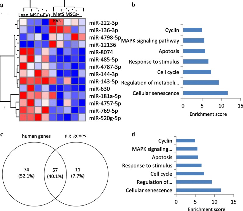
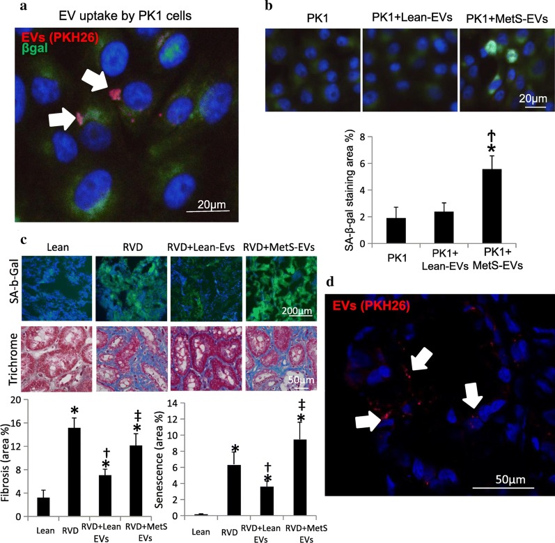
Following publication of the original article [1], the authors identified an error in Figs. 2 and 3. The two figures have been mistakenly transposed. The correct Figs. 2 and 3 have been presented below:
Fig. 2.
MicroRNA (miRNA) profile in MSC-EVs in human subjects and functional pathway analysis of the common SA-genes. a Heat map showed four upregulated (top) and nine downregulated (bottom) miRNAs in MetS compared with Lean MSC-EVs in human subjects. b Enrichment of functional pathway of the 131 miRNA-targeted senescence genes using DAVID 6.7 in human. *p < 0.05 vsersu Lean MSC-EVs. c 57 common SA-genes targeted by differentially expressed miRNAs in human and swine MetS-MSCs. d Enrichment of functional pathway of the 57 SA-genes using DAVID 6.7
Fig. 3.
Effects of MSC-derived EVs in PK1 cells and pig kidney. a Co-cultured with MetS MSC-EVs, PK1 cells showed higher senescence.*p < 0.05 vs PK1, †p < 0.05 vs PK1 + Lean-EVs. b PKH-26-labeled EVs (red) were detected in PK1 cells. c Representative kidney staining with immunofluorescent SA-b-Gal (left top) and trichrome (left bottom), and respective quantification. Lean EVs attenuated cellular senescence and fibrosis in vivo in injured kidneys, whereas MetS EVs failed to blunt them. d Pkh-26-labeled EVs (red) were detected in frozen section in the RVD kidney. *p < 0.05 versus Lean, †p < 0.05 vsersu RVD, ‡p< 0.05 versus RVD + Lean-EVs
Footnotes
Publisher's Note
Springer Nature remains neutral with regard to jurisdictional claims in published maps and institutional affiliations.
Reference
- 1.Li Y, Meng Y, Zhu X, et al. Metabolic syndrome increases senescence-associated micro-RNAs in extracellular vesicles derived from swine and human mesenchymal stem/stromal cells. Cell Commun Signal. 2020;18:124. doi: 10.1186/s12964-020-00624-8. [DOI] [PMC free article] [PubMed] [Google Scholar]


