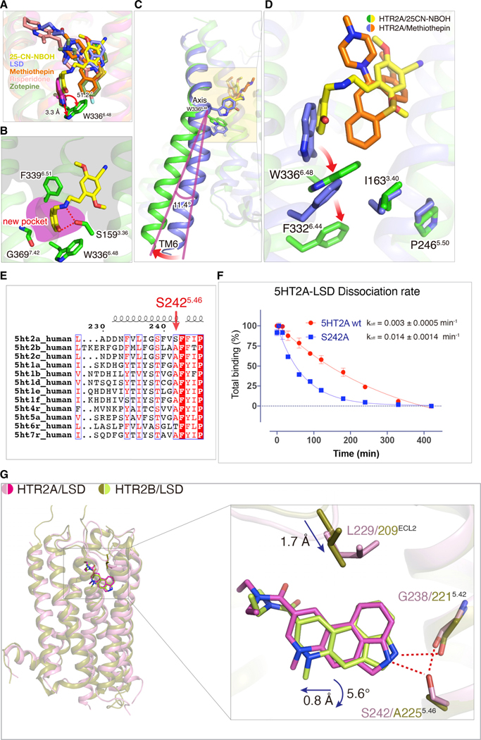
Figure 4. Differential 25-CN-NBOH and LSD Binding Modes.
(A) The superimposed structures of HTR2A with 25CN-NBOH (yellow), LSD (light blue), methiothepin (orange), risperidone (pink, PDB: 6A93), and zotepine (green, PDB: 6A94).
(B) S159, W336, and G369 form a binding pocket that is important for 25CN-NBOH’s agonist activity.
(C) W3366.48 acts as a pivot for the outward movement of TM6.
(D) Conformational displacement of side-chain of W3366.48, followed by F3326.44 in the P-I-F motif. See Figure S4.
(E)The sequence alignment of the serotonin receptor family with an HTR2A specific residue S2425.46 is highlighted.
(F)The S242A5.46 mutation accelerates LSD dissociation from HTR2A; data represent mean ± SEM of n = 3 biological replicates.
(G)The overall structural comparison of HTR2A/LSD (pink/magenta color) and HTR2B/LSD (olive/lime color) and inset shows side view of LSD (magenta)-bound HTR2A (pink) crystal structure overlaid with the LSD (lime)-bound HTR2B (olive) structure. Hydrogen-bond interactions are highlighted by red dash lines.

