Supplemental Digital Content is available in the text.
Keywords: Impella 5.5, Centrimag, hemolysis, platelet activation, microparticles, von Willebrand factor degradation, device hemocompatibility, mechanical circulatory support
Abstract
Despite growing use of mechanical circulatory support, limitations remain related to hemocompatibility. Here, we performed a head-to-head comparison of the hemocompatibility of a centrifugal cardiac assist system—the Centrimag, with that of the latest generation of an intravascular microaxial system—the Impella 5.5. Specifically, hemolysis, platelet activation, microparticle (MP) generation, and von Willebrand factor (vWF) degradation were evaluated for both devices. Freshly obtained porcine blood was recirculated within device propelled mock loops for 4 hours, and alteration of the hemocompatibility parameters was monitored over time. We found that the Impella 5.5 and Centrimag exhibited low levels of hemolysis, as indicated by minor increase in plasma free hemoglobin. Both devices did not induce platelet degranulation, as no alteration of β-thromboglobulin and P-selectin in plasma occurred, rather minor downregulation of platelet surface P-selectin was detected. Furthermore, blood exposure to shear stress via both Centrimag and Impella 5.5 resulted in a minor decrease of platelet count with associated ejection of procoagulant MPs, and a decrease of vWF functional activity (but not plasma level of vWF-antigen). Greater MP generation was observed with the Centrimag relative to the Impella 5.5. Thus, the Impella 5.5 despite having a lower profile and higher impeller rotational speed demonstrated good and equivalent hemocompatibility, in comparison with the predicate Centrimag, with the advantage of lower generation of MPs.
A growing trend has emerged of increased utilization of acute mechanical circulatory support (MCS) in clinical management strategies for acute and chronic heart failure patients, beyond traditional implantation of long-term durable systems.1 In recent years, cardiovascular medicine has witnessed a significant rise in the use of temporary ventricular assist devices (VADs) driven by societal guidelines, adoption of best practices and protocols leading to improved patient survival, and advancements in the device technologies. In advanced heart failure, patients are increasingly placed on acute MCS systems while awaiting transplantation.2 Similarly, in the treatment of ischemic heart disease, temporary support is being used to a greater degree for high-risk percutaneous coronary interventions.3 In cardiogenic shock, acute MCS has moved forward as the primary hemodynamic augmentation approach, with the intra-aortic balloon pumps failing to demonstrate efficacy in large randomized control trials.4,5 Most recently, with the emergence of pulmonary and cardiac compromise in COVID-19, acute MCS use has risen dramatically to support failing patients.6 The increased utilization and availability of acute MCS devices drives interest in understanding the differences in their performance and hemocompatibility to aid physicians in device selection.
Presently, two primary modes of blood propulsion are employed in acute MCS systems—either centrifugal propulsion or high-speed micro-axial, impeller-driven, rotary drive.1 A common misconception in rotary blood pumps is equating the device rotational speed to shear stress. Although the rotational speed is influential, the components in the devices which impart clinically relevant shearing force under normal operating conditions are the combination of the stationary surface, or pump housing, and rotating surfaces, or pumping blade. The smallest passage for blood flow between these two surfaces is located at the blade tip, often referred to as tip clearance. This passage is the location of the highest level of shearing forces as the rotating blade “pulls” viscous blood against the stationary pump housing surface. The resulting shearing force on blood from this “pull” is affected by the speed of the blade tip, or tip speed. The pumps’ rotational speed is the number of rotations the blade completes in a given time; however, the tip speed is the linear velocity at which the blade tip moves past the pump housing. The distance from the pump central axis of rotation to the blade tip and the pump’s rotational speed determine the tip speed. Most rotary blood pumps have different rotational speeds, but very similar tip speeds. The tip speed of a micro-axial impeller rotating at 46,000 r/min can be the same as in a large centrifugal pump rotating at 8,000 r/min.7 While both are effective in moving blood at high flow rates, intrinsically by virtue of the physics of design, each imparts differing levels of energy and shear to traversing blood. As a result, though these devices are approved by regulatory agencies based on evidence demonstrating both safety and efficacy, discussions and reports have emerged by practitioners inquiring as to the comparative hemocompatibility of each approach.8 Here, we directly address these questions using a well-defined system allowing for direct comparison of each under identical testing conditions.9
We hypothesized that despite blood propulsion through a smaller profile, high rotational speed, micro-axial catheter system, by virtue of intrinsic aspects of the fluid mechanics associated with its design, a micro-axial impeller pump would demonstrate similar hemocompatibility to a larger profile centrifugal propulsion system. We compared the hemocompatibility of the new Impella 5.5 (Abiomed, Danvers, MA) with that of the Centrimag (Abbott, Chicago, IL), serving as a reference device. Specifically, hemolysis, platelet activation, microparticle (MP) generation, and von Willebrand factor (vWF) degradation were evaluated for both devices under identical test conditions.
Methods
The study protocol was approved by the University of Arizona Institutional Animal Care and Use Committee (Protocol number 19-522, from May 21, 2019). Mock loop design, assembly and operation, blood collection, and sample processing were conducted following recommendations of the recent updates of ASTM F1841 and ISO 10993-4 standards.10,11 Assays to quantify hemolysis and platelet activation markers were chosen as suggested by ASTM F1841-2019 and ISO 10993-4: 2017, respectively.
Blood collection
Fresh porcine blood was collected via sacrificial exsanguination. A domestic swine (40 kg) was anticoagulated with 200 U/kg heparin before vessel catheterization. Blood was collected in JorVet blood collection bags (Jorgensen Laboratories, Loveland, CO) containing CPDA solution via catheterization of jugular and femoral veins (total blood volume collected from a single animal was approximately 2 L sufficient to fill in two loops).
Design and assembly of Impella 5.5 and Centrimag propelled circulatory loops
To objectively evaluate and fairly compare the hemocompatibility of Impella 5.5 and Centrimag devices in vitro, two types of mock circulatory loops were assembled. Key components of the two loops are depicted in Figure 1. Both mock loops consist of non-thrombogenic ½ inch Tygon tubing, a reservoir for blood de-airing, a plastic mesh filter, a straight connector with a luer port for blood sampling, a series of clamps to create required pressure difference, and a non-contact flow sensor. In addition, the Impella 5.5 loop included an extra Y-connector to accommodate Impella 5.5 catheter which was connected to the Automated Impella Controller. The outflow cannula of the Impella 5.5 was enclosed within a 1 inch tube region, mimicking its position in vivo within the ascending aorta. The 1 inch tube region was assembled from 1 inch Tygon tubing and two ½ to 1 inch adapters designed and three-dimensional (3D)-printed in-house using stereolithography 3D-printer (Formlabs Inc., Somerville, MA) with dental resin FLDMBE02 (Formlabs Inc.; Figure 1A). The Centrimag inflow and outflow cannulas were facing a 3/8 inch region connected to the loop via a 3/8–1/2 inch adapter (Figure 1B). Before every experiment, new loops were assembled incorporating an Impella 5.5 or Centrimag. Three brand-new clinical grade Impella 5.5 devices were used for comparison with one brand-new clinical grade Centrimag system, extensively cleaned with Tergazyme detergent and DI-water before the next run.
Figure 1.
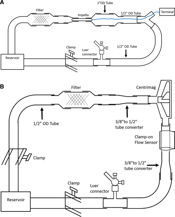
Design of the Impella 5.5 (A) and Centrimag (B) propelled circulatory loops.
Mock loop operation and blood sample processing
Before blood runs, mock loops were recirculated with saline, and a pressure difference was established at 60 mmHg for Impella 5.5 and 350 mmHg for Centrimag, as indicated by two pressure sensors (Omega Engineering Inc., Norwalk, CT) located at inflow and outflow pump regions.12 The Impella 5.5 pump was purged with 5% glucose solution containing 50 unit/ml heparin (Sigma-Aldrich, St. Louis, MO) according to the Impella Device Instructions for Use Product manual. The flow rate for both devices was maintained at 4.5 L/min. Then, saline was fully drained from the loops, and 750 ml of prewarmed blood collected from the same animal donor was loaded in each loop. Loops were de-aired and positioned in two water baths controlled at 37°C. Separately, a 50 ml aliquot of blood was kept at 37°C undisturbed as a “still” control. The pumps were started, and blood was recirculated through the loops for 5 minutes, which was consider as 0 hour time point. Then, blood was recirculated for 4 hours, and timing samples were collected every 30 minutes; in addition, “still” control samples were collected at 0 and 4 hour time points.11,13 Timing blood samples were processed immediately as described below. A 5 ml blood sample was centrifugated at 2,000g for 10 minutes at room temperature to obtain platelet-poor plasma. Plasma aliquots were collected and snap-frozen at –80°C to be examined for hemolysis and soluble markers of platelet activation. Other 5 ml of blood was fixed by adding 1% paraformaldehyde (PFA) in PBS with 0.1% BSA in a volume ratio 1:1 and incubated for 1 hour at room temperature as recommended elsewhere.14,15 PFA-fixed blood was then centrifuged at 150g for 15 minutes at room temperature to obtain PFA-fixed platelet rich plasma (PRP). Fixed PRP samples were stained with fluorescein-conjugated antibodies and proteins for flow cytometry detection of surface markers of platelet activation.
Hemolysis
The extent of hemolysis from exposure of porcine blood to shear stress in the device-operated mock loops was examined by determining the level of plasma free hemoglobin (pf-Hb) and lactate dehydrogenase (LDH) released in plasma from damaged red blood cells. Pf-Hb was measured on a HemoCue Plasma/Low Hb photometer (Ängelholm, Sweden) and using a colorimetric assay (CAYMAN Chemicals, Ann Arbor, MI) following the manufacturer’s instructions. LDH activity was tested using the colorimetric assay LDH-Cytotox (BioLegend, San Diego, CA).
Flow cytometry detection of surface markers of platelet activation and microparticles
Flow cytometric detection of platelet surface glycoproteins was performed following published recommendations.16,17 A 20 µL aliquot of PFA-fixed PRP was mixed with 80 µL of PBS containing 1% BSA and PE-conjugated anti-CD62P (1:100, clone Psel.KO2.5; eBioscience, San Diego, CA) staining P-selectin exposed on platelets as a result of α-granule secretion, or FITC-annexin V (1:20; Invitrogen, Waltham, MA) staining negatively charged phospholipids appearing on the platelet surface as a result of membrane depolarization after platelet activation. Samples were incubated in the dark for 30 minutes at room temperature and 900 µL of PBS with 1% BSA was added to each sample. Flow cytometry was conducted on FACSCanto II (BD Biosciences, San Jose, CA). Single platelets were distinguished from MPs based on their forward/side scatter characteristics, and 10,000 gated events were acquired. Flow cytometry data were analyzed using FCS Express 3 software (De Novo Software, Pasadena, CA). Marker-positive platelets or MPs were identified within platelet- or microvesicle-sized population based on their median fluorescence intensity (MFI) as compared with a nonstained control. The arbitrary number of platelets and MPs (expressed in % from 10,000 gated events) and their MFI were calculated.
Enzyme-linked immunosorbent assay detection of soluble markers of platelet activation and vWF
The concentration of soluble markers of platelet activation, β-thromboglobulin (β-TG), and soluble P-selectin (sP-selectin) was quantified using commercial enzyme-linked immunosorbent assay (ELISA) kits: Pig β-TG ELISA kit (MyBioSource, San Diego, CA) and Porcine sP-selectin ELISA kit (Biotang, Inc., Albuquerque, NM). As the concentration of soluble markers of platelet activation in porcine plasma largely varies, each kit was titrated to identify the optimal dilution of the plasma sample within 1× to 100× dilution range. Titration results revealed that no dilution of the sample was required. ELISAs were performed according to the manufacturer’s instructions. Optical density (λ = 450 nm) was measured on Versa MAX microplate reader (Molecular Devices Corp., Sunnyvale, CA). Calibration curves and protein concentration calculations were made using SoftMax Pro6 software (Molecular Devices Corp.).
vWF antigen (vWF:Ag) concentration and collagen binding were tested using in-house ELISA18 and vWF collagen binding assay (vWF:CBA)19 at Cornell University Animal Health Diagnostic Center (Ithaca, NY). Results were reported as % vWF:Ag or vWF:CBA of an internal assay standard (canine pooled plasma vWF:Ag = 100%).
Statistical analysis
Results from three independent experiments using blood from three different animal donors were summarized in plots. Pf-Hb, LDH, ELISA, and flow cytometry samples were run in duplicate. The statistical significance of hemocompatibility parameters’ alterations over time was evaluated using one-way repeated measures analysis of variance (ANOVA) with Dunnett’s multiple comparison test from GraphPad 8 Prism software (GraphPad Software Inc., San Diego, CA). In addition, the difference between the Impella 5.5 and Centrimag performance was evaluated using two-tail paired t-test, as recommended by ATFM F1841-19.10 Averages are reported as the mean ± standard deviation (SD). The level of statistical significance is indicated on figures as *,#p < 0.05 and **,##p < 0.01.
Results
Our in vitro hemocompatibility study of the Impella 5.5 and Centrimag aimed at objectively comparing the effect of device operational performance on red blood cell, platelet, and protein integrity in hemodynamic conditions similar to the worse clinical use scenarios in vivo. Specifically, we investigated the impact of each system and its means of propulsion on red blood cells—examining their damage (hemolysis) via pf-Hb and LDH activity in plasma, and on platelets—measuring activation markers (soluble and membrane bound) and quantifying platelet-derived microvesicles. We evaluated the effect of Impella 5.5 and Centrimag on vWF degradation by screening alteration of its concentration and intensity of collagen binding over the time of blood recirculation in the device-propelled loops.
Impella 5.5 and Centrimag induced minor hemolysis with similar trends of plasma free hemoglobin increment with no increase in LDH activity
The level of hemolysis induced by Impella 5.5 and Centrimag was evaluated by measuring pf-Hb concentration and LDH activity, both standard markers of red blood cell damage.10,11 We found that both Impella 5.5 and Centrimag induced mild hemolysis, with similar trends of pf-Hb elevation. Thus, 4-hour recirculation of whole blood in the Impella 5.5 loop led to a 2- and 2.5-fold increase of pf-Hb as indicated by colorimetric test and Hemocue photometer, respectively (Figure 2, A and B). A statistically significant increase of pf-Hb was detected after 120 minutes (colorimetric assay) and 210 minutes (HemoCue) of recirculation. Similarly, 4-hour recirculation within the Centrimag loop resulted in 1.7- and 2.5-fold increase of pf-Hb, as indicated by colorimetric test and Hemocue photometer, correspondingly. A statistically significant increment was registered at 150 minute (HemoCue) and 210 minute (colorimetric assay) time points (Figure 2, A and B).
Figure 2.
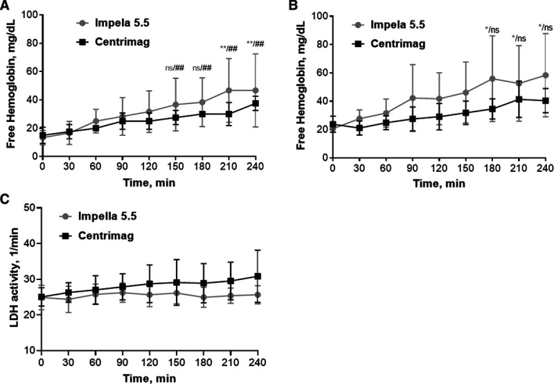
The Impella 5.5 and Centrimag induced minor hemolysis showing similar trend of plasma free hemoglobin (pfHb) increment and no increase of LDH activity over time. A and B, pfHb concentration detected by HemoCue Plasma/Low Hb photometer and colorimetric assay (CAYMAN Chemicals) respectively; C, plasma LDH activity (reported as OD405/min). Data of three experiments are reported as mean ± SD, one-way repeated measures ANOVA with Dunnett’s multiple comparison test: ns or no mark: p > 0.05, *,#p < 0.05, **,##p < 0.01 as compared with “0” time point. No statistically significant difference was found between devices for pfHb and LDH (two-tailed paired t-test, p > 0.05). LDH, lactate dehydrogenase; SD, standard deviation.
Interestingly, neither Impella 5.5 nor Centrimag caused a significant increase of plasma LDH activity (Figure 2C). After 4-hour recirculation in the Impella 5.5 loop, plasma LDH activity remained constant (25.65 ± 2.54 vs. 24.87 ± 3.41/min at 0 minute). The Centrimag tended to increase baseline plasma LDH activity, though no significant difference was detected under our experimental conditions (25.04 ± 2.57 vs. 30.83 ± 7.31/minute at 0 minute, ANOVA: p = 0.49).
The Impella 5.5 and Centrimag caused minor or no alterations of platelet activation markers
We found that neither Impella 5.5 nor Centrimag induced alteration of plasma levels of soluble markers of platelet activation, β-TG, and sP-selectin (Figures 3A; see Figure S1, Supplemental Digital Content, http://links.lww.com/ASAIO/A548). Thus, after 4 hour recirculation of porcine blood via Impella 5.5 and Centrimag propelled loops, concentration of β-TG remained essentially unchanged: 0.24 ± 0.10 ng/ml for baseline vs. 0.21 ± 0.03 ng/ml and 0.19 ± 0.09 ng/ml for 4 hour Impella 5.5 and Centrimag, respectively (Figure 3A). Similarly, the level of sP-selectin in porcine plasma was below the detection limit of the commercial kit (0.2 ng/ml) at baseline and remained undetectable even after 4 hour recirculation within the tested loops (Figure S1, Supplemental Digital Content, http://links.lww.com/ASAIO/A548). Furthermore, the level of membrane-bound P-selectin on platelet surface, as quantified by flow cytometry, was also not increased after exposure to shear stress generated by tested devices. On the contrary, the recirculation of blood in both device propelled loops led to a minor but continuous decline of platelet surface P-selectin over shear exposure time (Figure 3B). The MFI of P-selectin-positive platelets in nonsheared control was very low and further decreased significantly after 2 hours and reached 252.4 ± 3.8 and 246.2 ± 3.3 arbitrary units (AU) after 4 hours of shear exposure for Impella 5.5 and Centrimag, respectively, as compared with 272.9 ± 6.3 AU for nonsheared control “0 Still” (ANOVA: p < 0.01). Interestingly, in the nonsheared control sample kept undisturbed for 4 hours, the level of P-selectin MFI significantly increased (272.9 ± 6.3 AU for 0 Still vs. 309.7 ± 19.8 AU for 240 Still; ANOVA: p < 0.01), indicating mild P-selectin exposure over storage time (Figure 3B).
Figure 3.
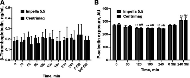
Alteration of platelet activation markers, β-thromboglobulin (A) and P-selectin (B), after blood recirculation in the Impella 5.5 and Centrimag circulatory loops. β-Thromboglobulin level in blood plasma was assessed by ELISA. P-selectin exposure on platelet surface was detected by flow cytometry and reported as MFI of marker-positive platelets. Data of three experiments are reported as mean ± SD, one-way repeated measures ANOVA with Dunnett’s multiple comparison test: **,##p < 0.01 as compared with nonsheared control “0 Still” for Impella 5.5 and Centrimag, respectively. No statistically significant difference was found between devices for β-thromboglobulin and P-selectin (two-tailed paired t-test, p > 0.05). ANOVA, analysis of variance; ELISA, enzyme-linked immunosorbent assay.
Externalization of anionic phospholipids on platelet surface, as a specific marker of shear-mediated platelet activation,20 was examined using annexin V binding assay and quantified as MFI of annexin V–positive population. We showed that platelet annexin V binding gradually increased over the shear exposure time for both devices (Figure S2, Supplemental Digital Content, http://links.lww.com/ASAIO/A548). For Impella 5.5, the statistically significant increase of annexin V MFI was registered even after 1 hour of recirculation striking its maximum at 4 hour time point (4,799.0 ± 908.4 AU and 10,000.0 ± 1,640.0 AU, respectively, vs. 3,582.0 ± 332.0 AU at nonsheared control “0 Still”). For Centrimag, annexin V binding also tended to increase over shear exposure time, but notable augmentation of MFI was only detected after 4 hour recirculation (Figure S2, Supplemental Digital Content, http://links.lww.com/ASAIO/A548, “0 Still” vs. “240 min”; ANOVA: p < 0.01). Of note, the dramatic increase of annexin V binding after 4 hour shear exposure was not only identical for both devices but was also replicated in the nonsheared control sample. Thus, after 4 hour incubation of the undisturbed blood sample at 37°C, the annexin V fluorescence escalated from 3,582.0 ± 332.0 AU at baseline up to 10,817.0 ± 2,156.0 AU at 240 minutes (Figure S2, Supplemental Digital Content, http://links.lww.com/ASAIO/A548, “0 Still” vs. “240 Still”; ANOVA: p < 0.01).
The Impella 5.5 and Centrimag slightly decreased platelet number and promoted generation of microparticles
In our study, MPs were analyzed in PFA-fixed PRP obtained from sheared blood samples immediately after shear exposure in the device-propelled loops. Illustrative dot diagrams of platelet and microparticle distribution after whole blood exposure to device-related shear are reported in Figure S3, Supplemental Digital Content, http://links.lww.com/ASAIO/A548. We found that after 4 hour recirculation of blood within the Impella 5.5 and Centrimag-propelled circulatory loops the number of platelets was slightly but statistically significantly reduced. In both cases, the platelet count drop occurred within the 1 hour of shear exposure and then largely remained unaltered (Figure 4A). The number of MPs was also increased in both Impella 5.5 and Centrimag-sheared samples as compared with nonsheared control (Figure 4B). Similar to the platelet count drop, for both devices, the microparticle increase occurred by the first hour of shear exposure and then remained steady for Impella 5.5, though gradually increased for Centrimag reaching, respectively, 27.4 ± 4.8% and 35.8 ± 9.0%, after 4 hour recirculation time, as compared with 18.3 ± 1.8% in the nonsheared control. The Impella 5.5 was shown to be less damaging, demonstrating a lower degree of platelet count decrease (p < 0.01) and MP generation (p < 0.05), as compared with Centrimag.
Figure 4.
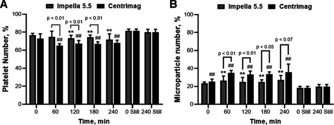
The number of platelets slightly decreased while the number of microparticles barely increased after exposure of whole blood to shear stress in the Impella 5.5 and Centrimag circulatory loops. A, Arbitrary number of platelets; B, arbitrary number of microparticles captured by flow cytometry. Data of three experiments are reported as mean ± SD, one-way repeated measures ANOVA with Dunnett’s multiple comparison test: **,##p < 0.01 as compared with nonsheared control “0 Still” for Impella 5.5 and Centrimag, respectively. Statistically significant difference was found between devices for number of platelets and microparticles (two-tailed paired t-test). ANOVA, analysis of variance; SD, standard deviation.
When phenotyping the microparticle pool ejected as a result of blood recirculation within device-propelled loops, we have found that the P-selectin-positive microparticle population remained steady over the shear exposure time in the case of Centrimag (14.5 ± 4.5% vs. 15.25 ± 3.76 in nonsheared control) or even slightly decreased with Impella 5.5 (9.85 ± 0.7%, ANOVA: p < 0.05). In the nonsheared control, P-selectin-positive microparticle number tended to drop, though no significant difference was detected (Figure 5A). The MFI of P-selectin-positive MPs also remained unaltered (data not shown). Yet, the number of annexin V–positive MPs, indicating a procoagulant MP population, incrementally increased over shear exposure time reaching its maximum at the 4 hour time point (Figure 5B). The MFI of the annexin V–positive MPs was also nearly 1.5-fold increased, reaching 2,148.3 ± 571.8 AU and 1,818.4 ± 292.0 AU for the Impella 5.5 and Centrimag, respectively, as compared with 1,148.7 ± 384.4 AU for the nonsheared control. Admittedly, the Centrimag shear exposure led to higher numbers of annexin V–positive MPs than Impella 5.5 (Figure 5B; ANOVA: p < 0.01).
Figure 5.
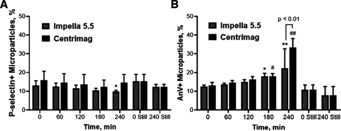
The Impella 5.5 and Centrimag did not promote ejection of P-selectin positive microparticles, while increasing the number of procoagulant annexin V–positive microparticles. A, Arbitrary number of P-selectin-positive microparticles; B, arbitrary number of annexin V–positive microparticles, both reported as % of all captured events. Data of three experiments are reported as mean ± SD, one-way repeated measures ANOVA with Dunnett’s multiple comparison test: *,#p < 0.05, **,##p < 0.01 as compared with non-sheared control “0 Still” for Impella 5.5 and Centrimag, respectively. No statistically significant difference was found between devices for P-selectin+ and annexin V+ microparticles, other than annexin V+ microparticles at “240” time point (two-tailed paired t-test, p = 0.006). ANOVA, analysis of variance; SD, standard deviation.
The Impella 5.5 and Centrimag did not affect vWF concentration while notably decreased its collagen binding
Premature degradation of vWF multimers during MCS and resultant quantitative and functional vWF deficiency have been long recognized as major contributors to pathophysiology of device-related bleeding complications.21,22 Thus, we examined the effect of the Impella 5.5 and Centrimag on plasma concentration of vWF antigen (vWF:Ag), as well as its collagen binding (vWF:CBA), indicating quantity and functional activity of the vWF pool. We showed that within 4 hours of blood recirculation in device-propelled circulatory loops neither Impella 5.5 nor Centrimag resulted in a significant decrease of plasma vWF:Ag (Figure 6A). Of note, a minor spike of vWF concentration could be noticed at 180 minutes of recirculation in the Centrimag (but not Impella 5.5) loop.
Figure 6.
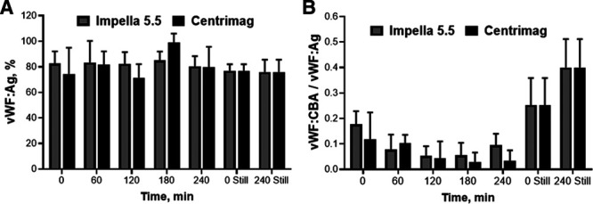
Neither Impella 5.5 nor Centrimag significantly altered plasma level of von Willebrand factor antigen (vWF:Ag) while facilitating a notable decrease of its collagen binding ability (vWF:CBA) over shear exposure time. vWF:Ag and vWF:CBA were measured using in-house ELISA. Results are reported as % vWF:Ag of an internal assay standard (canine pooled plasma vWF:Ag = 100%). vWF:CBA/vWF:Ag index indicates functional activity of vWF. Data of three experiments are reported as mean ± SD, one-way repeated measures ANOVA with Dunnett’s multiple comparison test: no mark: p > 0.05 as compared with nonsheared control “0 Still.” No statistically significant difference was found between devices for vWF:Ag and vWF:CBA (two-tailed paired t-test, p > 0.05). ANOVA, analysis of variance; ELISA, enzyme-linked immunosorbent assay; SD, standard deviation.
However, a significant drop of vWF:CBA, indicating a decrease in functional activity as a result of structural degradation, was registered for both devices. We found that collagen binding of porcine vWF in the nonsheared “0 Still” sample was initially low (26.3 ± 12.7% as compared with an internal assay standard, canine pooled plasma with vWF:CBA = 100%). Then, during blood recirculation in the Impella 5.5 loop, the vWF:CBA dropped to 5.5 ± 6.3% at 210 minutes, but then recovered to 11.7 ± 1.5% at 240 minutes. For the Centrimag loop, the vWF:CBA dropped irreversibly to 3.3 ± 3.6% at 180 minute. Resultant fluctuations of vWF:CBA/vWF:Ag index, a routine clinical indicator of vWF functional activity, are reported in Figure 6B. When vWF:CBA of sheared samples were normalized to the nonsheared control “0 Still,” a statistically significant drop was detected in Impella 5.5 loop after 1 hour recirculation with subsequent recovery to almost 50% of initial level (see Supplemental Material, Figure S4, Supplemental Digital Content, http://links.lww.com/ASAIO/A548). Conversely, the Centrimag caused an immediate statistically significant drop of vWF:CBA which steadily decreased to barely detectable levels.
Discussion
Continued improvement of MCS design has led to the advance of increasingly effective, miniaturized catheter devices for large volume, rapid hemodynamic restoration. We aimed to examine the hemocompatibility of the new Impella 5.5, in comparison with a reference device, the Centrimag. In our study, we examined hemolytic potential—a “must-have” marker of device hemocompatibility as per FDA requirements. Beyond this, we also evaluated the effect of the Impella 5.5 and Centrimag on platelet function, microparticle generation, and degradation of vWF, all vital components of hemostatic system which are rarely evaluated with standard preclinical testing of MCS systems.
Analyzing the hemolytic potential of the compared devices, we found that both the Impella 5.5 and Centrimag caused minor increases in pf-Hb, though with no increase in LDH activity. Multiple in vitro studies testing Centrimag hemocompatibility showed similar levels of hemolysis as indicated by an increase in pf-Hb.23–25 Poor correlation of LDH with pf-Hb in clinical cases of hemolysis of Impella 5.0–supported patients has been reported previously.26 Plasma levels of LDH in patients with cardiogenic shock may already be elevated due to myocardial injury suggesting low specificity of LDH as a hemolysis marker. Yet, given the low specificity and questionable sensitivity, LDH remains a popular clinical indicator of hemolysis partially due to the limited access of some centers to benchtop pf-Hb assays. Of note, here we demonstrated that pump-related increase of pf-Hb can be easily detected by the point-of-care photometer HemoCue Plasma/Low Hb. Although the Hemocue photometer demonstrated lower sensitivity in low range pf-Hb concentration, as compared with the bench top colorimetric assay, underestimating baseline pf-Hb, it showed almost identical pf-HB levels at the 4 hour end-point (Figure 2A versus Figure 2B; 0 and 240 minutes) and detected statistically significant pf-Hb increment associated with device-related shear exposure. Considering the short loop operation time in vitro (4 hours) as compared with several days/weeks for patient on MCS, we believe that Hemocue could be used to efficiently monitor pump-related hemolysis at the bedside.
Platelets are active participants and regulators of hemostasis, angiogenesis, and as recently recognized, inflammation and the immune response.27 When activated with biochemical or mechanical stimuli, platelets release a number of proteins and inflammatory factors required to manifest clotting and inflammation while limiting fibrinolysis and vascular leakage. To monitor platelet degranulation, we employed a robust approach such as testing surface bound markers of platelet degranulation, using immunostaining and flow cytometry, as well as soluble proteins released in plasma, using ELISA. We found that Impella 5.5 and Centrimag did not promote platelet degranulation, as no increase of plasma β-TG and sP-selectin, conventional markers of platelet activation and α-granule release, was detected. It is noteworthy that baseline levels of β-TG and sP-selectin in porcine plasma were much lower than those described for humans28,29 while correlating with recently reported porcine ones.30 We also showed that surface expression of P-selectin (CD62P) on platelets, as monitored by flow cytometry, was not elevated after blood recirculation within the device-propelled loops. Conversely, a mild downregulation of P-selectin surface expression occurred during long-term shear exposure (Figure 3B). The observed decrease of P-selectin surface expression is unlikely explained by the shedding of this protein, as no elevation of plasma sP-selectin was detected. Rather, we speculate that shear-mediated platelet dysfunction is associated with shear-compromised platelet granule release. As a relevant example, the study of Geisen et al.31 evaluating the pathophysiology of bleeding complications in LVAD-supported patients (HMII and HMIII), showed that 91% of patients developed impaired granule secretion as indicated by decreased surface expression of CD62P and CD63 on their platelets. Along with acquired vWF deficiency, impaired granule secretion was named as one of the risk factors of device-related bleeding complications.31 Similarly, Dewald et al.32 reported a decrease of platelet CD62P expression in patients supported with the Novacor VAD or Berlin Heart, though “no relation between expression of platelet activation markers and bleeding time ex vivo was found.”
MCS and ECMO systems are associated with a decrease of platelet count not related to heparin administration.33,34 Thrombocytopenia requiring platelet transfusion was also noted in patients supported with Impella systems.35,36 Therefore, we examined whether Impella 5.5 and Centrimag induce alteration of platelet number and generation of MPs; in addition, the surface marker phenotype of generated MPs was analyzed. We found that both devices induced a minor platelet count drop occurring within first hours of recirculation; the Centrimag showed more pronounced platelet count decline than the Impella 5.5 (Figure 4A). Simultaneously, we observed a gradual increase of the annexin V binding in the platelet population over the shear exposure time. Annexin V binding is usually regarded as a marker of membrane depolarization during platelet apoptosis.37,38 Yet, we have previously shown that externalization of anionic phospholipids (as indicated by annexin V binding) is a characteristic signature of shear-mediated platelet activation as opposed to platelet activation by biochemical agonists, for example, ADP and thrombin.20 Indeed, Mondal et al.39,40 reported that markers of platelet apoptosis and oxidative stress were elevated in bleeders versus non-bleeders among patient cohort supported by CF-VADs. However, we noted that annexin V binding was also elevated in the nonsheared control (see Figure S2, Supplemental Digital Content, http://links.lww.com/ASAIO/A548, “240 Still” sample) suggesting that the shear-mediated increase of platelet annexin V binding observed in our study could be at least partially attributed to in vitro platelet “aging” occurring in blood samples during long-term storage and handling.41,42 Thus, Block et al. showed that alterations of platelet function in freshly collected heparin-anticoagulated human blood occurred even within four hours of storage, which reiterates the importance of utilization of freshly collected blood for hemocompatibility testing.13
Shear-mediated alterations of platelet morphology result in platelet fragmentation and ejecting of MPs.43 Microparticles are submicron membrane vesicles generated by platelets and other blood cells, recently been declared to play a major role in promoting coagulation, inflammation, and immune response.44 We found that the number of circulating MPs slightly increased with exposure time in both devices, with Centrimag showing significantly higher rate of MP generation (Figure 4B). As depicted in Figure 5B, the majority of shear-ejected MPs were annexin V positive which indicated their procoagulant phenotype, an ability to bind coagulation factors and usher thrombin generation. The number of procoagulant MPs ejected in the Centrimag propelled loop were nearly two times greater as in the Impella 5.5 loop. Our in vitro findings suggest that the increase of procoagulant MPs and associated intensification of thrombin generation might be considered as predecessors of MCS-related thrombotic complications in device-supported patients. Recent clinical reports indeed validate our claim demonstrating a strong correlation between the number of circulating MPs and occurrence of adverse events in patients implanted with VADs.45–48 Using mass spectrometry, McClane et al.47 identified unique correlations between MPs and MP procoagulant activity in plasma from VAD-supported patients, suggesting their ability to predict thrombotic events and differentiate them from other hemostatic events. Nascimbene et al.49 found that increased levels of procoagulant MPs was associated with the high risk of adverse events even 3 months after the VAD implantation. In their review, Ivak et al.45 concluded that quantification and phenotyping of circulating MPs, as correlated with clinical data, could be used for monitoring of vascular status in patients with VADs to detect and manage short-term and long-term adverse events.
Along with the shear-compromised platelet count and function, acquired deficiency of vWF is another well-recognized contributor to the etiology of MCS-related bleeding complications.21,22,34 Therefore, we have analyzed the effect of shear stress generated by the Impella 5.5 and Centrimag on plasma levels of vWF:Ag and its collagen binding as indicators of intact vWF structure and function. We observed no significant decrease of vWF antigen concentration over the shear exposure time, rather a slight increase of vWF was noted at 180 minutes of recirculation for Centrimag. A similar spike of vWF concentration was reported in a previous in vitro study testing the hemocompatibility of HMIII using heparinized porcine blood.50 We speculate that such an increase could be attributed to vWF release from platelet α-granules. Why did we observe shear-mediated increase of plasma vWF concentration but not other α-granule proteins? The content of vWF in platelet α-granules is reportedly higher than other proteins, including β-TG and P-selectin.51 Furthermore, it has been shown that porcine granules have even higher load of vWF multimers than humans.52 Another explanation for the observed phenomena could be selective release of platelet α-granules storing various coagulation factors and cytokines in response to different activation stimuli.53,54
Also, we found that both tested devices promote notable and rapid degradation of vWF multimers as indicated by the decrease of vWF collagen binding activity. Thus, significant decline of vWF:CBA and correspondent vWF:CBA/vWF:Ag index (see Figure S4, Supplemental Digital Content, http://links.lww.com/ASAIO/A548; Figure 6B) was observed within the first hour indicating fast-track of vWF disruption. The greater extent of vWF:CBA decrease was registered in the case of Centrimag, while Impella 5.5 showed less steep decline. Similar dynamics of human vWF degradation in a mock circulatory loop propelled with the Impella-CP or 5.0 was described by Vincent et al.55 Thus, the rapid time-dependent decrease of vWF multimers capable to bind collagen (>60% at 5 minutes and >90% at 30 minutes of recirculation) coincided with the dramatic loss of vWF collagen binding activity, as indicated by decrease of vWB:CBA/vWB:Ag index (0.5 at 5 minutes and 0.35 at 30 minutes vs. 1 at the baseline).55 Whether shear-mediated vWF degradation during MCS occurs due to direct mechanical damage of the molecule or ADAMTS13-mediated proteolysis remains a topic of debate.56,57 Nevertheless, in vitro studies reveal that unfolding of vWF molecule, necessary for proteolysis, and further cleavage by ADMATS13 could occur within 200 s in response to acute changes in shear conditions.58
We recognize a few methodological limitations which however do not diminish the significance of this study. Freshly obtained porcine blood was anticoagulated with heparin (in vivo) and CPDA. Such dual anticoagulation is expected to significantly inhibit shear-mediated thrombin generation59 and therefore thrombin-mediated platelet activation and clotting. Thus, we believe that the phenomena reported in our study are largely caused by device-generated shear stress and not due to paracrine platelet activation by thrombin. The number of experiments was limited by three repetitions, which however represent good reproducibility. Three brand-new clinical grade Impella 5.5 disposable devices were used for comparison with one brand-new clinical grade Centrimag system. Due to the lack of commercially available fluorescein-bound antiporcine CD41 antibodies, that specifically stain platelets and platelet-derived MPs, the specificity of flow cytometric detection of platelet-derived MPs in swine is somewhat limited. To overcome this limitation, in our study we have applied size gating and two immunofluorescent stains, anti-human CD62P, clone Psel.KO2.5 showing significant cross reactivity with swine23 and annexin V showing multispecies cross reactivity. Therefore, circulating MPs monitored in our study, though largely platelet-derived, might contain a small portion of other blood cell derivatives.
Conclusion
The Impella 5.5 and Centrimag showed low hemolytic potential as indicated by minor increment of pf-Hb with no alteration of plasma LDH activity. Thus, pf-Hb but not LDH is recommended as a sensitive and specific marker of hemolysis induced by these devices. Furthermore, our study shows that device-related hemolysis can be successfully measured with the point-of-care photometer HemoCue Plasma/Low Hb. In addition, both tested devices did not induce platelet degranulation, as assessed by soluble and surface bound protein markers, rather minor downregulation of platelet surface P-selectin was observed suggesting impaired degranulation of sheared platelets. Blood exposure to shear stress via both Centrimag and Impella resulted in a minor decrease of platelet count over time, with MP release and a decrease of vWF functional activity. Interestingly, significantly greater MP generation was observed overtime with the Centrimag relative to the Impella 5.5. Thus, the Impella 5.5 despite having a lower profile and higher rotation speed demonstrated good and equivalent hemocompatibility, in comparison with Centrimag, with the advantage of lower MP generation.
Acknowledgment
The authors would like to express their gratitude to Dr. Marjory Brooks, Animal Health Diagnostic Center, Cornell University for testing vWF:Ag and vWF:CBA; to Dr. Cynthia J. Doane from University Animal Care and Alice McArthur from Resuscitation Research Laboratory, Sarver Heart Center, University of Arizona, for animal surgery and blood collection; and to John J. Jackson Sr. from ACABI, University of Arizona, for administrative support of the project.
Footnotes
Disclosure: The authors have no conflicts of interest to report.This research was funded by the research grant from Abiomed Inc., and Arizona Center for Accelerated Biomedical Innovation (ACABI) of the University of Arizona.
Supplemental digital content is available for this article. Direct URL citations appear in the printed text, and links to the digital files are provided in the HTML and PDF versions of this article on the journal’s Web site (www.asaiojournal.com).
References
- 1.Bartoli CR, Dowling RD: Next-generation mechanical circulatory support devices in different phases of development and clinical use are anticipated to improve outcomes and quality of life in patients with advanced heart failure. The next wave of mechanical circulatory support. Dev Card Interv Today. 201913:53–59. [Google Scholar]
- 2.Esmailian F, Dimbil S, Levine R, et al. : Temporary MCS versus durable MCS as a bridge to heart transplantation. J Hear Lung Transplant. 201938:S344 [Google Scholar]
- 3.Gilotra NA, Stevens GR: Temporary mechanical circulatory support: A review of the options, indications, and outcomes. Clin Med Insights Cardiol. 20158:75–85. [DOI] [PMC free article] [PubMed] [Google Scholar]
- 4.Prondzinsky R, Lemm H, Swyter M, et al. : Intra-aortic balloon counterpulsation in patients with acute myocardial infarction complicated by cardiogenic shock: The prospective, randomized IABP SHOCK Trial for attenuation of multiorgan dysfunction syndrome. Crit Care Med. 201038:152–160. [DOI] [PubMed] [Google Scholar]
- 5.Thiele H, Zeymer U, Neumann FJ, et al. ; IABP-SHOCK II Trial Investigators: Intraaortic balloon support for myocardial infarction with cardiogenic shock. N Engl J Med. 2012367:1287–1296. [DOI] [PubMed] [Google Scholar]
- 6.Bartlett RH, Ogino MT, Brodie D, et al. : Initial ELSO guidance document. ASAIO J. 202066:472–474 [DOI] [PMC free article] [PubMed] [Google Scholar]
- 7.Roberts N, Chandrasekaran U, Das S, et al. : Hemolysis associated with Impella heart pump positioning: In vitro hemolysis testing and computational fluid dynamics modeling. Int J Artif Organs. 2020391398820909843. [DOI] [PubMed] [Google Scholar]
- 8.Goldstein DJ, Meyns B, Xie R, et al. : Third annual report from the ISHLT mechanically assisted circulatory support registry: A comparison of centrifugal and axial continuous-flow left ventricular assist devices. J Hear Lung Transplant. 201938:352–3632019. [DOI] [PubMed] [Google Scholar]
- 9.Li M, Walk R, Roka-Moiia Y, et al. : Circulatory loop design and components introduce artifacts impacting in vitro evaluation of ventricular assist device thrombogenicity: A call for caution. Artif Organs. 202044:E226–E237. [DOI] [PMC free article] [PubMed] [Google Scholar]
- 10.ASTM:F1841-19: Standard Practice for Assessment of Hemolysis in Continuous Flow Blood Pumps, 2019.
- 11.ISO 10993-4:2017: Biological evaluation of medical devices—Part 4: Selection of tests for interactions with blood (ISO 10993-4:2017) 2017.
- 12.Zhang J, Gellman B, Koert A, et al. : Computational and experimental evaluation of the fluid dynamics and hemocompatibility of the Centrimag blood pump. Artif Organs. 200630:168–177. [DOI] [PubMed] [Google Scholar]
- 13.Blok SL, Engels GE, van Oeveren W: In vitro hemocompatibility testing: The importance of fresh blood. Biointerphases. 201611:029802. [DOI] [PubMed] [Google Scholar]
- 14.Schmidt V, Hilberg T: ThromboFix platelet stabilizer: Advances in clinical platelet analyses by flow cytometry? Platelets. 200617:266–273. [DOI] [PubMed] [Google Scholar]
- 15.Hu H, Daleskog M, Li N: Influences of fixatives on flow cytometric measurements of platelet P-selectin expression and fibrinogen binding. Thromb Res. 2000100:161–166. [DOI] [PubMed] [Google Scholar]
- 16.Schmitz G, Rothe G, Ruf A, et al. : European working group on clinical cell analysis: Consensus protocol for the flow cytometric characterisation of platelet function. Thromb Haemost. 199879:885–896. [PubMed] [Google Scholar]
- 17.Pieper IL, Radley G, Thornton CA: Multidimensional flow cytometry for testing blood-handling medical devices, in Multidimensional Flow Cytometry Techniques for Novel Highly Informative Assays., vol 2018, pp. 2 InTech 63–79. [Google Scholar]
- 18.Benson RE, Catalfamo JL, Dodds WJ: A multispecies enzyme-linked immunosorbent assay for von Willebrand’s factor. J Lab Clin Med. 1992119:420–427. [PubMed] [Google Scholar]
- 19.Sabino EP, Erb HN, Catalfamo JL: Development of a collagen-binding activity assay as a screening test for type II von Willebrand disease in dogs. Am J Vet Res. 200667:242–249. [DOI] [PubMed] [Google Scholar]
- 20.Roka-Moiia Y, Walk R, Palomares DE, et al. : Platelet activation via shear stress exposure induces a differing pattern of biomarkers of activation versus biochemical agonists. Thromb Haemost. 2020120:776–792. [DOI] [PMC free article] [PubMed] [Google Scholar]
- 21.Crow S, Chen D, Milano C, et al. : Acquired von Willebrand syndrome in continuous-flow ventricular assist device recipients. ATS. 201090:1263–1269. [DOI] [PubMed] [Google Scholar]
- 22.Rauch A, Susen S, Zieger B: Acquired von Willebrand Syndrome in patients with ventricular assist device. Front Med. 20196:7. [DOI] [PMC free article] [PubMed] [Google Scholar]
- 23.Chan CH, Pieper IL, Hambly R, et al. : The Centrimag centrifugal blood pump as a benchmark for in vitro testing of hemocompatibility in implantable ventricular assist devices. Artif Organs. 201539:93–101. [DOI] [PMC free article] [PubMed] [Google Scholar]
- 24.Sobieski MA, Giridharan GA, Ising M, Koenig SC, Slaughter MS: Blood trauma testing of centrimag and rotaflow centrifugal flow devices: A pilot study. Artif Organs. 201236:677–682. [DOI] [PubMed] [Google Scholar]
- 25.Chan CHH, Pieper IL, Robinson CR, Friedmann Y, Kanamarlapudi V, Thornton CA: Shear stress-induced total blood trauma in multiple species. Artif Organs. 201741:934–947. [DOI] [PubMed] [Google Scholar]
- 26.Esposito ML, Morine KJ, Annamalai SK, et al. : Increased plasma-free hemoglobin levels identify hemolysis in patients with cardiogenic shock and a trans valvular micro-axial flow pump. Artif Organs. 201943:125–131. [DOI] [PubMed] [Google Scholar]
- 27.Koupenova M, Clancy L, Corkrey HA, Freedman JE: Circulating platelets as mediators of immunity, inflammation, and thrombosis. Circ Res. 2018122:337–351. [DOI] [PMC free article] [PubMed] [Google Scholar]
- 28.Blann AD, Lip GYH, Islim IF, Beevers DG: Evidence of platelet activation in hypertension. J Hum Hypertens. 199711:607–609. [DOI] [PubMed] [Google Scholar]
- 29.Cella G, Scattolo N, Girolami A, Sasahara AA: Are platelet factor 4 and beta-thromboglobulin markers of cardiovascular disorders? Ric Clin Lab. 198414:9–18. [DOI] [PubMed] [Google Scholar]
- 30.Akeho K, Nakata H, Suehiro S, et al. : Hypothermic effects on gas exchange performance of membrane oxygenator and blood coagulation during cardiopulmonary bypass in pigs [published online ahead of print, 2020 Feb 3] Perfusion. 2020. 026765912090141, doi: 10.1177/0267659120901413. [DOI] [PubMed] [Google Scholar]
- 31.Geisen U, Brehm K, Trummer G, et al. : Platelet secretion defects and acquired von Willebrand syndrome in patients with ventricular assist devices. J Am Heart Assoc. 20187:e006519. [DOI] [PMC free article] [PubMed] [Google Scholar]
- 32.Dewald O, Schmitz C, Diem H, et al. : Platelet activation markers in patients with heart assist device. Artif Organs. 200529:292–299. [DOI] [PubMed] [Google Scholar]
- 33.Subramaniam AV, Barsness GW, Vallabhajosyula S, Vallabhajosyula S: Complications of temporary percutaneous mechanical circulatory support for cardiogenic shock: An appraisal of contemporary literature. Cardiol Ther. 20198:211–228. [DOI] [PMC free article] [PubMed] [Google Scholar]
- 34.Petricevic M, Milicic D, Boban M, et al. : Bleeding and thrombotic events in patients undergoing mechanical circulatory support: A review of literature. Thorac Cardiovasc Surg. 201563:636–646. [DOI] [PubMed] [Google Scholar]
- 35.Nelson D, Mohammed A, Klein E, Joyce D: Impella 5.0 as a bridge to clinical decision making. J Am Coll Cardiol. 201973:705 [Google Scholar]
- 36.Konstantinou K, Keeble TR, Kelly PA, et al. : Protected percutaneous coronary intervention with Impella CP in a patient with left main disease, severe left ventricular systolic dysfunction and established hemolysis. Cardiovasc Diagn Ther. 20199:194–199. [DOI] [PMC free article] [PubMed] [Google Scholar]
- 37.Leytin V, Mykhaylov S, Allen DJ, et al. : Chemical agonists and high shear stress induce apoptosis in human platelets. Blood. 2015104:3875 LP-3875 [Google Scholar]
- 38.Gyulkhandanyan AV, Mutlu A, Freedman J, Leytin V: Markers of platelet apoptosis: Methodology and applications. J Thromb Thrombolysis. 201233:397–411. [DOI] [PubMed] [Google Scholar]
- 39.Mondal NK, Sorensen EN, Hiivala NJ, et al. : Intraplatelet reactive oxygen species, mitochondrial damage and platelet apoptosis augment non-surgical bleeding in heart failure patients supported by continuous-flow left ventricular assist device. Platelets. 201526:536–544. [DOI] [PubMed] [Google Scholar]
- 40.Mondal NK, Li T, Chen Z, et al. : Mechanistic insight of platelet apoptosis leading to non-surgical bleeding among heart failure patients supported by continuous-flow left ventricular assist devices. Mol Cell Biochem. 2017433:125–137. [DOI] [PMC free article] [PubMed] [Google Scholar]
- 41.Mohebali D, Kaplan D, Carlisle M, Supiano MA, Rondina MT: Alterations in platelet function during aging: Clinical correlations with thromboinflammatory disease in older adults. J Am Geriatr Soc. 201462:529–535. [DOI] [PMC free article] [PubMed] [Google Scholar]
- 42.Pleines I, Lebois M, Gangatirkar P, et al. : Intrinsic apoptosis circumvents the functional decline of circulating platelets but does not cause the storage lesion. Blood. 2018132:197–209. [DOI] [PubMed] [Google Scholar]
- 43.Leytin V, Allen DJ, Mykhaylov S, et al. : Pathologic high shear stress induces apoptosis events in human platelets. Biochem Biophys Res Commun. 2004320:303–310. [DOI] [PubMed] [Google Scholar]
- 44.Burnouf T, Goubran HA, Chou ML, Devos D, Radosevic M: Platelet microparticles: Detection and assessment of their paradoxical functional roles in disease and regenerative medicine. Blood Rev. 201428:155–166. [DOI] [PubMed] [Google Scholar]
- 45.Ivak P, Pitha J, Netuka I: Circulating microparticles as a predictor of vascular properties in patients on mechanical circulatory support; hype or hope? Physiol Res. 201665:727–735. [DOI] [PubMed] [Google Scholar]
- 46.Diehl P, Aleker M, Helbing T, et al. : Enhanced microparticles in ventricular assist device patients predict platelet, leukocyte and endothelial cell activation. Interact Cardiovasc Thorac Surg. 201011:133–137. [DOI] [PubMed] [Google Scholar]
- 47.McClane N, Jeske W, Walenga JM, et al. : Identification of novel hemostatic biomarkers of adverse clinical events in patients implanted with a continuous-flow left ventricular assist. Device Clin Appl Thromb. 201824:965–972. [DOI] [PMC free article] [PubMed] [Google Scholar]
- 48.Jeske WP, Walenga JM, Menapace B, Schwartz J, Bakhos M: Blood cell microparticles as biomarkers of hemostatic abnormalities in patients with implanted cardiac assist devices. Biomark Med. 201610:1095–1104. [DOI] [PubMed] [Google Scholar]
- 49.Nascimbene A, Hernandez R, George JK, et al. : Association between cell-derived microparticles and adverse events in patients with nonpulsatile left ventricular assist devices. J Hear Lung Transplant. 201433:470–477. [DOI] [PMC free article] [PubMed] [Google Scholar]
- 50.Woelke E, Klein M, Mager I, et al. : Miniaturized test loop for the assessment of blood damage by continuous-flow left-ventricular assist devices. Ann Biomed Eng. 202048:768–779. [DOI] [PubMed] [Google Scholar]
- 51.Blair P, Flaumenhaft R: Platelet alpha-granules: Basic biology and clinical correlates. Blood Rev. 200923:177–189. [DOI] [PMC free article] [PubMed] [Google Scholar]
- 52.Wilbourn B, Harrison P, Lawrie A, Savariau E, Savidge G, Cramer EM: Porcine platelets contain an increased quantity of ultra-high molecular weight von Willebrand factor and numerous alpha-granular tubular structures. Br J Haematol. 199383:608–615. [DOI] [PubMed] [Google Scholar]
- 53.Sehgal S, Storrie B: Evidence that differential packaging of the major platelet granule proteins von Willebrand factor and fibrinogen can support their differential release. J Thromb Haemost. 20075:2009–2016. [DOI] [PubMed] [Google Scholar]
- 54.Italiano JE, Jr, Richardson JL, Patel-Hett S, et al. : Angiogenesis is regulated by a novel mechanism: Pro- and antiangiogenic proteins are organized into separate platelet alpha granules and differentially released. Blood. 2008111:1227–1233. [DOI] [PMC free article] [PubMed] [Google Scholar]
- 55.Vincent F, Rauch A, Loobuyck V, et al. : Arterial pulsatility and circulating von willebrand factor in patients on mechanical circulatory support. J Am Coll Cardiol. 201871:2106–2118. [DOI] [PubMed] [Google Scholar]
- 56.Bartoli CR, Restle DJ, Zhang DM, Acker MA, Atluri P: Pathologic von Willebrand factor degradation with a left ventricular assist device occurs via two distinct mechanisms: Mechanical demolition and enzymatic cleavage. J Thorac Cardiovasc Surg. 2015149:281–289. [DOI] [PubMed] [Google Scholar]
- 57.Bortot M, Ashworth K, Sharifi A, et al. : Turbulent flow promotes cleavage of VWF (von Willebrand Factor) by ADAMTS13 (a disintegrin and metalloproteinase with a thrombospondin type-1 Motif, member 13) Arterioscler Thromb Vasc Biol. 201939:1831–1842. [DOI] [PMC free article] [PubMed] [Google Scholar]
- 58.Zhang X, Halvorsen K, Zhang CZ, Wong WP, Springer TA: Mechanoenzymatic cleavage of the ultralarge vascular protein von Willebrand factor. Science. 2009324:1330–1334. [DOI] [PMC free article] [PubMed] [Google Scholar]
- 59.Fallon AM, Marzec UM, Hanson SR, Yoganathan AP: Thrombin formation in vitro in response to shear-induced activation of platelets. Thromb Res. 2007121:397–406. [DOI] [PubMed] [Google Scholar]


