

Abstract
The outer membrane is a key virulence determinant of gram‐negative bacteria. In Yersinia pestis, the deadly agent that causes plague, the protein Ail and lipopolysaccharide (LPS)6 enhance lethality by promoting resistance to human innate immunity and antibiotics, enabling bacteria to proliferate in the human host. Their functions are highly coordinated. Here we describe how they cooperate to promote pathogenesis. Using a multidisciplinary approach, we identify mutually constructive interactions between Ail and LPS that produce an extended conformation of Ail at the membrane surface, cause thickening and rigidification of the LPS membrane, and collectively promote Y. pestis survival in human serum, antibiotic resistance, and cell envelope integrity. The results highlight the importance of the Ail–LPS assembly as an organized whole, rather than its individual components, and provide a handle for targeting Y. pestis pathogenesis.
Keywords: Ail, lipopolysaccharide, LPS, MD, nanodisc, nuclear magnetic resonance, structure, Yersinia pestis
We identify mutually constructive interactions between the outer membrane protein Ail and lipopolysaccharide from Yersinia pestis that produce an extended conformation of Ail at the membrane surface, cause thickening and rigidification of the outer membrane, and collectively promote Y. pestis survival in human serum, antibiotic resistance and cell envelope integrity. The results highlight the importance of the Ail‐LPS assembly as an organized whole, rather than its individual components, and provide a handle for targeting Y. pestis pathogenesis.

Abbreviations
- Ail
attachment invasion locus
- CFU
colony forming unit
- DMPC
dimyristoyl‐phosphatidyl‐choline
- DMPG
dimyristoyl‐phosphatidyl‐glycerol
- EL1‐4
extracellular loops 1‐4
- HIB
heart infusion broth
- HIS
heat‐inactivated serum
- HSQC
heteronuclear single‐quantum correlation
- LPS
lipopolysaccharide
- MD
molecular dynamics
- MIC
minimum inhibitory concentration
- MSP
membrane scaffold protein
- NHS
normal human serum
- NMR
nuclear magnetic resonance
- OD620
optical density at 620 nm
- PMB
polymyxin B
- PMSF
phenylmethylsulfonyl fluoride
- SDS‐PAGE
sodium‐dodecyl sulfate polyacrylamide gel electrophoresis
- T1‐4
intracellular turns 1‐4
- TBA base
tryptose blood agar base
- TLR4
Toll‐like receptor 4
- TMH
thoroughly modified Higuchi's media
1. INTRODUCTION
The outer membrane of gram‐negative bacteria functions as a critical barrier against the external environment and as a key mediator of first line of interactions with the host. In the case of Yersinia pestis, the deadly pathogen responsible for precipitating massive human pandemics of plague throughout history, the surface protein Ail and lipopolysaccharide (LPS) have co‐evolved to confer resistance to human innate immunity (Parkhill et al., 2001; Deng et al., 2002; Knirel and Anisimov, 2012; Kolodziejek et al., 2012). Y. pestis depends on this property to survive in the blood meal of its flea vector and produce high‐level septicemia in a wide range of mammalian hosts, including humans (Hinnebusch et al., 2017). Ail and LPS are the two major building blocks of the outer membrane and represent key virulence factors.
Ail has established roles in promoting cell adhesion/invasion, biofilm formation, and resistance to the immune defenses present in human serum (Miller et al., 1990; Bartra et al., 2008; Felek and Krukonis, 2009; Kolodziejek et al., 2010; Hinnebusch et al., 2011). Notably, the upregulation of ail gene expression and the downregulation of genes responsible for LPS acylation are synchronized: at the mammalian infection temperature of 37°C, Y. pestis produces both Ail, to a level well over 30% of the outer membrane proteome (Sebbane et al., 2006; Anisimov et al., 2007; Bartra et al., 2008; Kolodziejek et al., 2010; Chauvaux et al., 2011), and a tetra‐acylated (lipid IVA) form of LPS that is poorly recognized by TLR4 and enables high titers of bacteria to accumulate in the blood stream (Rebeil et al., 2004; Knirel et al., 2005). Y. pestis mutants possessing different LPS core lengths or altered sugar structures have altered sensitivity to human serum and antibiotics (Knirel et al., 2007; Felek et al., 2010) and ail deletion mutants of Y. pestis also appear to be less resistant to cell wall stress.
The inactivating mutation of five genes involved in LPS biosynthesis was a critical event in the evolution of Y. pestis from its mildly entero‐pathogenic ancestor (Parkhill et al., 2001; Knirel and Anisimov, 2012). As a consequence, Y. pestis produces a rough‐type LPS (Figure 1a) that lacks an extended O‐antigen polysaccharide and is limited to a core octa‐saccharide. This modification is thought to provide a selective advantage by enabling exposure of the four extracellular loops of Ail at the cell surface. Indeed, while Ail is expressed by all three pathogenic yersiniae (Kolodziejek et al., 2012), its role is most significant in Y. pestis, partly because it does not express the other adhesion/invasion factors YadA and Inv, and also because its rough LPS is much shorter than the smooth O‐antigen LPS found on the surface of the less virulent entero‐pathogens Y. enterocolitica and Y. pseudotuberculosis, and hence does not mask the extracellular loops of Ail.
FIGURE 1.

Effect of Y. pestis LPS on the 1H/15N correlation NMR spectrum of Ail in nanodiscs. (a) Structure of the major form of LPS produced by Y. pestis at 37°C. (b) 1H/15N HSQC (heteronuclear single‐quantum correlation) spectra of 15N‐labeled Ail in nanodiscs containing DMPC/DMPG (75/25 molar; black) or DMPC/DMPG/Yp‐LPS (68/23/3; red). (c) Map of combined perturbations (Δδ) in 1H (ΔH) and 15N (ΔN) chemical shifts observed for each Ail residue (Δδ = [(ΔH)2 + (ΔN/5)2]1/2). The protein secondary structure is depicted above the graph. (d) Snapshot (taken at 1.4 µs) of a MD simulation of Ail in Y. pestis outer membrane. Ail colors reflect the magnitude of NMR chemical shift perturbation from 0 ppm (gray) to 0.2 ppm (red). LPS (cyan) in the outer membrane leaflet and phospholipids (pink) in the inner leaflet are shown as lines and spherical surface [Colour figure can be viewed at wileyonlinelibrary.com]
These lines of evidence point to coordination of Ail and LPS functionalities. Here we describe the molecular basis for this phenomenon. Using a multidisciplinary approach that includes nuclear magnetic resonance (NMR) with nanodiscs that incorporate Y. pestis Ail and LPS, molecular dynamics (MD) simulations, and functional Y. pestis cell assays, we identify an LPS‐recognition motif on the surface of Ail, describe how it establishes mutually reinforcing interactions between Ail and LPS, and show its importance in promoting phenotypes that are critical for bacterial virulence. Y. pestis is a category A select agent due to its high lethality and potential for engineered antibiotic resistance. This study highlights the importance of the Ail–LPS assembly as an organized whole, rather than its individual components, and provides a handle for targeting a devastating human pathogen.
2. RESULTS AND DISCUSSION
2.1. Y. pestis LPS and Ail interact specifically in the membrane
Because Y. pestis Ail and LPS have co‐evolved, we sought to study the Ail–LPS membrane assembly as a whole. To avoid the deleterious effects of detergent, we devised a way to incorporate native Y. pestis Ail and LPS in detergent‐free nanodiscs—nanometer‐size discoidal patches of phospholipid bilayer membrane that are stabilized by two copies of a membrane scaffold protein (MSP) derived from the apolipoprotein ApoA1 (Bayburt et al., 2002). We prepared nanodiscs using the short‐scaffold MSP1D1Δh5 (Hagn et al., 2013), with a 75/25 molar mixture of the phospholipids, dimyristoyl‐phosphatidyl‐choline (DMPC) and dimyristoyl‐phosphatidyl‐glycerol (DMPG) plus one of two types of LPS (Figure S1): Yp‐LPS obtained by purification from Y. pestis grown at 37°C (Knirel and Anisimov, 2012), or Ec‐LPS purified from an E. coli F583 Rd rough mutant (Sigma L6893). LPS nanodiscs were prepared by replacing nine molecules of phospholipid with three molecules of LPS to reflect the ratio of phospholipid/LPS surface areas (~60 Å2 for DMPC or DMPG and ~170 Å2 for Yp‐LPS). The final nanodisc samples contained Ail, MSP, DMPC, DMPG, and LPS at 1/2/68/23/3 molar ratio.
Analysis by size‐exclusion chromatography (Figure S2a) demonstrates that Ail nanodiscs supplemented with either LPS type each elute as a single homogeneous fraction, with a profile similar to LPS‐free nanodiscs and within a range of the 8‐nm estimate for their Stokes radius (Ding et al., 2015; Dutta et al., 2017). The nanodiscs yield 1H/15N NMR spectra with high resolution and line shape homogeneity, similar to those observed for LPS‐free Ail nanodiscs (Figure 1b; Figure S2). The overall spectral features are similar for all three samples of LPS‐free, Yp‐LPS, and Ec‐LPS nanodiscs, indicating that the core fold of the protein, an eight‐stranded transmembrane β‐barrel with four extracellular loops (EL1–EL4) and three intracellular turns (T1‐T3) (Yamashita et al., 2011; Marassi et al., 2015; Dutta et al., 2017), is maintained in all three environments.
The incorporation of either type of LPS also induces distinct perturbations in select NMR peaks (Figure 1b,c). The most prominent map to Ail sites in the extracellular loops (G19 in EL1; D59, G60, and G66 in EL2; G105 in EL3; L140 and G146 in EL4) and near the extracellular membrane–water interface (W148, Q48, Q49, A50, and H95). Some perturbations are also observed for two intracellular N‐terminal residues (G2 and E3). These observations can be explained in two ways: either (i) the LPS molecules are evenly distributed across both bilayer leaflets of the nanodisc membrane and the LPS polar groups interact with both extracellular and intracellular regions of the protein; or (ii) LPS incorporates unidirectionally into the nanodisc membrane, guided by specific interactions of its lipid A and oligosaccharide polar groups with the extracellular sites of Ail. The lack of chemical shift perturbations at Ail intracellular turns (W41 in T1; R80‐L87 in T2; and Q122‐N124 in T3) argues against the first possibility.
All‐atom MD simulations provide structural context for the NMR data (Figure 1d; Figure S3). Four independent MD simulations of Ail in the Y. pestis outer membrane (Table S1) resulted in a protein conformation that optimizes hydrophobic match with the membrane (Figure S4). Ail sites most perturbed by LPS map to the base and extremities of the extracellular loops and are found associated with the LPS membrane leaflet in the MD simulations. The MD data reveal that the Ail N‐terminus is more water exposed in the Y. pestis membrane than a symmetric bilayer. Combined with an analysis of the membrane's MD properties (see below), this indicates that the chemical shift perturbations observed at G2 and E3 reflect a global physical/chemical change in the protein environment caused by incorporation of LPS in the opposite bilayer leaflet, rather than direct LPS–Ail interactions with the N‐terminus. Overall, the data reflect the presence of specific interactions between LPS and Ail extracellular sites, suggesting that these drive the incorporation of an Ail–LPS complex in the lipid bilayer and result in nanodisc preparations with asymmetric bilayer leaflets. Interactions with LPS molecules have been observed for E. coli FhuA (Ferguson et al., 2000) and OmpE36 (Arunmanee et al., 2016) in crystalline samples, and for P. aeruginosa OprH (Kucharska and Tamm, 2017) in detergent micelles. In this study, specific LPS–Ail interactions are observed in detergent‐free membranes where the open configuration of the nanodisc platform enables the formation of asymmetric bilayer leaflets that more closely resemble the bacterial outer membrane.
2.2. The extracellular surface of Ail contains an LPS‐recognition motif
To explore the basis for the Ail–LPS interaction specificity, we examined the NMR signals of the eight Arg side chains of Ail (Figure 2; Figure S5). While R34, R80, and R155 are buried in the barrel interior near the intracellular end of the protein, the rest either are located in EL2 and EL3 (R52, R110) or radiate from the β‐barrel at the extracellular membrane surface (R14, R27, R51), and therefore, may be expected to interact with LPS polar groups.
FIGURE 2.

Ail Arg side chains interact with LPS. (a–c) NMR 1H/15N HSQC spectra acquired at 45°C, for 15N‐labeled Ail in DMPC/DMPG nanodiscs (black), DMPC/DMPG/Yp‐LPS nanodiscs (red), or DMPC/DMPG/Yp‐LPS nanodiscs after exchange of H2O for D2O (green). Asterisks denote NMR peaks that disappear (black) or appear (red) in the presence of Yp‐LPS [Colour figure can be viewed at wileyonlinelibrary.com]
The 1H/15N spectrum of Ail in LPS‐free nanodiscs (Figure 2a) displays eight NHε resonances, one from each of the eight Arg side chains, but none of the NHη signals. This is consistent with NHη peak attenuation through Nε–Cζ and Cζ–Nη bond rotation and/or rapid hydrogen exchange with water (Henry and Sykes, 1995; Yamazaki et al., 1995). Inclusion of either Ec‐LPS or Yp‐LPS in the nanodisc membrane, however, has two distinct effects on the Arg spectrum (Figure 2b; Figure S5): a change in the NHε peak pattern with two NHε peaks moving downfield (Figure 2b, black/red asterisks) and the appearance of pronounced NHη signal intensity. Although the new NHη signal is poorly resolved, its appearance indicates that LPS slows the rates of NHη proton exchange (with water and/or through bond rotation) for at least some of the Arg side chains. This is confirmed by acquiring a 1H/15N spectrum of Ail in Yp‐LPS nanodiscs after exchange into 2H2O buffer (Figure 2c): while many peaks disappear due to 1H/2H exchange, a significant portion of the NHη signal and two NHε peaks resist exchange and persist in the spectrum. We attribute these observations to the formation of hydrogen bonds with membrane‐incorporated LPS. We conclude that the NMR spectra of Ail in LPS nanodiscs reflect a direct interaction of LPS polar groups with one or more Arg side chains.
Basic side chains produce two clusters of positive charge (denoted here I and II) at opposite poles of the extracellular rim of the Ail β‐barrel (Figure 3a). Cluster I (R14, K16, K144, and K139) is tightly localized to the base of the two short extracellular loops, EL1 and EL4 (Figure 3b), whereas cluster II occupies a broader region on the barrel surface, with a ladder‐like arrangement that extends from the base (R27, R51, H95) to the outer extremities (K69, K97, K99) of the two long loops, EL2 and EL3 (Figure 3c). Cluster II is highly reminiscent of the LPS‐recognition motif that has been identified on the surface of the outer membrane siderophore FhuA (Figure 3d), where eight basic residues provide most of the hydrogen bond and electrostatic interactions with bound LPS (Ferguson et al., 2000). Residues in cluster II are similarly arranged on the surface of the Ail β‐barrel, suggesting that they too play a role in mediating LPS binding. While the LPS‐recognition motif of FhuA is composed solely of Arg and Lys, the locations of FhuA K430 and R382 are analogous to those of Ail Y71 and H95, which are also capable of forming hydrogen bonds with LPS though the Tyr Oε and His imidazole Nδ and Nε groups.
FIGURE 3.

Ail contains an LPS‐recognition motif. (a) Side view of the structure of Ail showing the orthogonal locations of the two clusters (I and II) of basic residues (yellow sticks). The surface representation is colored by electrostatic potential from −5 kT/e (red) to +5 kT/e (blue). (b,c) Orthogonal views of the structure of Ail showing side chains in cluster I (b) and cluster II (c). (d) Structure of FhuA (PDB: 1qff) (Ferguson et al., 2000) showing side chains (yellow sticks) of its LPS‐recognition motif [Colour figure can be viewed at wileyonlinelibrary.com]
MD simulations of Ail in a native Y. pestis outer membrane shed light on the contributions of these side chains to the interaction of Ail with LPS. The simulations reveal multiple interactions between LPS and Ail (Figure S6). Cluster I and II residues cooperate to form extensive hydrogen bond interactions with the phosphate and hydroxyl groups of LPS (Figure 4a). While the interactions of LPS with cluster I are mostly confined to the tetra‐acylated lipid IVA moiety, the ladder‐like formation of cluster II also enables EL2 and EL3 to establish multiple interactions with the core oligosaccharide groups. The imidazole NH groups of R51 and R27, for example, form a two‐pronged clasp of hydrogen bonds that coordinates an LPS pyrophosphate group, as do the NH groups of the H95/K97, R14/K16, and K139/K144 side chain pairs, while Y71, K97, and K99 form a network of hydrogen bonds with the oligosaccharide hydroxyls.
FIGURE 4.
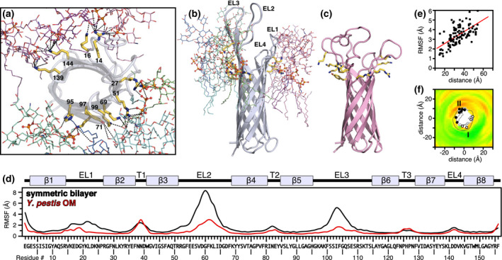
Association of LPS with Ail. Data are shown for MD simulations initiated from one structural model of the NMR structure of Ail. (a,b) Snapshot of Ail obtained after 1.5 µs of MD in a Y. pestis outer membrane. Residues in the LPS‐recognition motif (R27, R51, K69, F71, H95, K97, K99) and in cluster I (K14, K16, K139, K144) establish multiple interactions with the PO4 2‐ and OH groups of LPS. For clarity, only LPS molecules that interact directly with Ail are shown. (c) Snapshot of Ail obtained after 1 µs of MD in a DMPC/DMPG symmetric lipid bilayer. (d) Time‐averaged RMSF of Ail residues in Y. pestis outer membrane (red) or symmetric DMPC/DMPG lipid bilayer (black). RMSF was calculated for heavy atoms over the last 1 µs of MD trajectories. The protein secondary structure is depicted above the graph. (e) Time‐averaged RMSF of individual LPS molecules (calculated for phosphate groups) relative to their distance from the Ail center of mass. (f) Thickness topology of the Y. pestis outer membrane around Ail mapped onto the XY plane. The outer membrane is viewed from the extracellular surface with the membrane normal along the Z direction. The positions of cluster I (white circles) and cluster II (black circles) residues are marked on the surface of the Ail β‐barrel (central black outline). The map is colored by hydrophobic thickness, from 16 Å (green) to 28 Å (red) [Colour figure can be viewed at wileyonlinelibrary.com]
2.3. The Ail–LPS interactions are mutually constructive
In a previous solid‐state NMR study (Dutta et al., 2017) we showed that Ail acquires conformational order when embedded in LPS‐containing liposomes. Here, the MD simulations indicate that the Ail–LPS interaction alters the conformation and dynamics of both Ail and the LPS outer membrane. The extracellular loops of Ail acquire β secondary structure resulting in an extension of the β‐barrel length by ~10 Å compared to the structure embedded in a symmetric phospholipid bilayer (Figure 4b,c; Figure S7). The loops also experience a marked reduction in conformational flexibility, in line with the experimental results. The time‐averaged root mean‐squared fluctuations (RMSF) calculated for heavy atoms over the last 1 µs of 1.5‐µs MD trajectories (Figure 4d; Figure S7) show that the flexible regions of Ail are reduced in both length (number of residues) and amplitude of fluctuation when Ail is embedded in the Y. pestis outer membrane. The effect is particularly marked for EL2 and EL3, which contain the LPS‐recognition motif and interact extensively with the LPS core oligosaccharide groups.
In addition to its ordering effect on the protein, Ail–LPS association also confers dynamic order to LPS in the periphery of Ail. The profile of RMSF calculated for individual LPS molecules is distinctly asymmetric: LPS molecules neighboring Ail have dramatically reduced mobility compared to more peripheral LPS molecules (Figure 4e; Figure S8). This parameter, which reflects the time‐averaged fluctuations of the LPS phosphate groups relative to the protein's center of mass, includes terms for lateral and rotational diffusion, as well as internal dynamics of LPS. Compared to the rapid rotational and lateral diffusion rates (~10‐3 nm2/ns) of the phospholipids in the inner membrane leaflet, LPS molecules are highly immobilized (~10‐11 nm2/ns) on the timescale of the simulations (Muhlradt et al., 1974; Wu et al., 2013). The MD results indicate that direct association with Ail contributes to the reduced mobility of LPS molecules, and the effect appears most prominent for LPS molecules associated with the LPS‐recognition motif in cluster II.
Finally, the outer membrane adopts a pronounced thickness asymmetry around the Ail β‐barrel: thicker on the side of cluster II and thinner on the side of cluster I (Figure 4f; Figure S8). Cluster II sites along the extended barrel wall that is formed by EL2 and EL3 appear to be responsible for this effect, with Ail–LPS interactions on this side of the barrel effectively “stretching” the bound LPS molecules along the direction of the membrane normal axis. The high abundance of Ail at 37°C (~30% of the outer membrane surface) suggests that a pronounced thickening of the Y. pestis outer membrane may be associated with human infection, a feature that would further buttress the bacterial cells against human host defenses.
2.4. The Ail LPS‐recognition motif is important for Y. pestis serum resistance, antibiotic resistance, and outer membrane integrity
The NMR and MD structural data demonstrate that Ail and LPS interact specifically in vitro and have mutual ordering effects. These are likely to have important consequences for the virulence phenotypes that rely on Ail and LPS. The serum resistance function of Ail is thought to depend on its ability to bind elements of the human innate immune system, such as complement components and the blood protein Vitronectin (Bartra et al., 2015; Krukonis and Thomson, 2020), and these binding events may be expected to rely on the presentation of a competent conformation of Ail at the bacterial cell surface. Moreover, the high abundance of Ail and LPS at the bacterial cell surface suggests that the Ail–LPS interaction network plays an important role in maintaining outer membrane integrity, with implications for resistance to antibiotics and other environmental stressors. What roles do the Ail–LPS interactions play in serum resistance, antibiotic resistance, and outer membrane integrity? To address this question, we generated mutant Y. pestis bacterial cells with Ail cluster I and/or cluster II residues converted to Ala and examined their viability.
First, we assessed outer membrane integration and cell surface accessibility of these Ail mutants by means of protease accessibility experiments. Whole cells of wild‐type and ail‐mutant Y. pestis were incubated with and without proteinase K, as described (Song et al., 2008). Periplasmic DsbA was clearly protected from proteinase K degradation (Figure S9e,f) demonstrating that the outer membrane of Y. pestis is intact and provides a permeability barrier in all cases. Ail, by contrast, is degraded by Proteinase K in both wild‐type and mutant Y. pestis cells (Figure S9a–d). Thus, we conclude that all Ail mutants as wild‐type Ail are properly inserted in the bacterial outer membrane with the extracellular loops exposed to the cell surface. Notably, cluster II and combined cluster I/II mutants are more susceptible to Proteinase K. In line with the structural data, we propose that enhanced loop disorder, caused by the disruption of proper Ail–LPS interactions in these mutants, may enhance their susceptibility to extracellular protease.
Analysis by SDS‐PAGE provides further confirmation that the mutants adopt the proper transmembrane β‐barrel conformation of wild‐type Ail. The SDS‐PAGE migration profiles of unfolded and folded β‐barrels, including Ail (Yamashita et al., 2011), are dramatically different, with the unfolded state migrating more slowly, at distinctly higher apparent molecular weight, due to entanglement of the randomly disordered polypeptide chain with the polyacrylamide network of the gel. Wild‐type and Ail mutants have similar SDS‐PAGE migration patterns, consistent with a similar, intact β‐barrel fold (Figure S9a,c). The slight mobility increase (toward lower apparent molecular weight) observed for cluster II mutants may be attributed to altered protein charge, altered interaction with LPS, or the loop disorder defect associated with defective LPS binding described above.
Next we assessed the ability of Ail mutants to survive in normal human serum (NHS) compared to heat‐inactivated serum (HIS). While wild‐type Y. pestis cells survive in NHS, the ail deletion mutant (Δail) was completely defective for survival (Figure 5a). Survival was restored by engineering the Δail strain to express plasmid‐encoded FLAG‐tagged Ail (Δ+pF‐Ail), but not FLAG tag alone (Δ+ pF) or FLAG‐tagged OmpX (Δ+pF‐OmpX), another Y. pestis outer membrane protein that has no known virulence activity. Notably, serum survival was highly suppressed for both cluster II and combined cluster I/II mutant cells. By contrast, levels of survival similar to Δ+pF‐Ail were observed for cluster I mutant cells, and for two independent mutants each comprising residues at the base (R27, R51, H95) or outer extremities (K69, K97, K99) of cluster II.
FIGURE 5.
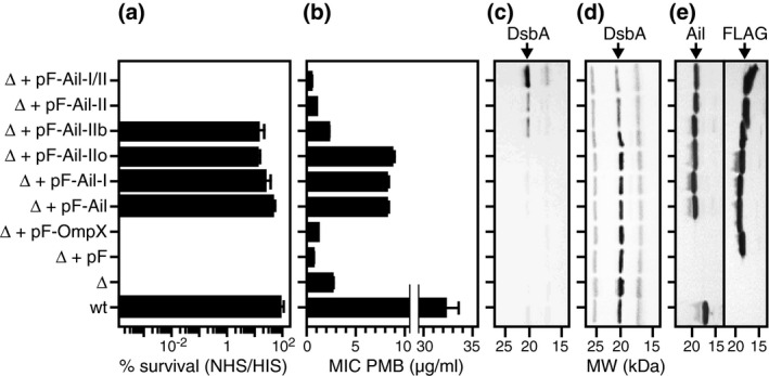
Effects of mutations on Y. pestis cell viability. (a) Survival in NHS relative to HIS was assayed by incubating cells with serum (NHS or HIS), then plating on agar and counting the surviving bacterial colonies. Survival is reported as percent serum resistance relative to the number of surviving colonies in HIS. (b) Resistance to polymyxin B (PMB) antibiotic was assayed by growing the cells at 30°C in media supplemented with increasing concentrations of PMB and measuring OD620 after 5 hr of growth. The apparent minimal inhibitory concentration (MIC) of PMB was derived from the intersect of the baseline plateau of OD620 and the projection of the inflection point for log‐phase growth (Figure S10). (c,d) Leakage across the outer membrane was assayed by probing cell culture supernatants (c), or pelleted whole cells (d), for the presence of the periplasmic protein DsbA by SDS‐PAGE and immunoblotting with anti‐DsbA antibody. (e) Expression of Ail, FLAG‐tagged Ail or FLAG‐tagged‐OmpX was assayed by SDS‐PAGE and immunoblotting with anti‐Ail or anti‐FLAG antibodies. The following Y. pestis cells were tested: wild type‐expressing native Ail (wt); Δail deletion mutant (Δ); Δail complemented with plasmid‐expressing FLAG tag (Δ+pF), FLAG‐tagged OmpX (Δ+pF‐OmpX), FLAG‐tagged Ail (Δ+pF‐Ail), or FLAG‐tagged Ail mutants for cluster I (Δ+pF‐Ail‐I; R14A, K16A, K139A, and K144A), outer cluster II (Δ+pF‐Ail‐IIo; K69A, K97A, K99A), base cluster II (Δ+pF‐Ail‐IIb; R27A, R51A, Y71A, H95A), cluster II (Δ+pF‐Ail‐II; R27A, R51A, Y71A, H95A, K69A, K97A, K99A), or cluster I plus cluster II combined (Δ+pF‐Ail‐I/II). Error bars represent standard deviation for triplicate experiments. Each data point represents the average of three independent measurements. Each immunoblot is representative of triplicate experiments
We further tested the mutants' resistance to polymyxin B, an effective Y. pestis antibiotic (Felek et al., 2010). Since polymyxin B directly binds to LPS and induces outer membrane permeabilization, leading to disruption of the bacterial cell membrane, we reasoned that it would be a good tool for evaluating the importance of the Ail–LPS interaction motifs for membrane integrity. Deletion of the ail gene reduces the minimum inhibitory concentration (MIC) of polymyxin B from 32 µg/ml in wild‐type bacteria to 2.5 µg/ml (Figure 5B; Figure S10a). Resistance can be restored to a level of 9 µg/ml by plasmid expression of Ail (Δ + pF‐Ail), but not empty plasmid or OmpX. Mutations of either cluster I or outer cluster II residues have little effect on polymyxin B resistance, but base cluster II, cluster II, and combined cluster I/II mutants have highly suppressed resistance. We also tested resistance to vancomycin and nisin, which do not have the capability to cross an intact outer membrane and act, instead, by inhibiting peptidoglycan synthesis (Figure S10b,c). All of our strains were resistant to both these antibiotics at the levels we used (0.25 to 64 µg/ml). Moreover, the mutants appear to have a similar growth rate as wild‐type (Figure S10d). These combined results point to a relevant role for the Ail–LPS interaction complex in promoting outer membrane integrity.
Finally, we tested the effects of the mutants on outer membrane leakage, by measuring the release of protein from the cell periplasmic space into the culture supernatant (Figure 5c). Periplasmic DsbA is effectively undetectable in the supernatants of either wild‐type or mutant Y. pestis cells, except for base cluster II, cluster II, and combined cluster I/II cells, where moderate to very high levels are released in the cell culture medium. The levels of DsbA found in the cells (Figure 5d) correlate inversely with those from the culture supernatants, validating the finding of enhanced outer membrane leakage for these three mutants. Alternatively, these mutants may promote an enhanced release of outer membrane vesicles which could also account for the presence of DsbA in the supernatant. The data indicate that these mutants have an actively destructive toxic effect on outer membrane integrity, since neither ail deletion nor expression of OmpX appear to promote a comparable level of leakage. Taken together these assays demonstrate the importance of cluster I/II residues for outer membrane integrity as well as resistance to human serum and membrane‐permeabilizing antibiotics.
2.5. Conclusions
Ail and LPS are key factors contributing to Y. pestis virulence. Collectively, our data show that Ail and LPS interact in a specific manner and have reciprocal ordering effects. Interactions of LPS with cluster I and cluster II sites predispose Ail to adopt an ordered conformation within the outer membrane, while Ail causes a reduction in LPS dynamics and a thickening of the outer membrane in proximity of its β‐barrel. Deletion of the Ail binding sites for LPS reduces Y. pestis survival in serum, enhances antibiotic susceptibility, and compromises cell envelope integrity.
LPS‐induced order has been predicted for other outer membrane proteins (Straatsma and Soares, 2009; Piggot et al., 2013; Wu et al., 2014b; Balusek and Gumbart, 2016; Lee et al., 2017), but the reciprocal effects of proteins on the membrane have been less appreciated. The data highlight the importance of Ail for shaping the physical properties of the outer membrane and the importance of LPS for guiding specific presentation of Ail at the bacterial membrane surface.
The acquisition of conformational order could have important implications for the various virulence attributes that rely on the interactions of Ail with human host proteins, specifically, boosting fibrinolysis to create a permissive environment for microbial circulation, promoting host cell adhesion, and evading immune defenses to promote survival in blood by interfering with the assembly of the terminal complement complex (Bartra et al., 2015; Krukonis and Thomson, 2020). The extracellular loops of Ail, particularly EL2 and EL3, are important for recruiting a range of human factors to the bacterial cell surface (Thomson et al., 2019), and LPS‐induced conformational order in the Ail loops could have a substantially positive effect on the binding affinity of Ail for its other human host partners by minimizing the conformational entropy loss incurred upon binding. In turn, Ail‐induced rigidity and thickening of the outer membrane could play a parallel constructive role in reinforcing the bacterial cell envelope against human host defenses and antibiotics. The results show how Ail and LPS cooperate to construct a Y. pestis outer membrane that is primed for resistance to the human host defenses. They emphasize the functional importance of Ail and LPS as a coordinated assembly and offer a way for targeting Y. pestis pathogenesis.
3. EXPERIMENTAL PROCEDURES
3.1. Sample preparation
Purified Ail and Ail nanodiscs were prepared as described previously (Ding et al., 2015; Dutta et al., 2017), with the short‐membrane scaffold protein MSP1D1Δh5 (Hagn et al., 2013) and the phospholipids DMPC and DMPG (Avanti). Nanodiscs without LPS contained a 1:2:75:25 molar ratio of Ail:MSP:DMPC:DMPG. LPS‐containing nanodiscs contained Ail, MSP, DMPC, DMPG, and LPS at 1/2/68/23/3 molar ratio. Yp‐LPS was purified from Y. pestis grown at 37°C, as described (Knirel and Anisimov, 2012). Ec‐LPS from E. coli F583 Rd rough mutant was obtained commercially (Sigma cat. L6893). Briefly, folded and purified Ail was dissolved in 170 mM n‐decylphosphocholine (Avanti) and added dropwise to a 20 mM sodium cholate solution of the phospholipids, LPS and MSP1D1Δh5. Detergent was removed by overnight treatment with Bio‐Beads SM‐2 (Bio‐Rad), followed by dialysis. Nanodiscs were isolated by size exclusion chromatography (Superdex 75 10/300 GL column, GE Healthcare) in 20 mM Tris–Cl, pH 7.5, 100 mM NaCl, 1 mM EDTA). For NMR studies, the bacterial growth medium was supplemented with (15NH4)2SO4 (Cambridge Isotopes) to produce 15N‐labeled protein. Nanodiscs were transferred to 25 mM sodium phosphate, pH 6.5, 5 mM NaCl, 1 mM EDTA supplemented with 10% 2H2O. The final concentration of Ail in the NMR samples was 0.5–0.6 mM.
3.2. NMR spectroscopy
Two‐dimensional 1H/15N HSQC NMR spectra were recorded at 45°C on a Bruker Avance 600 MHz NMR spectrometer equipped with a 1H/15N/13C triple‐resonance cryoprobe. Spectra were acquired at 318K with 128 transients and 256 or 512 t1 points. Backbone assignments were determined previously (Dutta et al., 2017).
3.3. MD simulations
All‐atom MD simulations were performed as described (Lee et al., 2017), using the CHARMM36 force fields for protein, lipids, carbohydrate, and LPS (Guvench et al., 2008; Brooks et al., 2009; Hatcher et al., 2009; Klauda et al., 2010) with the TIP3P water model (Jorgensen et al., 1983). Briefly, all systems were prepared using CHARMM‐GUI Membrane Builder (Jo et al., 2008; 2009; Wu et al., 2014a; Lee et al., 2019) and equilibrated with the CHARMM‐GUI standard protocol. Planar and dihedral harmonic restrains were applied to the LPS, phospholipid, and water molecules to prevent structural distortion during the equilibration steps, and were gradually reduced to zero for the production runs. van der Waals interactions were switched off between 10 and 12 Å by a force‐based switching method (Steinbach and Brooks, 1994), and the particle‐mesh Ewald method (Essmann et al., 1995) was used for long‐range electrostatic interactions. A 2‐fs time step was used for all simulations with the SHAKE algorithm (Ryckaert et al., 1977) by fixing all bonds including hydrogen atoms. The temperature was maintained at 310.15 K using Langevin dynamics, and pressure was maintained at 1 bar using the semi‐isotropic Monte‐Carlo barostat method (Chow and Ferguson, 1995; Åqvist et al., 2004) with a 5 ps−1 of coupling frequency. MD production simulations were conducted with OpenMM (Eastman et al., 2013) for 1.5 µs, and the last 1 µs of trajectories were used for analysis.
A total of eight independent MD simulations were performed, with four structural models of Ail simulated in either a symmetric phospholipid lipid bilayer containing DMPC and DMPG, or an asymmetric Y. pestis outer membrane (Table S1). The starting structure included the first three models of the NMR structure (PDB: 2n2l) (Marassi et al., 2015) and model A from the crystal structure (PDB 3qra) (Yamashita et al., 2011). The initial orientation and location of Ail and lipid molecules are different in the different replicas; hence, each MD run represents a distinct independent experiment.
The outer leaflet of the Y. pestis outer membrane contained 36 LPS molecules. The structure of Y. pestis LPS (Figure 1a) includes a lipid A moiety with one 4‐amino‐4‐deoxyarabinose (Ara4N) residue and two 2‐amino‐2‐deoxyglucose (GlcN) residues with tetra‐acylated fatty acid tails (Rebeil et al., 2004; Knirel et al., 2005; Knirel and Anisimov, 2012). The LPS core contains αdd‐Hep(1 7)‐αld‐Hep(1 7)[βd‐GlcNAc(1 3)]‐αld‐Hep(1 3)[βd‐Glc(1 4)]‐αld‐Hep(1 5)[αd‐Kdo(2 4)]‐αd‐Kdo(2 , where dd‐Hep is for d‐glycero‐d‐manno‐Heptose, ld‐Hep is for l‐glycero‐d‐manno Heptose, d‐Glc is for d‐glucose, d‐GlcNAc is for d‐glucosamine, and d‐Kdo is for d‐3‐deoxy‐d‐manno‐oct‐2‐ulosonic acid (Knirel et al., 2005). The inner leaflet of the Y. pestis outer membrane contained a 15:4:1 molar ratio of PPPE (1‐palmitoyl(16:0)‐2‐palmitoleonyl(16:1 cis‐9)‐phosphatidylethanolamine), PVPG (1‐palmitoyl(16:0)‐2‐vacenoyl(18:1 cis‐11)‐phosphatidylglycerol), and PVCL2 (1,1’‐palmitoyl‐2,2’‐vacenoyl cardiolipin) with a net charge of −2e (Vance and Vance, 2008). Ca2+ counterions were added to neutralize the LPS charges, and bulk 150 mM KCl was added to mimic the biological ionic strength.
3.4. Y. pestis cell assays
Whole‐cell assays were performed essentially as described (Bartra et al., 2015). Y. pestis KIM8‐E (pPCP1‐; pCD1 + ΔyopE–sycE:: dhfr) (Bartra et al., 2006) (wt), KIM8‐E Δail (Bartra et al., 2008), and KIM8 Δail carrying plasmids pFLAG‐ATS (Sigma), pFLAG‐OmpX, or pFLAG‐Ail (Bartra et al., 2015) were routinely grown in heart infusion broth (HIB) or on tryptose blood agar (TBA) base plates at 30°C (Difco) containing appropriate antibiotics. Expression of FLAG‐OmpX, FLAG‐Ail, and FLAG‐Ail alanine point mutants was induced by the addition of IPTG (0.05 mM final concentration). The lysates and cell culture supernatants were subjected to SDS‐PAGE and immunoblot analysis with anti‐Ail (Bartra et al., 2015), anti‐FLAG M2 (Sigma), or anti‐DsbA (Sigma) antibodies.
To assay protease accessibility, Y. pestis cell growths in TMH media (Straley and Bowmer, 1986) (containing 2.5 mM CaCl2 and 0.05 mM IPTG) were started from overnight cultures and grown at 30°C with shaking. After 4 hr of growth, when optical density at 620 nm (OD620) had reached 0.5, chloramphenicol was added to a final concentration of 20 µg/ml, and 2 ml of culture was taken for Proteinase K digestion. Proteinase K in 50% glycerol (NEB; final concentration 1,000 µg/ml) or an equivalent volume of 50% glycerol (control) was added to the bacteria and the cells incubated for 1 hr at 37°C. Digestion was terminated by adding 2 mM PMSF and placing the cells on ice. The OD620 of each digestion reaction was measured, and the bacterial cells were harvested and resuspended, based on the final OD620, in 1X sample buffer for SDS‐PAGE and immunoblot analysis.
Survival in NHS relative to HIS was assayed by incubating cells, washed 1X in PBS, with serum (80% NHS or 80% HIS) in triplicate for 1 hr with shaking at 37°C. The number of surviving bacteria (CFU) in each sample was determined by dilution and plating on TBA plates at 30°C for 48 hr. The percent survival was calculated as (CFUNHS/ CFUHIS) x 100, where CFUNHS and CFUHIS are the average number of bacteria that survived exposure to NHS or HIS at 1 hr.
Resistance to polymyxin B antibiotic was assayed by growing the cells at 30°C in heart infusion broth (HIB; Difco) media supplemented with increasing concentrations of polymyxin B (Sigma) for 5 hr, and measuring OD620 at various time points during growth. The data at 5 hr of growth were used to derive the minimal inhibitory concentration (MIC) from plots of the change in bacterial cell density (ΔOD620) as a function of polymyxin B concentration, as described (Lambert and Pearson, 2000). The MIC was taken as the intersect of the baseline plateau of OD620 and the projection of the inflection point for log‐phase growth (Figure S10). It reflects the minimum concentration of antibiotic where growth was less than one doubling of OD620 after 5 hr at 30°C.
Outer membrane leakage was assayed by probing cell culture supernatants or pelleted whole cells for the presence of the periplasmic protein DsbA following growth in HIB at 30°C by SDS‐PAGE and immunoblotting.
Cell‐based experiments were performed at 30°C because the additional calcium required to suppress growth restriction associated with Type III Secretion System function is known to dramatically alter the polymyxin B sensitivity of gram‐negative bacteria. Nevertheless, we note that the expression level of Ail at 30°C is only marginally lower than at 37°C, and Y. pestis produces the same type of LPS lipid IVA at 30 and 37°C. The cell‐based studies, therefore, are representative of the mammalian infection state of Y. pestis.
CONFLICT OF INTEREST
Authors declare no conflict of interest.
AUTHOR CONTRIBUTIONS
C.S., H.L., S.S.B., L.M.F., S.A.I., and R.Z.S. performed research. C.S., H.L., Y.T., S.S.B., S.H., Y.Y., A.P.A., G.V.P., W.I., and F.M.M. analyzed data. A.P.A., G.V.P., W.I., and F.M.M. designed research. F.M.M. wrote the paper.
Supporting information
Supplementary Material
ACKNOWLEDGMENTS
This study was supported by grants from the National Institutes of Health (GM 118186 to FMM; AI 130009 to GVP), the National Science Foundation (MCB‐1727508, MCB‐1810695 and XSEDE MCB‐070009 to WI), and the Ministry of Science and Higher Education of the Russian Federation (Agreement 075‐15‐2019‐1671 to SAI., RZS. and APA). It utilized the Structural Biology Resource supported by grant P30 CA030199.
Singh C, Lee H, Tian Y, et al. Mutually constructive roles of Ail and LPS in Yersinia pestis serum survival. Mol Microbiol. 2020;114:520–530. 10.1111/mmi.14530
DATA AVAILABILITY STATEMENT
All data needed are present in the paper and/or the Supplementary Materials. Additional information is available upon request.
REFERENCES
- Anisimov, A.P. , Shaikhutdinova, R.Z. , Pan'kina, L.N. , Feodorova, V.A. , Savostina, E.P. , Bystrova, O.V. , et al. (2007). Effect of deletion of the lpxM gene on virulence and vaccine potential of Yersinia pestis in mice. Journal of Medical Microbiology, 56, 443–453. [DOI] [PubMed] [Google Scholar]
- Åqvist, J. , Wennerström, P. , Nervall, M. , Bjelic, S. and Brandsdal, B.O. (2004). Molecular dynamics simulations of water and biomolecules with a Monte Carlo constant pressure algorithm. Chemical Physics Letters, 384, 288–294. [Google Scholar]
- Arunmanee, W. , Pathania, M. , Solovyova, A.S. , Le Brun, A.P. , Ridley, H. , Basle, A. , et al. (2016). Gram‐negative trimeric porins have specific LPS binding sites that are essential for porin biogenesis. Proceedings of the National Academy of Sciences of the United States of America, 113, E5034–E5043. [DOI] [PMC free article] [PubMed] [Google Scholar]
- Balusek, C. and Gumbart, J.C. (2016). Role of the native outer‐membrane environment on the transporter BtuB. Biophysical Journal, 111, 1409–1417. [DOI] [PMC free article] [PubMed] [Google Scholar]
- Bartra, S.S. , Ding, Y. , Miya Fujimoto, L. , Ring, J.G. , Jain, V. , Ram, S. , et al. (2015). Yersinia pestis uses the Ail outer membrane protein to recruit vitronectin. Microbiology, 161, 2174–2183. [DOI] [PMC free article] [PubMed] [Google Scholar]
- Bartra, S.S. , Jackson, M.W. , Ross, J.A. and Plano, G.V. (2006). Calcium‐regulated type III secretion of Yop proteins by an Escherichia coli hha mutant carrying a Yersinia pestis pCD1 virulence plasmid. Infection and Immunity, 74, 1381–1386. [DOI] [PMC free article] [PubMed] [Google Scholar]
- Bartra, S.S. , Styer, K.L. , O'Bryant, D.M. , Nilles, M.L. , Hinnebusch, B.J. , Aballay, A. , et al. (2008). Resistance of Yersinia pestis to complement‐dependent killing is mediated by the Ail outer membrane protein. Infection and Immunity, 76, 612–622. [DOI] [PMC free article] [PubMed] [Google Scholar]
- Bayburt, T.H. , Grinkova, Y.V. and Sligar, S.G. (2002). Self‐assembly of discoidal phospholipid bilayer nanoparticles with membrane scaffold proteins. Nano Letters, 2, 853–856. [Google Scholar]
- Brooks, B.R. , Brooks, C.L. 3rd , Mackerell, A.D. Jr , Nilsson, L. , Petrella, R.J. , Roux, B. , et al. (2009). CHARMM: the biomolecular simulation program. Journal of Computational Chemistry, 30, 1545–1614. [DOI] [PMC free article] [PubMed] [Google Scholar]
- Chauvaux, S. , Dillies, M.A. , Marceau, M. , Rosso, M.L. , Rousseau, S. , Moszer, I. , et al. (2011). In silico comparison of Yersinia pestis and Yersinia pseudotuberculosis transcriptomes reveals a higher expression level of crucial virulence determinants in the plague bacillus. International Journal of Medical Microbiology, 301, 105–116. [DOI] [PubMed] [Google Scholar]
- Chow, K.‐H. and Ferguson, D.M. (1995). Isothermal‐isobaric molecular dynamics simulations with Monte Carlo volume sampling. Computer Physics Communications, 91, 283–289. [Google Scholar]
- Deng, W. , Burland, V. , Plunkett, G. 3rd , Boutin, A. , Mayhew, G.F. , Liss, P. , et al. (2002). Genome sequence of Yersinia pestis KIM. Journal of Bacteriology, 184, 4601–4611. [DOI] [PMC free article] [PubMed] [Google Scholar]
- Ding, Y. , Fujimoto, L.M. , Yao, Y. , Plano, G.V. and Marassi, F.M. (2015). Influence of the lipid membrane environment on structure and activity of the outer membrane protein Ail from Yersinia pestis . Biochimica et Biophysica Acta, 1848, 712–720. [DOI] [PMC free article] [PubMed] [Google Scholar]
- Dutta, S.K. , Yao, Y. and Marassi, F.M. (2017). Structural insights into the Yersinia pestis outer membrane protein ail in lipid bilayers. The Journal of Physical Chemistry B, 121, 7561–7570. [DOI] [PMC free article] [PubMed] [Google Scholar]
- Eastman, P. , Friedrichs, M.S. , Chodera, J.D. , Radmer, R.J. , Bruns, C.M. , Ku, J.P. , et al. (2013). OpenMM 4: a reusable, extensible, hardware independent library for high performance molecular simulation. Journal of Chemical Theory and Computation, 9, 461–469. [DOI] [PMC free article] [PubMed] [Google Scholar]
- Essmann, U. , Perera, L. , Berkowitz, M.L. , Darden, T. , Lee, H. and Pedersen, L.G. (1995). A smooth particle mesh Ewald method. The Journal of Chemical Physics, 103, 8577–8593. [Google Scholar]
- Felek, S. and Krukonis, E.S. (2009). The Yersinia pestis Ail protein mediates binding and Yop delivery to host cells required for plague virulence. Infection and Immunity, 77, 825–836. [DOI] [PMC free article] [PubMed] [Google Scholar]
- Felek, S. , Muszynski, A. , Carlson, R.W. , Tsang, T.M. , Hinnebusch, B.J. and Krukonis, E.S. (2010). Phosphoglucomutase of Yersinia pestis is required for autoaggregation and polymyxin B resistance. Infection and Immunity, 78, 1163–1175. [DOI] [PMC free article] [PubMed] [Google Scholar]
- Ferguson, A.D. , Welte, W. , Hofmann, E. , Lindner, B. , Holst, O. , Coulton, J.W. , et al. (2000). A conserved structural motif for lipopolysaccharide recognition by procaryotic and eucaryotic proteins. Structure, 8, 585–592. [DOI] [PubMed] [Google Scholar]
- Guvench, O. , Greene, S.N. , Kamath, G. , Brady, J.W. , Venable, R.M. , Pastor, R.W. , et al. (2008). Additive empirical force field for hexopyranose monosaccharides. Journal of Computational Chemistry, 29, 2543–2564. [DOI] [PMC free article] [PubMed] [Google Scholar]
- Hagn, F. , Etzkorn, M. , Raschle, T. and Wagner, G. (2013). Optimized phospholipid bilayer nanodiscs facilitate high‐resolution structure determination of membrane proteins. Journal of the American Chemical Society, 135, 1919–1925. [DOI] [PMC free article] [PubMed] [Google Scholar]
- Hatcher, E. , Guvench, O. and Mackerell, A.D. (2009). CHARMM additive all‐atom force field for aldopentofuranoses, methyl‐aldopentofuranosides, and fructofuranose. The Journal of Physical Chemistry B, 113, 12466–12476. [DOI] [PMC free article] [PubMed] [Google Scholar]
- Henry, G.D. and Sykes, B.D. (1995). Determination of the rotational dynamics and pH dependence of the hydrogen exchange rates of the arginine guanidino group using NMR spectroscopy. Journal of Biomolecular NMR, 6, 59–66. [DOI] [PubMed] [Google Scholar]
- Hinnebusch, B.J. , Jarrett, C.O. and Bland, D.M. (2017). “Fleaing” the plague: adaptations of Yersinia pestis to its insect vector that lead to transmission. Annual Review of Microbiology, 71, 215–232. [DOI] [PubMed] [Google Scholar]
- Hinnebusch, B.J. , Jarrett, C.O. , Callison, J.A. , Gardner, D. , Buchanan, S.K. and Plano, G.V. (2011). Role of the Yersinia pestis Ail protein in preventing a protective polymorphonuclear leukocyte response during bubonic plague. Infection and Immunity, 79, 4984–4989. [DOI] [PMC free article] [PubMed] [Google Scholar]
- Jo, S. , Kim, T. , Iyer, V.G. and Im, W. (2008). CHARMM‐GUI: a web‐based graphical user interface for CHARMM. Journal of Computational Chemistry, 29, 1859–1865. [DOI] [PubMed] [Google Scholar]
- Jo, S. , Lim, J.B. , Klauda, J.B. and Im, W. (2009). CHARMM‐GUI Membrane Builder for mixed bilayers and its application to yeast membranes. Biophysical Journal, 97, 50–58. [DOI] [PMC free article] [PubMed] [Google Scholar]
- Jorgensen, W.L. , Chandrasekhar, J. , Madura, J.D. , Impey, R.W. and Klein, M.L. (1983). Comparison of simple potential functions for simulating liquid water. The Journal of Chemical Physics, 79, 926–935. [Google Scholar]
- Klauda, J.B. , Venable, R.M. , Freites, J.A. , O'Connor, J.W. , Tobias, D.J. , Mondragon‐Ramirez, C. , et al. (2010). Update of the CHARMM all‐atom additive force field for lipids: validation on six lipid types. The Journal of Physical Chemistry B, 114, 7830–7843. [DOI] [PMC free article] [PubMed] [Google Scholar]
- Knirel, Y.A. and Anisimov, A.P. (2012). Lipopolysaccharide of Yersinia pestis, the cause of plague: structure, genetics, biological properties. Acta Naturae, 4, 46–58. [PMC free article] [PubMed] [Google Scholar]
- Knirel, Y.A. , Dentovskaya, S.V. , Bystrova, O.V. , Kocharova, N.A. , Senchenkova, S.N. , Shaikhutdinova, R.Z. , et al. (2007). Relationship of the lipopolysaccharide structure of Yersinia pestis to resistance to antimicrobial factors. Advances in Experimental Medicine and Biology, 603, 88–96. [DOI] [PubMed] [Google Scholar]
- Knirel, Y.A. , Lindner, B. , Vinogradov, E.V. , Kocharova, N.A. , Senchenkova, S.N. , Shaikhutdinova, R.Z. , et al. (2005). Temperature‐dependent variations and intraspecies diversity of the structure of the lipopolysaccharide of Yersinia pestis . Biochemistry, 44, 1731–1743. [DOI] [PubMed] [Google Scholar]
- Kolodziejek, A.M. , Hovde, C.J. and Minnich, S.A. (2012). Yersinia pestis Ail: multiple roles of a single protein. Frontiers in Cellular and Infection Microbiology, 2, 103. [DOI] [PMC free article] [PubMed] [Google Scholar]
- Kolodziejek, A.M. , Schnider, D.R. , Rohde, H.N. , Wojtowicz, A.J. , Bohach, G.A. , Minnich, S.A. , et al. (2010). Outer membrane protein X (Ail) contributes to Yersinia pestis virulence in pneumonic plague and its activity is dependent on the lipopolysaccharide core length. Infection and Immunity, 78, 5233–5243. [DOI] [PMC free article] [PubMed] [Google Scholar]
- Krukonis, E.S. and Thomson, J.J. (2020). Complement evasion mechanisms of the systemic pathogens Yersiniae and Salmonellae . FEBS Letters. [DOI] [PubMed] [Google Scholar]
- Kucharska, I. and Tamm, L.K. (2017). Solution NMR provides new insight into lipid‐protein interaction. Biochemistry, 56, 4291–4292. [DOI] [PMC free article] [PubMed] [Google Scholar]
- Lambert, R.J. and Pearson, J. (2000). Susceptibility testing: accurate and reproducible minimum inhibitory concentration (MIC) and non‐inhibitory concentration (NIC) values. Journal of Applied Microbiology, 88, 784–790. [DOI] [PubMed] [Google Scholar]
- Lee, J. , Patel, D.S. , Kucharska, I. , Tamm, L.K. and Im, W. (2017). Refinement of OprH‐LPS interactions by molecular simulations. Biophysical Journal, 112, 346–355. [DOI] [PMC free article] [PubMed] [Google Scholar]
- Lee, J. , Patel, D.S. , Stahle, J. , Park, S.J. , Kern, N.R. , Kim, S. , et al. (2019). CHARMM‐GUI membrane builder for complex biological membrane simulations with glycolipids and lipoglycans. Journal of chemical theory and computation, 15, 775–786. [DOI] [PubMed] [Google Scholar]
- Marassi, F.M. , Ding, Y. , Schwieters, C.D. , Tian, Y. and Yao, Y. (2015). Backbone structure of Yersinia pestis Ail determined in micelles by NMR‐restrained simulated annealing with implicit membrane solvation. Journal of Biomolecular NMR, 63, 59–65. [DOI] [PMC free article] [PubMed] [Google Scholar]
- Miller, V.L. , Bliska, J.B. and Falkow, S. (1990). Nucleotide sequence of the Yersinia enterocolitica ail gene and characterization of the Ail protein product. Journal of Bacteriology, 172, 1062–1069. [DOI] [PMC free article] [PubMed] [Google Scholar]
- Muhlradt, P.F. , Menzel, J. , Golecki, J.R. and Speth, V. (1974). Lateral mobility and surface density of lipopolysaccharide in the outer membrane of Salmonella typhimurium . European Journal of Biochemistry, 43, 533–539. [DOI] [PubMed] [Google Scholar]
- Parkhill, J. , Wren, B.W. , Thomson, N.R. , Titball, R.W. , Holden, M.T. , Prentice, M.B. , et al. (2001). Genome sequence of Yersinia pestis, the causative agent of plague. Nature, 413, 523–527. [DOI] [PubMed] [Google Scholar]
- Piggot, T.J. , Holdbrook, D.A. and Khalid, S. (2013). Conformational dynamics and membrane interactions of the E. coli outer membrane protein FecA: a molecular dynamics simulation study. Biochimica et Biophysica Acta, 1828, 284–293. [DOI] [PubMed] [Google Scholar]
- Rebeil, R. , Ernst, R.K. , Gowen, B.B. , Miller, S.I. and Hinnebusch, B.J. (2004). Variation in lipid A structure in the pathogenic yersiniae. Molecular Microbiology, 52, 1363–1373. [DOI] [PubMed] [Google Scholar]
- Ryckaert, J.‐P. , Ciccotti, G. and Berendsen, H.J.C. (1977). Numerical integration of the Cartesian equations of motion of a system with constraints: molecular dynamics of n‐alkanes. Journal of Computational Physics, 23, 327–341. [Google Scholar]
- Sebbane, F. , Lemaitre, N. , Sturdevant, D.E. , Rebeil, R. , Virtaneva, K. , Porcella, S.F. , et al. (2006). Adaptive response of Yersinia pestis to extracellular effectors of innate immunity during bubonic plague. Proceedings of the National Academy of Sciences of the United States of America, 103, 11766–11771. [DOI] [PMC free article] [PubMed] [Google Scholar]
- Song, H. , Sandie, R. , Wang, Y. , Andrade‐Navarro, M.A. and Niederweis, M. (2008). Identification of outer membrane proteins of Mycobacterium tuberculosis . Tuberculosis, 88, 526–544. [DOI] [PMC free article] [PubMed] [Google Scholar]
- Steinbach, P.J. and Brooks, B.R. (1994). New spherical‐cutoff methods for long‐range forces in macromolecular simulation. Journal of Computational Chemistry, 15, 667–683. [Google Scholar]
- Straatsma, T.P. and Soares, T.A. (2009). Characterization of the outer membrane protein OprF of Pseudomonas aeruginosa in a lipopolysaccharide membrane by computer simulation. Proteins, 74, 475–488. [DOI] [PMC free article] [PubMed] [Google Scholar]
- Straley, S.C. and Bowmer, W.S. (1986). Virulence genes regulated at the transcriptional level by Ca2+ in Yersinia pestis include structural genes for outer membrane proteins. Infection and Immunity, 51, 445–454. [DOI] [PMC free article] [PubMed] [Google Scholar]
- Thomson, J.J. , Plecha, S.C. and Krukonis, E.S. (2019). Ail provides multiple mechanisms of serum resistance to Yersinia pestis . Molecular Microbiology, 111, 82–95. [DOI] [PMC free article] [PubMed] [Google Scholar]
- Vance, D.E. and Vance, J.E . (2008). Biochemistry of Lipids, Lipoproteins and Membranes, p. xii, 631 p., 638 p. of Plates. Amsterdam: Elsevier. [Google Scholar]
- Wu, E.L. , Cheng, X. , Jo, S. , Rui, H. , Song, K.C. , Davila‐Contreras, E.M. , et al. (2014a). CHARMM‐GUI Membrane Builder toward realistic biological membrane simulations. Journal of Computational Chemistry, 35, 1997–2004. [DOI] [PMC free article] [PubMed] [Google Scholar]
- Wu, E.L. , Engstrom, O. , Jo, S. , Stuhlsatz, D. , Yeom, M.S. , Klauda, J.B. , et al. (2013). Molecular dynamics and NMR spectroscopy studies of E. coli lipopolysaccharide structure and dynamics. Biophysical Journal, 105, 1444–1455. [DOI] [PMC free article] [PubMed] [Google Scholar]
- Wu, E.L. , Fleming, P.J. , Yeom, M.S. , Widmalm, G. , Klauda, J.B. , Fleming, K.G. , et al. (2014b). E. coli outer membrane and interactions with OmpLA. Biophysical Journal, 106, 2493–2502. [DOI] [PMC free article] [PubMed] [Google Scholar]
- Yamashita, S. , Lukacik, P. , Barnard, T.J. , Noinaj, N. , Felek, S. , Tsang, T.M. , et al. (2011). Structural insights into Ail‐mediated adhesion in Yersinia pestis . Structure, 19, 1672–1682. [DOI] [PMC free article] [PubMed] [Google Scholar]
- Yamazaki, T. , Pascal, S.M. , Singer, A.U. , Forman‐Kay, J.D. and Kay, L.E. (1995). NMR pulse schemes for the sequence‐specific assignment of arginine guanidino 15N and 1H chemical shifts in proteins. Journal of the American Chemical Society, 117, 3556–3564. [Google Scholar]
Associated Data
This section collects any data citations, data availability statements, or supplementary materials included in this article.
Supplementary Materials
Supplementary Material
Data Availability Statement
All data needed are present in the paper and/or the Supplementary Materials. Additional information is available upon request.