Abstract
Memory B cells (MBCs) are long-lived and produce high-affinity, generally, class-switched antibodies. Here, we use a multiparameter approach involving CD27 to segregate naïve B cells (NBC), IgD+ unswitched (unsw)MBCs and IgG+ or IgA+ class-switched (sw)MBCs from humans of different age, sex and race. Conserved antibody variable gene expression indicates that MBCs emerge through unbiased selection from NBCs. Integrative analyses of mRNAs, miRNAs, lncRNAs, chromatin accessibility and cis-regulatory elements uncover a core mRNA-ncRNA transcriptional signature shared by IgG+ and IgA+ swMBCs and distinct from NBCs, while unswMBCs display a transitional transcriptome. Some swMBC transcriptional signature loci are accessible but not expressed in NBCs. Profiling miRNAs reveals downregulated MIR181, and concomitantly upregulated MIR181 target genes such as RASSF6, TOX, TRERF1, TRPV3 and RORα, in swMBCs. Finally, lncRNAs differentially expressed in swMBCs cluster proximal to the IgH chain locus on chromosome 14. Our findings thus provide new insights into MBC transcriptional programs and epigenetic regulation, opening new investigative avenues on these critical cell elements in human health and disease.
Subject terms: Epigenomics, Class switch recombination, Immunological memory, B-2 cells
Human memory B cells differentiate from naïve B cells and can express different immunoglobulin (Ig) isotypes resulted from class-switch recombination. Here the authors describe, using transcriptional and epigenetic data from human memory B cells and integrated multi-omics analyses, the differentiation regulation and trajectory of IgG+, IgA+ and IgD+ memory B cells.
Introduction
B cell maturation in the bone marrow, peripheral differentiation, and survival is dictated by genetic programs that drive critical pathways, giving rise to plasma cells and memory B cells (MBCs). Upon activation by antigen, mature naïve B cells (NBCs) enter the germinal center (GC) reaction to undergo activation-induced deaminase (AID)-mediated somatic hypermutation (SHM) and class switch DNA recombination (CSR)1–4, leading to plasma cell or MBC differentiation. MBCs are central to the adaptive immune response, establishing long-term antigen-specific immunity. Their generation and function are critical for the development of anamnestic antibody responses induced by vaccines or natural infections. Effective anamnestic immune response to foreign antigens, such as those on bacteria and viruses5, is required for the establishment of “herd immunity” and eradication of an infectious agent in a population as a whole. MBCs generally express somatically mutated, affinity mature B cell surface receptors (BCRs) for antigen and persist in a quiescent state until re-encountering their cognate antigen6. While many MBCs express class-switched BCRs, such as immunoglobulin (Ig)G or IgA, “unswitched” IgD+IgM+ (unsw)MBCs also exist, with human switched (sw)MBCs and unswMBCs bearing somatically mutated BCRs and expressing surface CD27, a marker of “antigen-experience”7–10. Upon reactivation, MBCs rapidly proliferate and differentiate into plasma cells to secrete large amounts of high-affinity antibodies6,11,12. Alternatively, MBCs re-enter the GC reaction to undergo additional rounds of SHM/CSR13,14. Thus, enhanced responses against previously experienced antigens are contingent upon functional MBCs, which possess intrinsic advantages over NBCs, including longer lifespan and accelerated antibody response15.
Research to date, primarily in the mouse, has attempted to attribute intrinsic differences between MBCs and NBCs to select gene expression programs in MBC differentiation, survival, and reactivation16–21. IL-9 signaling has been suggested to drive MBC development within GCs as well as mediate antibody recall responses16,17. High Bach2 expression biases GC B cells towards MBC differentiation, while reduced Bach2 expression skews MBCs towards plasma cell differentiation upon reactivation18,19. Divergent transcriptional programming has been tentatively identified in MBCs, with mouse IgG2a+ and IgA+MBCs utilizing T-bet and Rorα, respectively, to maintain their distinct identities and functions20. As recently reported, the transcription factor (TF) Hhex interacts with the corepressor Tle3 to promote MBC differentiation, but not maintenance of mouse MBCs22. Finally, MBCs accelerated and heightened response has been attributed to reduced expression of quiescence factors, such as ZBTB16, KLF4, and KLF9, as well as enhanced intrinsic signaling of class-switched BCR Igγ chain. This carries tail tyrosine ITT motifs that reduce IgG+MBCs activation threshold21,23.
The human MBC transcriptome and its regulation, particularly by epigenetic mechanisms, need better understanding24,25. As we and others have shown, coordinated regulation of gene networks is critical in human and mouse B cell differentiation and antibody responses26–33. Such coordinated regulation is mediated by epigenetic modifications and factors, including DNA methylation, histone post-translational modifications, and non-coding RNAs (ncRNAs), such as microRNAs (miRNAs) and long non-coding RNAs (lncRNAs)34,35. These layers of epigenetic regulation synergize with TFs and chromatin accessibility to outline distinct gene expression programs, thereby dictating cell functions26,36. Although different miRNA profiles have been reported in different B cell subsets, including MBCs37, and lncRNAs have been associated with different stages of B cell development38, no comprehensive analysis of the protein-coding and non-coding transcriptome and chromatin accessibility has been reported in human MBCs.
Integrative analysis of differentially expressed mRNAs, miRNAs, lncRNAs, chromatin landscape, and cis-regulatory elements reveals how gene expression intersects with epigenetic regulation to segregate distinct MBCs. Analysis of CD27–IgD+ NBCs, CD27+IgD+ unswMBCs, CD27+IgG+ swMBCs and CD27+IgA+ swMBCs as well as total CD27− NBCs and CD27+ MBCs from healthy human subjects defines a differential transcriptional signature that distinguishes CD27+IgG+ swMBCs and CD27+IgA+ swMBCs from CD27–IgD+ NBCs, stratifying CD27+IgD+ unswMBCs between the two. The integration of RNA-Seq with ATAC-Seq data uncovers distinct profiles of cis-regulatory elements in human MBCs. MIR181 downregulation with concomitant upregulation of this microRNA’s target genes indicates an important role for MIR181 in MBC function(s). Further, the inverse correlation of upregulated MIR181 sponge, lncRNA MIAT, with downregulated MIR181a/MIR181b in swMBCs points to MIAT as a regulator that, along with chromatin accessibility, reduces MIR181 expression and promotes swMBC-specific gene expression. Overall, our findings provide evidence for overlapping layers of regulation, including chromatin remodeling, cis-regulatory elements, and distinct sets of miRNAs and lncRNAs, which integrate to dictate gene expression profiles and activation pathways characteristic of human MBCs.
Results
Identification and purification of human swMBCs, unswMBCs, and NBCs
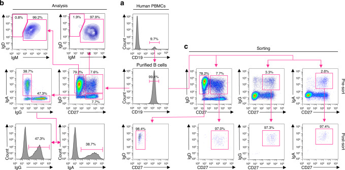
To elucidate the transcriptional landscape of human MBCs, we set up to identify and isolate MBCs and their NBC counterparts. We purified B cells (>99% CD19+) from PBMCs of 7 healthy human subjects (age 20–39, 4 males, 3 females of different race and ethnic backgrounds) (Supplementary Table 1) by immunomagnetic negative selection (Fig. 1a). CD19+ cells were analyzed for surface expression of CD27, a marker for antigen-experienced B cells, as well as IgD, IgM, IgG, and IgA. This allowed for the identification of four distinct subsets: CD27–IgD+ NBCs (63.9 ± 14.3%), CD27+IgD+ unswMBCs (9.7 ± 8.3%), CD27+IgG+ swMBCs (6.5 ± 3.1%) and CD27+IgA+ swMBCs (4.9 ± 2.0%). A fifth subset identified “double negative” CD27–IgD– accounted for the remaining 15.0% of B cells. CD27– B cells were IgD+IgM+, while CD27+ B cells comprised comparable proportions of IgD+ and IgD– B cells. CD27+IgD+ B cells were mostly IgM+ with only 3.8 ± 3.0% IgD+IgM– B cells. CD27+IgD– B cells were primarily either IgG+ or IgA+ (Fig. 1b). CD19+ cells from subjects B, C and G were sorted into 4 fractions: CD27–IgD+ NBCs (70.2 ± 15.2%), CD27+IgD+ unswMBCs (10.1 ± 5.1%), CD27+IgG+ swMBCs (6.6 ± 5.2%) and CD27+IgA+ swMBCs (4.3 ± 2.7%). Reanalysis of the sorted fractions confirmed the identity and purity of the 4 discrete B cell subsets (Fig. 1c). Finally, CD19+ cells from the remaining four subjects (A, D, E, and F) were analyzed and isolated as CD27− (NBCs) and CD27+ total (MBCs) (Supplementary Table 1).
Fig. 1. Identification and purification of CD27−IgD+, CD27+IgD+, CD27+IgG+, and CD27+IgA+ B cells.
a Purification of CD19+ B cells from PBMCs by immunomagnetic negative selection. b FACS analysis of CD19+ B cells for surface expression of CD27, IgD, IgM, IgG, and IgA. c FACS sorting of CD19+ cells (subjects B, C. and G) into four subsets: CD27−IgD+ NBCs, CD27+IgD+ unswMBCs, CD27+IgG+ swMBCs, and CD27+IgA+ swMBCs using the indicated gating strategy and two distinct staining panels. Three independent experiments. The purity of the sorted four B cell subsets as analyzed by FACS. Data are from subject G.
IgVH, JH, and VL, JL gene expression in human NBCs and MBCs
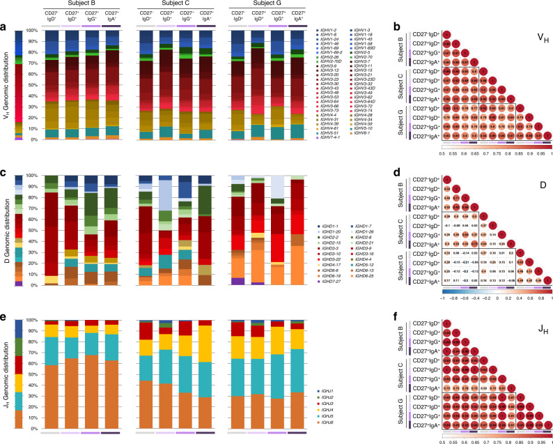
We analyzed the expression of Ig VH, D, JH, and Vκ, Jκ, as well as Vλ, Jλ genes in CD27–IgD+ NBCs, CD27+IgD+ unswMBCs, CD27+IgG+ swMBCs, and CD27+IgA+ swMBCs. The human IgH locus comprises 36–49 functional VH genes segregated into 7 families39. In all four subsets within each subject, VH family expression was stochastic (rs = 0.98), reflecting the genomic representation of the 7 VH families, with VH3 genes being the most represented (37.8-59.9%), followed by VH4 (9.7–26.4%) and VH1 (11.3–23.7%) (Fig. 2a). Expression of individual VH genes was conserved in each VH family, across the 4 subsets in each subject (rs = 0.88) and across the three subjects (rs = 0.84) (Fig. 2b). As expected, D gene expression was diverse across the four subsets in each subject (rs = 0.49) and across the three subjects (rs = 0.17) (Fig. 2c, d). JH gene usage was conserved across the four subsets within each subject (rs = 0.90), with a preponderant JH5 (19.2–40.5%) and JH6 (27.8–67.7%) utilization in all three subjects, albeit to differing degrees (Fig. 2e, f).
Fig. 2. Expressed Ig VH, D, and JH repertoire in human CD27−IgD+, CD27+IgD+, CD27+IgG+, and CD27+IgA+ B cells.
a, c, e Ig VH (a), D (c), and JH (e) gene expression in CD27−IgD+, CD27+IgD+, CD27+IgG+, and CD27+IgA+ B cells, depicted by stacked column for each subject (subjects B, C, G). Ig gene family members grouped within each Ig VH, D, JH family or subgroup are depicted in shades of the respective family/group color. VH family: VH1 (blue), VH2 (green), VH3 (red), VH4 (yellow), VH5 (teal), VH6 (orange), VH7 (purple). D family: D1 (blue), D2 (green), D3 (red), D4 (yellow), D5 (teal), D6 (orange), D7 (purple). JH family: JH1 (blue), JH2 (green), JH3 (red), JH4 (yellow), JH5 (teal), JH6 (orange). b, d, f Correlation matrix showing the relationship of individual Ig VH (b), D (d), and JH (f) gene expression between B cell subsets in the three subjects analyzed. Color bars depict the correlation coefficients on a scale from 0.5 (white) to 1 (red) (b, f), or from −1 (blue) to 0 (white) to 1 (red) (d), with color, circle size, and value all indicating the overall strength of the correlation. B cell subsets are repeated at the bottom, left to right in CD27−IgD+ (gray), CD27+IgD+ (lavender), CD27+IgG+ (purple), and CD27+IgA+ (dark purple).
The human Igκ locus comprises 39 functional Vκ and 5 Jκ genes39. Vκ and Jκ gene expression were comparable across the four subsets within each subject (rs = 0.88; 0.86, respectively) and across the three subjects (rs = 0.86; 0.82, respectively). Vκ gene usage was biased towards Vκ3 and Vκ4 and away from Vκ1 and Vκ2. Jκ gene usage was biased towards Jκ1, Jκ3, and Jκ5 (Supplementary Fig. 1a–d). The human Igλ locus comprises 30 functional Vλ genes segregated into 10 subgroups and 5 functional Jλ-Cλ clusters39. Vλ gene expression was comparable across the four subsets within each subject (rs = 0.82) and across the three subjects (rs = 0.83), with over-representation of Vλ3, Vλ2, and Vλ1 subgroups (Supplementary Fig. 1e, f). Jλ-Cλ usage was biased towards Jλ3-Cλ3 (35.2–57.5%) and Jλ2-Cλ2 (17.9–54.6%), with conserved distributions in all four subsets (rs = 0.87) (Supplementary Fig. 1g, h).
IgH CDR3 length and somatic point-mutations in human MBCs
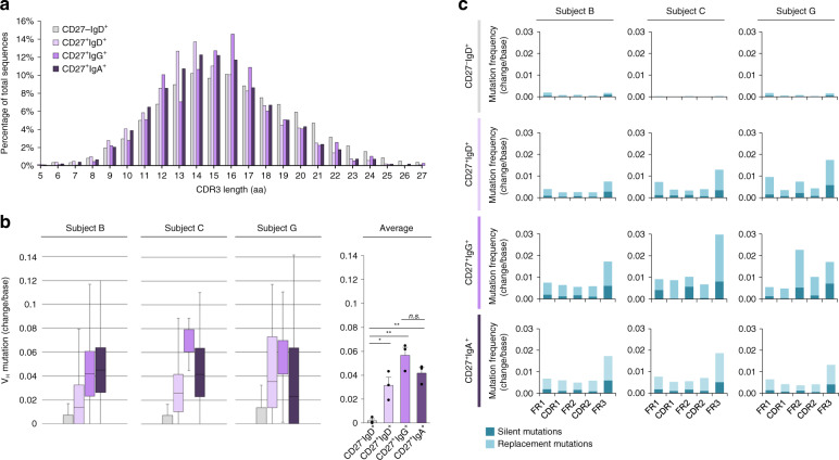
Somatic IgH VHDJH rearrangement determines the sequence and length of the complementary determining region 3 (CDR3), which is critical for BCR-antigen contact. To address IgH CDR3 length and nature as well as VH mutational load, swMBCs, unswMBCs, and NBCs, the recombined VHDJH-CH transcripts were amplified using forward (degenerate) primers for VH1, VH2, VH3, VH4, VH5, VH6, VH7 genes leader sequences in conjunction with reverse Cμ, Cδ, Cγ, or Cα isotype-specific primer and analyzed by IMGT/HighV-QUEST40. The distribution of CDR3 lengths was largely conserved among NBCs (16.05 ± 0.10 AA’s), unswMBCs (14.86 ± 0.11 AA’s), IgG+ swMBCs (15.53 ± 0.12 AA’s), and IgA+ swMBCs (15.08 ± 0.11 AA’s) (Fig. 3a). A salient feature of MBCs is the load of somatic point-mutations in expressed Ig V(D)J genes, a result of precursor B cells undergoing antigen-driven SHM and selection41. Consistent with their non-antigen-experienced status (CD27−), NBCs exhibited a negligible frequency of somatic point-mutations (0.0020 change/base). By contrast, consistent with their antigen-experienced status, CD27+ B cells, be they IgG+, IgA+ or IgD+ exhibited a higher frequency of somatic point-mutations (0.0314, 0.0566 and 0.0417 change/base, respectively), mostly replacement mutations, than NBCs (Fig. 3b, c), with IgG+ swMBCs and IgA+ swMBCs carrying greater mutational burdens than unswMBCs.
Fig. 3. CDR3 lengths and somatic point-mutations in recombined Ig VHDJH gene segments of human MBCs.
a Average percentage of total sequences at any given CDR3 length in recombined VHDJH transcripts expressed by CD27–IgD+, CD27+IgD+, CD27+IgG+, and CD27+IgA+ B cells. b Somatic point-mutations in recombined Ig VHDJH transcripts expressed by CD27–IgD+ (4302 transcripts), CD27+IgD+ (4705 transcripts), CD27+IgG+ (1564 transcripts), and CD27+IgA+ (13,549 transcripts) B cells with boxplots depicting the frequencies of point-mutations (change/base). Data are represented as boxplots where the middle line is the median, the lower and upper hinges correspond to the first and third quartiles, the upper whisker extends from the hinge to the largest value no further than 1.5 × IQR from the hinge (where IQR is the inter-quartile range) and the lower whisker extends from the hinge to the smallest value at most 1.5 × IQR of the hinge. *p < 0.05, **p < 0.01, ***p < 0.001, ns not significant (paired two-sided t-test). c Frequency of silent and replacement point-mutations in framework regions (FR) and complementarity-determining regions (CDRs) in all four sorted B cell subsets for each of the three healthy human subjects. Data are mean ± SEM of three independent experiments. B cell subsets are repeated at the bottom, left to right in CD27−IgD+ (gray), CD27+IgD+ (lavender), CD27+IgG+ (purple), and CD27+IgA+ (dark purple).
A core transcriptional signature distinguishes IgG+ swMBCs and IgA+ swMBCs from NBCs
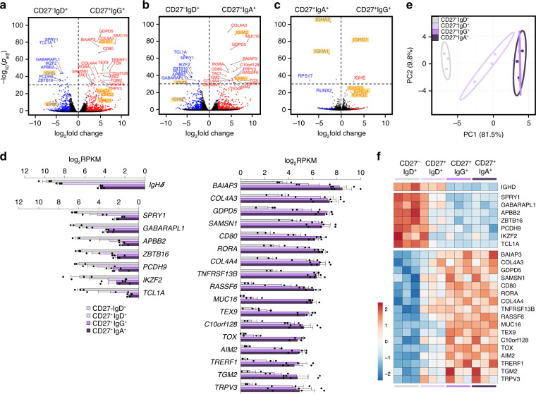
Using next-generation sequencing, we identified differentially expressed (DE) mRNA transcripts in CD27+IgG+ vs. CD27–IgD+ B cells and CD27+IgA+ vs. CD27–IgD+ B cells by pairwise comparisons. IgCγ and IgCα transcripts were significantly increased and IgCμ and IgCδ correspondingly decreased in CD27+IgG+ and CD27+IgA+ swMBCs (Fig. 4a, b). The higher IgCγ1 and IgCα2 expression reflected the peripheral blood predominance of IgG1 and IgA2 subclasses and respective B cells. Analysis at the highest level of significance of padj < 10 × 10–30 identified the same 24 (17 upregulated and 7 downregulated) DE mRNAs (not including IgCδ) in both IgG+ and IgA+ swMBCs as compared to their NBC counterparts (Fig. 4a, b and Supplementary Fig. 2a, b, Supplementary Table 2). RSP17, a ribosomal subunit protein, and RUNX2, a TF essential for CSR to IgA, were highly expressed in IgA+ swMBCs as compared to IgG+ swMBCs (Fig. 4c). IgCε expression in IgG+ swMBCs, but not IgA+ swMBCs, likely reflected the sequential CSR poise from IgG1 to IgE of such swMBCs42. Importantly, the “cancellation” of the 24 DE genes when comparing CD27+IgG+ vs. CD27+IgA+ B cells outlined a gene expression profile characteristic and distinctive of swMBCs, regardless of BCR isotype (Fig. 4c and Supplementary Table 2). This was strengthened by an equivalent level of normalized expression of the 24 transcripts in IgG+ swMBCs and IgA+swMBCs (Fig. 4d).
Fig. 4. Transcriptome analysis in human NBCs and swMBCs.
a–c Global transcriptional differences of mRNAs in CD27+IgG+ vs. CD27–IgD+ B cells (a), CD27+IgA+ vs. CD27–IgD+ B cells (b) and CD27+IgG+ vs. CD27+IgA+ B cells (c), as depicted by volcano plots. All genes annotated in human GENCODE v24 GRCh38 are shown, with each circle representing 1 mRNA. The −log10 FDR-adjusted p (padj) value is shown on the y axis (padj < 10 × 10–30 indicated above-dashed line). DE mRNAs at padj < 0.05 are highlighted in red (upregulated) or in blue (downregulated). DE mRNAs at padj < 10 × 10–30 and common to both CD27+IgG+ and CD27+IgA+ MBCs are annotated; IgH chain transcripts are annotated and highlighted. d Normalized (log2RPKM) expression of the 24 (IgHδ excluded) DE mRNAs at padj < 10 × 10–30 in swMBCs as compared to NBCs depicted by histogram for each B cell subset (left, downregulated and right, upregulated). Data are mean ± SEM from all three subjects. e Transcriptome clustering of the sorted subsets performed using the top 24 DE mRNAs depicted by principal component analysis (PCA) plot. Prediction ellipses define 95% confidence intervals. Each symbol represents an individually sorted subset (n = 3). f Relative expression profiles of swMBC core transcriptional signature mRNAs at padj < 10 × 10–30 compared by heatmap, depicting relative transcriptional changes across CD27–IgD+, CD27+IgD+, CD27+IgG+, and CD27+IgA+ B cell subsets in each subject (order; B,C,G). Data in a–f depict DE mRNA as determined by edgeR. B cell subsets are repeated at the bottom, left to right in CD27–IgD+ (gray), CD27+IgD+ (lavender), CD27+IgG+ (purple), and CD27+IgA+ (dark purple).
The transcriptional distance between the four B cell subsets, as measured by principal component analysis (PCA), yielded three discrete clusters, identifying NBCs, unswMBCs, and swMBCs (Fig. 4e and Supplementary Fig. 2c). The 24 DE genes discriminated IgG+ swMBCs and IgA+swMBCs from the other two B cell subsets across all three subjects (Fig. 4f). Further, the 24 DE genes discriminated total MBCs (CD27+ B cells) from NBCs (CD27− B cells) in the 4 additional healthy subjects (A, D, E, F) (Supplementary Table 1 and Supplementary Fig. 2c, d). Thus, at the highest level of significance, 24 DE mRNAs characteristically distinguish class-switched IgG+ swMBCs and IgA+ swMBCs from their NBC counterparts (swMBC core transcriptional signature).
Gene expression outlines distinct signaling and TF activities in swMBCs
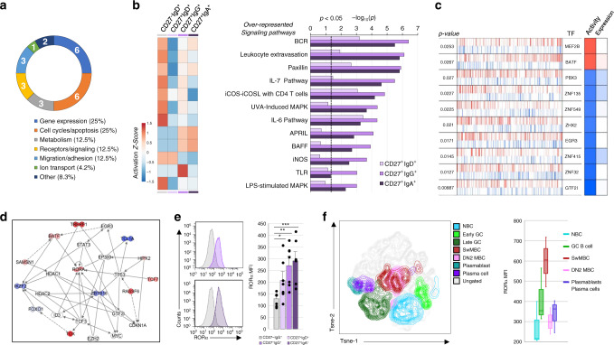
Among the 24 swMBC core transcriptional signature genes, six have a role in cell cycle and apoptosis, and six are implicated in gene expression (Fig. 5a). The aggregate mRNA profile (DE transcripts, padj < 0.05) in MBCs was explored by IPA and revealed significant over-representation in leukocyte extravasation as well as cytokine and MAP kinase signaling pathways. The activation status of such pathways differed significantly between swMBCs and NBCs (Z-score) (Fig. 5b), suggesting a contribution of such pathways to MBC function. As mRNA expression fails to capture post-transcriptional and post-translational events, we inferred the differential activity of TFs in human MBCs by applying a master regulator inference algorithm (MARINa) using a human B cell-specific Bayesian interactome31. In swMBCs, MEF2B and BATF were inferred to have increased activity, while GTF2I, EGR3, and multiple zinc-finger TFs were inferred to have decreased activity (Fig. 5c). Two DE genes, RORα, and ZBTB16, were central to a transcriptional network that incorporated MARINa-inferred TFs, nucleus-localized swMBC core transcriptional signature genes, and IPA-identified coactivators and corepressors (Fig. 5d). Probing deeper into MARINa TF analysis, ZBTB16 was found to be significant (p = 0.034), ranking 15th out of the 621 TFs in the regulon. RORα was not part of the transcriptional “regulon” object31 and, consequently, its activity was not addressed here. Analysis of RORα protein, however, showed it to be significantly expressed in swMBCs and unswMBCs across the seven human subjects (Fig. 5e). Further, RORα protein was analyzed in five discrete tonsil B cell subsets from three additional subjects and was highly expressed in such tonsil swMBCs (Fig. 5f). Thus, our integrative analysis identified signaling pathways and TF networks that are important to MBC identity and/or functions, suggesting a central role for TF RORα in the phenotype/function of human swMBCs.
Fig. 5. Functional characterizations of the swMBC core transcriptional signature.
a Functional gene ontology annotation of the 24 DE genes at padj < 10 × 10−30 in swMBCs vs. NBCs (swMBC core transcriptional signature). Numbers denote genes in each category. b Canonical pathways in swMBCs and unswMBCs displaying significant increases or decreases in activation status were determined by IPA, with −log10-transformed p shown on the x axis (p < 0.05 indicated by the dashed line). Activation Z-scores of pathways in each subset are shown by heatmap color-coded according to legend: increased activation, red; decreased activation, blue. c SwMBC transcriptional regulators as identified by the MARINa algorithm, which is measured using as differentially expressed target odds ratio90. Gene expression is rank-ordered from the most downregulated to the most upregulated gene in swMBCs. In each row, the transcription factor (TF) names are given; positive (activated; red bars) and negative (repressed; blue bars) targets of the regulator have plotted along with the gene expression signature in rank order. The first column on the right depicts inferred differential activity of the regulator (Activity), while the second shows the regulator differential expression (expression). d IPA network analysis indicating annotated interactions between MARINa-inferred TFs, nucleus-localized DE swMBC signature genes, and IPA-identified cofactors. CD27+IgG+ and CD27+IgA+ B cell data sets are overlaid on the IPA network, depicting upregulated genes in red and downregulated genes in blue. e RORα expression in CD27-IgD+, CD27+IgD+, CD27+IgG+, and CD27+IgA+ B cells from peripheral blood of seven healthy human donors, as analyzed by intracellular staining followed flow cytometry. *p < 0.05, **p < 0.01, ***p < 0.001, ns not significant (paired two-sided t-test). Data are mean ± SEM of three independent experiments. f RORα expression in B cell fractions from the tonsil of three human subjects, as analyzed by intracellular staining followed flow cytometry. Fractions analyzed comprise NBCs (CD19+CD38−CD27−IgD+CD138−; blue), GC B cells (CD19+CD38loCD27+/−IgD+/−CD138−; green), swMBCs (CD19+CD38−CD27+IgD−CD138−; red), DN2 MBCs (CD19+CD38−CD27−IgD−CD138−; pink), plasmablasts and plasma cells (CD19+/−CD38hiCD27+IgD−CD138+/−; purple). Data are represented as boxplots where the middle line is the median, the lower and upper hinges correspond to the first and third quartiles, the upper whisker extends from the hinge to the largest value no further than 1.5 × IQR from the hinge (where IQR is the inter-quartile range) and the lower whisker extends from the hinge to the smallest value at most 1.5 × IQR of the hinge. Quantification of RORα MFI in e (n = 7) and f (n = 3). B cell subsets; CD27–IgD+ (gray), CD27+IgD+ (lavender), CD27+IgG+ (purple), and CD27+IgA+ (dark purple).
unswMBCs display a transcriptional profile of transition between NBCs and swMBCs
Although the memory phenotype of unswMBC is an accepted notion, the nature of these CD27+IgD+ B cells remains a matter of debate. Virtually all CD27+IgD+ B cells retained expression of surface IgM (Fig. 1b), yet they were reported to express mutated Ig VH genes8. These unswMBCs carried significantly more somatic point-mutations than NBCs (Fig. 3b, c) and displayed (pairwise comparison) two DE upregulated transcripts, TFEC and ZBTB32, at padj < 10 × 10–30, as well as four swMBC core signature transcripts (TRPV3, TACI, COL4A4, and SAMSN1), upregulated at a significance of padj < 10 × 10–15 (Supplementary Fig. 3a, b and Fig. 4d). PCA showed unswMBCs clustering between NBCs and swMBCs (Fig. 4e) and displayed at lower levels signaling pathways characteristic swMBCs (Fig. 5b). PCA and hierarchical clustering analyses were expanded across the DE gene profile at differing levels of significance and consistently showed CD27+IgD+ unswMBCs as an independent cluster, slightly closer to swMBCs (Supplementary Fig. 4). Overall, CD27+IgD+ unswMBCs display a transcriptome of transition from NBCs to swMBCs.
Chromatin accessibility and active gene landscape in MBCs and NBCs
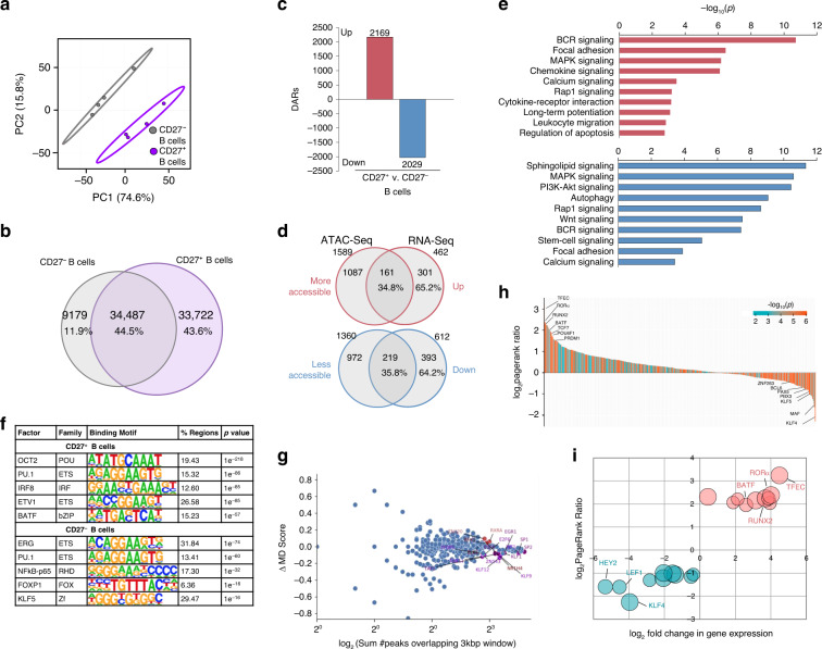
As RNA expression is dependent upon DNA accessibility, we sought to define the chromatin landscape of human MBCs by ATAC-Seq and characterize its intersection with the transcriptome in these B cells (subjects A, D, E, F). Of the 77,388 accessible loci identified, NBCs (CD27− B cells) and total MBCs (CD27+ B cells) displayed distinct patterns of chromatin accessibility, as indicated by component 1 of PCA (74.6% variance) (Fig. 6a). Overall, total MBCs trended towards higher chromatin accessibly, with 43.6% of identified peaks unique to them, 11.9% of peaks unique to NBCs, and 44.5% of peaks accessible in both total MBCs and NBCs (Fig. 6b). Differentially accessible regions (DARs) at p < 0.05 were identified in total MBCs vs. NBCs. Overall, 4198 loci were differentially accessible in total MBCs vs. NBCs, with 2169 (51.7%) DARs with increased accessibility and 2029 (48.3%) with decreased accessibility (Fig. 6c).
Fig. 6. The chromatin landscape of human MBCs.
a Clustering of CD27– NBCs and CD27+ total MBCs based on top differentially accessible loci as displayed by PCA (4198). Prediction ellipses define 95% confidence intervals. Each symbol represents an individually sorted subset (n = 4). b Overlap of accessible loci between NBCs and total MBCs depicted by Venn diagram. c Differential chromatin accessibility (p < 0.05) between total MBCs and NBCs as determined by DEseq2. Differentially accessible regions (DARs) with increased (red) or decreased (blue) accessibility depicted by histograms. d Proportion of DE genes that overlap with DARs, as depicted by Venn diagrams. Percentage of genes exhibiting corresponding increases/decreases in chromatin accessibility shown below the number of genes. e Statistically over-represented KEGG pathways associated with increased (red) or decreased (blue) DARs depicted by the histogram. f Significant TF- binding motifs enriched in NBC- or all MBC-specific open chromatin regions as determined by HOMER motif analysis. g For all motifs (dots), the changes in MD score between NBCs and total MBCs (y-axis) are (MA) plotted against the number of motifs within 1.5 kb of any ATAC-Seq peak center (x axis)—computed by DAStk94. Red points depict statistically significant increased MD-scores (p < 0.05). h, i RNA-Seq and ATAC-Seq data sets were integrated using the Taiji PageRank algorithm92 to generate PageRank activity scores and p-values for human TFs. h All 421 TFs (p < 0.05) plotted in rank order according to their log2PageRank score ratio between MBCs vs. NBCs. Each TF PageRank −log10(p) is denoted by color-coding according to legend: more significance, red; less significance, blue. i The top 10 TFs in NBCs and top 10 TFs in MBCs was plotted according to their log2PageRank score ratio and log2fold change in transcript expression in MBCs vs. NBCs. The bubble size of each TF is determined by the −log10(p) of Taiji analysis.
To determine whether cell-specific open chromatin regions identified by ATAC-Seq correlated with cell-specific gene expression, we integrated our NBC and MBC ATAC-Seq and mRNA-Seq data sets. Overall, 462 and 612 DE mRNAs were at significantly higher or lower levels, respectively, in both IgG+swMBCs and IgA+ swMBCs compared to NBCs at padj < 0.05. Of the 462 upregulated genes, 161 displayed increased chromatin accessibility nearby or within their loci. Of the 612 downregulated genes, 219 displayed decreased chromatin accessibility nearby and/or within their loci. Thus, of 1074 DE mRNAs, 35.4% displayed corresponding increases or decreases in chromatin accessibility near their genetic loci (Fig. 6d). Chromatin accessibility was altered in the exon and promoter regions of 5 of 7 downregulated and 9 of 17 upregulated genes in the swMBC core transcriptional signature (Supplementary Fig. 5a–c). Gains in chromatin accessibility were associated with genes encoding proteins involved in MAPK, BCR, and Ca2+ signaling and leukocyte migration, validated by pathway analysis of the mRNA transcriptome (Fig. 5b). Losses in chromatin accessibility were associated with genes involved in Sphingolipid, MAPK, and stem-cell signaling, focal adhesion, and autophagy (Fig. 6e).
As the transcriptional regulation of human MBCs is poorly understood, we sought to determine whether MBCs DARs were associated with given cis-regulatory elements. Significant enrichment in DNA-binding factors that poise B cells for proliferation and plasma cell differentiation were found in DARs with increased accessibility in total MBCs, such as OCT2, IRF8, and bZIP motifs43. Conversely, enrichment in DNA-binding factors involved in B cell development and cellular quiescence, such as ERG, RHD, and KLF motifs21,44, were found in decreased accessible DARs of total MBCs (Fig. 6f). To further assess changes in TF activity, we probed for the enrichment of cis-regulatory elements in all accessible loci found in total MBCs over NBCs (Δ-MD score). NR1H4- and RXRA-binding motifs were enriched in the open chromatin regions of total MBCs while ERG1-, KLF-, and ZF-binding motifs were enriched in the open chromatin regions NBCs (Fig. 6g). These differences in motif accessibility were supported by MARINa-inferred changes in related TF activity. For example, BATF activity was inferred to be increased in swMBCs based on patterns of mRNA expression, and BATF-binding motifs were enriched in increased DARs in total MBCs (Figs. 5c and 6f, g).
Taiji was used to develop an integrative transcriptional regulatory network and predict TF activity based off a personalized PageRank algorithm. Consistent with our previous analyses, Taiji identified TFEC, RORα, and BATF as the TFs with the highest activity in MBCs, while KLF4, HEY2, and LEF1 were identified as the TFs with the highest activity in NBCs (Fig. 6h, i). Consistent with the notion that MBCs are poised to differentiate into plasma cells, PageRank inferred increased activity of PRDM1 and decreased activity of BCL6 in MBCs (Fig. 6h). Thus, the profile of chromatin accessibility contrasted human MBCs with NBCs and outlined signaling pathways and cis-regulatory elements relevant to MBCs.
MicroRNAs in MBCs and identification of MIR181 as a major gene expression regulator
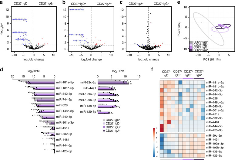
While chromatin accessibly is necessary for transcription, gene expression is subject to additional layers of regulation, mostly epigenetic, both at the DNA as well as post-transcriptional level. miRNAs are small ncRNAs, which target mRNA 3′ UTRs to post-transcriptionally repress gene expression45. As we have shown, miRNAs play a critical role in modulating B cell SHM, CSR, and plasma cell differentiation28,29,45. miRNAs role in MBCs is less understood. Our analysis of the miRNAome identified 19 DE miRNAs, 6 upregulated and 13 downregulated, in both IgG+ swMBCs and IgA+ swMBCs vs. NBCs at a significance of p < 0.05 (Fig. 7a, b). These 19 DE miRNAs “canceled out” when IgG+ swMBCs were compared with IgA+ swMBCs (Fig. 7c), being expressed at similar levels in IgG+swMBCs and IgA+ swMBCs (Fig. 7d and Supplementary Fig. 3c). Although also expressed in some cases in unswMBCs, they overall discriminated MBCs from their NBC counterparts (Fig. 7e, f). Among the 13 downregulated miRNAs in swMBCs, MIR181a and MIR181b, both MIR181 family members, were decreased by more than 60,000 and 3000 transcripts (Fig. 7d). MIR181a and MIR181b transcript downregulation were concomitant with decreased chromatin accessibility at these miRNA’s host–gene (HG) promoters (Supplementary Fig. 6a).
Fig. 7. Altered expression of the miRNA profile in human swMBCs.
a–c Global transcriptional differences of miRNAs in CD27+IgG+ vs. CD27–IgD+ B cells (a), CD27+IgA+ vs. CD27–IgD+ B cells (b), and CD27+IgG+ vs. CD27+IgA+ B cells (c) as depicted by volcano plot. All miRNAs annotated in human GENCODE v24 GRCh38 are shown, with each circle representing 1 miRNA. −Log10-transformed p is shown on the y axis (p < 0.05 indicated above dashed line). DE miRNAs at p < 0.05 are highlighted in red (upregulated) or blue (downregulated). DE miRNAs at p < 0.05 (with Δ abundance > 1000) shared by CD27+IgG+ and CD27+IgA+ are annotated. d Normalized (log2RPM) expression of the 19 DE miRNAs at p < 0.05 in swMBCs as compared to NBCs depicted by histogram for each B cell subset. Data are mean ± SEM of all three subjects. e Transcriptome clustering of the sorted subsets performed using the 19 DE miRNAs depicted by the PCA plot. Prediction ellipses define 95% confidence intervals. Each symbol represents an individually sorted subset (n = 3). f Relative expression profiles of swMBC core transcriptional signature miRNAs at p < 0.05 compared by heatmap, depicting relative transcriptional changes across CD27–IgD+, CD27+IgD+, CD27+IgG+ and CD27+IgA+ B cell subsets in each subject (order; B, C, G). Data in a–f depict DE miRNAs as determined by edgeR. B cell subsets; CD27–IgD+ (gray), CD27+IgD+ (lavender), CD27+IgG+ (purple), and CD27+IgA+ (dark purple).
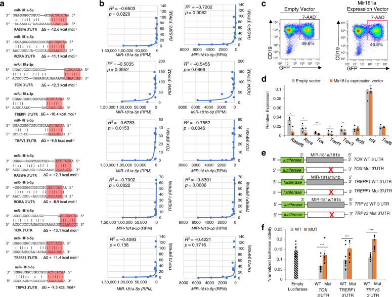
Downregulation of MIR181a and MIR181b was expected to relieve the silencing of MIR181 targeted mRNAs. Indeed, 5 of the 17 significantly upregulated mRNAs in swMBC core transcriptional signature, RASSF6, TOX, TRERF1, RORα, and TRPV3, were predicted targets of MIR181a and MIR181b, exhibiting 3’UTR sequences highly complementary with MIR181seed regions and favorable thermodynamics (Fig. 8a)—overall mRNA profile at padj < 0.05 showing 50 of the 462 mRNAs (10.8%) upregulated in both IgG+swMBCs and IgA+swMBCs to be predicted targets of MIR181 (Supplementary Fig. 6b). A significant inverse correlation between MIR181a/MIR181b and their 5 mRNA targets indicated the epigenetic release of these select mRNA transcripts in swMBCs (Fig. 8b). Indeed, enforced expression of Mir181a in retroviral-transduced mouse B cells significantly reduced the expression of these five genes by 32.3–72.4% (Fig. 8c, d and Supplementary Fig. 6c). Further, luciferase reporter assays involving (CD154 and IL-4) induced human CL-01 B cells expressing MIR181 and transfected with wildtype (WT) or mutant (Mut) reporter constructs confirmed TOX, TRERF1, and TRPV3 to be direct and specific targets of MIR181 (Fig. 8e, f)—RASSF6 was shown to be a direct target of MIR181 family members in the context of gastric cancer46; RORα was not tested because of the length and complexity of its 3′UTR, which, however, contains three MIR181 target sites. Thus, MIR181 releases select swMBC gene expression from epigenetic regulation.
Fig. 8. MIR181 as a key regulator of human swMBC-specific gene expression.
a Schematic of 3′UTRs of RASSF6, RORα, TOX, TRERF1, TRPV3 mRNAs, and their targeting by MIR181a-5p and MIR181b-5p. Canonical target site base-pairing with miRNA seed sequences are highlighted in red. Hybridization is depicted as the most thermodynamically favorable molecular interaction between the two RNA species, considering RNA secondary structure. Gibbs free energy (ΔG) of nucleic acid folding and hybridization calculated by Mfold. Watson–Crick base-pairing (“|”); wobble base-pairing (“:”). Dashes indicate looping in the secondary RNA structure. b Spearman rank correlations of miRNA expression (RPM) and target mRNA expression (RPKM) calculated across all sorted subsets (n = 12). c FACs sorting of MDH1-PGK-GFP (Empty Vector) or MDH1-miR-181a-1-PGK-GFP (miR181 Expression Vector) retrovirally transduced (7AAD-CD19+GFP+) B cells. d Target gene expression (qPCR) in sorted (7AAD−CD19+GFP+) B cells transduced by MDH1-PGK-GFP (gray) or MDH1-miR-181a-1-PGK-GFP (orange) retrovirus (three independent experiments yielding similar results). *p < 0.05, **p < 0.01, ***p < 0.001, ns not significant (paired two-sided t-test). Data are mean ± SEM of three independent experiments. e Schematic diagram of the wildtype (WT) 3′UTRs of TOX, TRERF1, and TRPV3 mRNAs and their mutant (Mut) counterparts cloned into the psiCHECK-2 reporter vector. Point-mutations in putative miRNA target sites are depicted by “X”. f Luciferase activity in human CL-01 cells transfected with constructs containing wildtype (gray) or mutated (orange) TOX, TRERF1, or TRPV3 3′UTRs (cloned into psiCHECK-2 luciferase reporter constructs) after 24 h stimulation with CD154 plus IL-4 (n = 10). Transfection efficiency was controlled for by normalization to Renilla reniformis signal encoded on the same psiCHECK-2 vector. *p < 0.05, **p < 0.01, ***p < 0.001, ns not significant (paired two-sided t-test). Data are mean ± SEM of three independent experiments.
Potential regulatory capacities of lncRNAs in human swMBCs
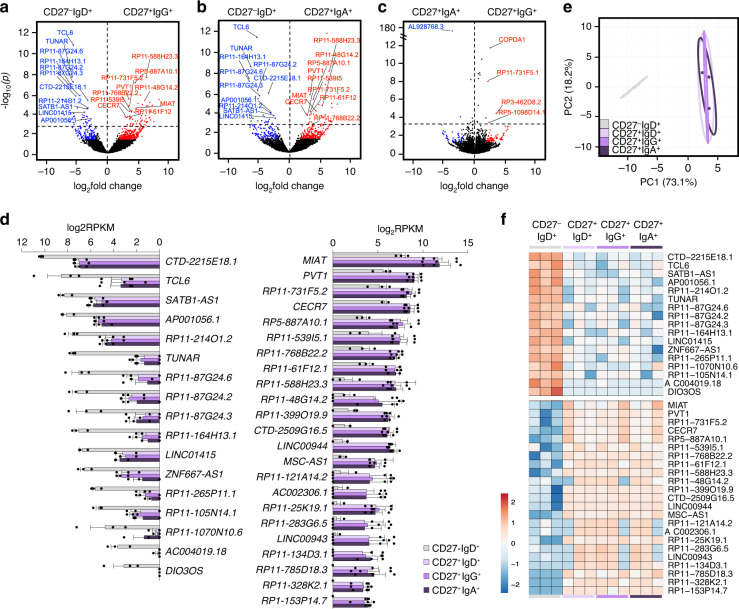
lncRNAs are greater than 200 nucleotides in length and can interact with DNA, RNA, and proteins, thereby mediating a layer of epigenetic regulation to positively or negatively modulate gene expression47. lncRNAs have been implicated in key processes of T cells and innate lymphocytes47. While profiled in B cells48, their functional significance in human MBCs has not been explored. A comprehensive analysis by pairwise comparisons at p < 0.001 of MBC lncRNA landscape identified the same 23 upregulated and 17 downregulated DE lncRNAs in IgG+ swMBCs and IgA+ swMBCs as compared to NBCs—among these 40 DE lncRNAs, 21 had a change in abundance of more than 100 transcripts (Fig. 9a, b). All 40 lncRNAs “canceled out” when comparing IgG+ swMBCs vs. IgA+ swMBCs (Fig. 9c). Such 40 lncRNAs were indeed expressed at similar levels in IgG+ swMBCs and IgA+ swMBCs as well as in unswMBCs, thereby discriminating total MBCs from NBCs (Fig. 9d–f and Supplementary Fig. 3d).
Fig. 9. lncRNA expression profile in human MBCs and NBCs.
a–c Global transcriptional differences of lncRNAs in CD27+IgG+ vs. CD27–IgD+ B cells (a), CD27+IgA+ vs. CD27–IgD+ B cells (b), as well as CD27+IgG+ vs. CD27+IgA+ B cells (c), as depicted by volcano plots. All lncRNAs annotated in human GENCODE v24 GRCh38 are shown, each circle representing 1 lncRNA. −Log10-transformed p is shown on the y axis (p < 0.001 indicated above dashed line). DE lncRNAs at p < 0.05 are highlighted in red (upregulated) or in blue (downregulated). DE lncRNAs at p < 0.001 (with Δ abundance > 100) common to both CD27+IgG+ and CD27+IgA+ are annotated. d Normalized (log2RPKM) expression of the top 40 DE lncRNAs at p < 0.001 in swMBCs as compared to NBCs depicted by histogram for each B cell subset. Data are mean ± SEM of all three subjects. e Transcriptome clustering of the sorted subsets performed using the 40 DE lncRNAs depicted by the PCA plot. Prediction ellipses define 95% confidence intervals. Each symbol represents an individually sorted subset (n = 3). f Relative expression profiles of swMBC core transcriptional signature lncRNAs at p < 0.001 compared by heatmap, depicting relative transcriptional changes across CD27–IgD+, CD27+IgD+, CD27+IgG+, and CD27+IgA+ B cell subsets in each subject (order; B, C, G). Data in a–f depict DE lncRNAs as determined by edgeR. B cell subsets; CD27–IgD+ (gray), CD27+IgD+ (lavender), CD27+IgG+ (purple), and CD27+IgA+ (dark purple).
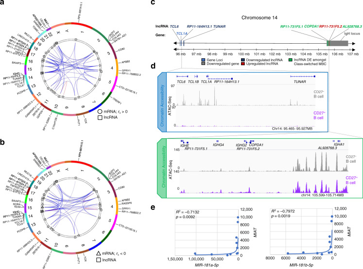
lncRNAs can modulate transcription of specific gene loci, generally in cis and nearby, including protein-coding loci (mRNA) and HGs of ncRNAs, such as miRNAs47. lncRNA functions have been inferred from lncRNA/mRNA co-expression analyses paired with functional and spatial enrichment. By integrated co-expression analysis, we identified significant positive and negative correlations between DE lncRNAs and DE mRNAs in swMBCs as compared to NBCs (Fig. 10a, b). One cluster of positively correlated transcripts found at the telomeric end of chromosome 14, upstream of the IgH locus contained 3 downregulated lncRNAs, TCL6, RP11-164H13.1, and TUNAR, together with 1 downregulated mRNA, TCL1A (Fig. 10c). The decreased expression of such lncRNAs and mRNA reflected the decreased chromatin accessibility at their respective loci (Fig. 10d). Interestingly, lncRNA AL928768.3 found to be expressed at 10.8-fold higher levels in IgA+MBCs than IgG+ MBCs is located 2.5 kB downstream of the IGHA1 locus. lncRNAs COPDA1 and RP11-731F5.1 were found to be expressed at 6.0 and 4.6-fold higher levels in IgG+ MBCs than IgA+ MBCs and are located in promoters of IGHG2 and IGHE loci, respectively (Fig. 10c, d). MIAT, a molecular sponge for MIR181b49, was consistently upregulated in swMBCs and unswMBCs of all three subjects analyzed and displayed significant negative correlations with MIR181a and MIR181b expression (Fig. 10e).
Fig. 10. Characterization of the human MBC lncRNA profile.
a, b lncRNA-to-mRNA co-expression correlations depicted as Circos plots with human chromosomal ideograms (various colors). lncRNA-to-mRNA correlations showing 21 DE lncRNAs (Δ abundance>100; depicted as squares) in swMBCs and the top three positively (a circles, rs > 0) correlated mRNAs and top three negatively (b triangles, rs < 0) correlated mRNAs for each lncRNA with p < 0.05 (Pearson correlation). c Schematic of the terminal end of chromosome 14 (q32.2–q32.3), depicted with DE mRNAs, lncRNAs, and critical B cell gene loci annotated. DE mRNAs and DE lncRNAs in swMBCs, as well as differences in genetic loci between CD27+IgG+ swMBCs, CD27+IgA+ swMBCs, and CD27–IgD+ NBCs are depicted. Downregulated lncRNAs (dark blue), upregulated lncRNAs (dark red), DE lncRNAs between swMBCs (green), downregulated mRNA (light blue), and the IgH locus (gray). d Chromatin accessibility upstream and in the human IgH loci is displayed by IGV gene track. Coverage includes gene and lncRNA introns, exons, promoter regions, and potential enhancer regions. ATAC-Seq signal is normalized for the window of interest, with NBCs depicted in gray and total MBCs in purple. e Correlations between expressed miRNAs and MIAT as calculated across all sorted subsets (n = 12) by Spearman’s rank correlation.
To outline potential lncRNA regulatory functions, trans and cis co-expression correlations of lncRNAs with mRNAs or miRNAs were analyzed in swMBC, unswMBCs, and NBCs (Supplementary Fig. 7a–d). Trans lncRNA-mRNA correlations trended positive (rs > 0.7, 3.29%) than negative (rs < −0.7, 2.60%), as did cis lncRNA-mRNA correlations (rs > 0.7, 3.97%; rs < −0.7, 3.05%) (Supplementary Table 3). lncRNAs with strong positive or negative cis correlations (rs > 0.65 or rs < −0.65) with mRNAs genes revealed enrichment in lipid metabolic genes and GPCR signaling (Supplementary Fig. 7e, f). Thus, a lncRNA expression profile is characteristic of all CD27+ MBCs as compared to NBC, with select lncRNAs proximal to the IgH locus displaying potential regulatory activity or acting as miRNA sponges and overall co-expression patterns of different RNA species indicating enrichment in distinct signaling pathways.
Discussion
mRNA, miRNA, and lncRNA transcriptomes revealed a common and unique transcriptional profile of human IgG+swMBCs and IgA+swMBCs, distinguishing them from NBCs. At the highest level of significance, swMBCs differed from NBCs in the expression of 24 genes, 17 increased and 7 decreased, with unswMBCs displaying a transcriptional signature of transition. The swMBC transcriptome was enriched in distinct MAPK, migratory and cytokine signaling pathways, likely influencing survival, homing and cell-to-cell interactions. In all (CD27+) MBCs, the chromatin landscape reflected changes in gene transcription and outlined differentially accessible cis-regulatory elements, potentially influencing cell gene expression and functions. As compared to NBCs, swMBCs non-coding transcriptome revealed a characteristic downregulation of MIR181a/MIR181b, pointing to this miRNA as a key modulator of swMBC gene expression. Also, lncRNA MIAT, a molecular sponge of MIR18149, was significantly increased in such swMBCs. MIAT likely released target genes from MIR181-mediated silencing, in concert with decreased accessibility of MIR181 HG promoters. Thus, when compared to NBCs, swMBCs exhibit a distinct mRNA, miRNA, and lncRNA transcriptome, dynamically interacting with a changing chromatin landscape to shape the identity and functions of these B cells.
CD27–IgD+, CD27+IgD+, CD27+IgG+, and CD27+IgA+ B cells displayed a pattern of IgVH gene expression conserved across NBCs, unswMBCs, and swMBCs within each subject and reflecting the genomic representation of the seven VH gene families. This was associated with a conserved distribution of CDR3 lengths in recombined VHDJH-CH transcripts, indicating an unbiased clonal differentiation of MBCs from their NBC progenitors. This was further supported by the conservation of Ig JH, Vκ, Jκ, Vλ and Jλ gene expression across NBCs, unswMBCs, and swMBCs, vastly overlapping across the subjects studied. Reflecting on a different degree of antigen experience, the mutational load in recombined transcripts was the greatest in swMBCs, intermediate in unswMBCs, and negligible in NBCs.
Our findings outlined a distinct gene expression profile characteristic of human swMBCs, regardless of their effector Ig class, be it IgG+ or IgA+, and possibly IgE+. They extend previous studies, which addressed select human MBCs, as conditioned by different antigens, microenvironments, and/or biased genetic programs41, and identified transcriptional programs not associated with MBCs of specific effector Ig classes25,50. In our seven subjects, NBCs accounted for two-thirds of the B cell repertoire, with unswMBCs and (IgG+ and IgA+) swMBCs making up for about one-tenth each. The 17 upregulated and seven downregulated mRNA transcripts distinguishing swMBCs from NBCs “canceled out” when comparing IgG+ MBCs with IgA+ MBCs, leaving the reciprocal IgH expression of the respective IgCγ and IgCα transcripts as the distinguishing difference. Expression of IgCε transcripts in IgG+ MBCs, but not in IgA+MBCs likely reflected the transition to IgE through sequential CSR from IgM to IgG1 and to IgE42.
The phenotype, transcriptome, functional cellular pathways, and gene loci accessibility yielded unifying information on MBCs of different isotypes, as different from NBCs in the same subjects. This was unexpected and per se remarkable, as we found a consistent homogeneity and concordance in the mRNAome, miRNAome, lncRNAome, chromatin accessibility, and inferred pathways of cell functions within MBCs—whether considered as CD27+IgG+ swMBCs and CD27+IgA+ swMBCs or total CD27+ B cells vs. NBCs, using two different B cell isolation approaches (isotype-specific MBCs and total MBCs) and despite the heterogeneous origin of the B cell samples: seven subjects of different age, sex, and race—a cohort quite different from any cohort of inbred mouse strains. Single-cell RNA-Seq may resolve further DE genes beyond this core transcriptional signature.
The identity and role of unswMBCs (CD27+IgD+) are a matter of debate. These B cells have been suggested to act as a reservoir of elements ready to supplement a recall antibody response, particularly in parasitic and bacterial infections24,51–53. Our findings suggest a transitional identity for human unswMBCs. These B cells expressed IgVH, JH, Vκ, Jκ, Vλ, and Jλ genes as well as a VHDJH-CH CDR3 length distribution similar to those of NBCs and swMBCs, and carried a moderate load of somatic point-mutations, intermediate between NBCs and swMBCs, consistent with unswMBCs emerging from NBCs and being potential precursors of swMBCs. This transitional identity is further emphasized by: (i) the intermediate expression of the 24 genes distinguishing swMBCs from NBCs; (ii) the unswMBCs hierarchical clustering pattern; (iii) the consistent downregulation of MIR181a and MIR181b, which is characteristic of swMBCs; and (iv), finally, the differential lncRNA profile nearly identical to that of swMBCs.
Of the 17 mRNAs upregulated in swMBCs at the highest level of significance, 6 transcripts, AIM2, CD80, RASSF6, RORα, TACI, and TOX, were reported to be upregulated in human CD27+B cells, mouse IgA+ MBCs or NP-specific GC-derived memory precursors, thereby emphasizing the importance of such genes also in human IgG+ MBCs and IgA+ MBCs17,20,50,54–57. Baff-R signaling would be important in the survival of mouse MBCs58. BAFFR was expressed in human NBCs and swMBC, while TACI, another BAFF receptor, was upregulated in swMBCs, thereby suggesting similar signaling requirements for mouse and human MBC survival. BAIAP3, TGM2, and TRPV3 encode proteins critical to ion channel Ca2+ flux, Ca2+ signal transduction, or Ca2+-dependent activity, likely playing a role in MBC reactivation59. TOX, TRERF1, and RORα, encode for TOX, a high-mobility group (HMG) box protein, which binds DNA architectural motifs and is critical in immune cell development60, TRERF1, which interacts with p300 and SF-1 to regulate P450scc gene expression, thereby impacting the cell cycle61, and RORα.
RORα was highly expressed in IgG+ MBCs and IgA+ MBCs, but not in NBCs, suggesting a central role for this TF in the identity of human swMBCs. RORα is a sequence-specific ligand-dependent TF implicated in mouse ILC2 and Th17 cell differentiation as well as mouse IgA+ MBC maintenance20,62. RORα was found to be central to the transcriptional network of MARINa-inferred TFs, nucleus-localized swMBC signature genes, and IPA-identified coactivators and corepressors. This indicates RORα’s importance in MBC identity and/or functions, as further supported by the integrative Taiji analysis. RORα’s high expression in peripheral blood and tonsil swMBCs further suggested an important role of this TF in MBCs maintenance. MUC16, which encodes a 22,000 amino acid membrane-associated adhesion molecule, was significantly upregulated in swMBCs. Through its binding partners galectin-1, galectin-3, and L-selectin, abundantly expressed by CD169+ macrophages63, Mucin-16 would stabilize cell-to-cell contact between MBCs and CD169+ subcapsular sinus macrophages to promote antigen subsequent B cell reactivation64. Mesothelin, another Mucin-16 binding partner, potentially home MBCs to lungs, consistent with reports of influenza-specific lung-resident MBCs in the mouse65,66. Thus, alongside changes in leukocyte migratory signaling and positional cues, the upregulation of MUC16 could suggest unique trafficking patterns that promote antigen surveillance by MBCs in the respiratory tract.
The downregulation at the highest level of significance of seven mRNAs in swMBCs suggested these genes are not essential for differentiation or maintenance of these B cells. Such genes included ZBTB1621 (ZBTB16’s activity was inferred by MARINa to be significantly decreased in swMBCs as compared to NBCs) as well as six other genes. Of these, IKZF2 codes for a key TF in early B cell development67 and double negative self-reactive T cells68, while TCL1A, an AKT signal transduction molecule, is downregulated in the transition from NBCs to GC B cells69. PCDH9, is a Ca2+ dependent cell-adhesion protein70 and SPRY1 a negative regulator of cell proliferation, migration, and promoter of apoptosis71.
At a lower level of significance (padj < 0.05), swMBCs’ gene expression profile comprised transcripts that were previously reported, including upregulated CD86, CXCR3, EBI2, FAS, and ZBTB32, and downregulated KLF4 and KLF921,50,63,72. IgG+ swMBCs and IgA+ swMBCs displayed decreased expression of BACH2, a gene that skews mouse MBCs toward plasma cell differentiation, upon reactivation18. In unswMBC and swMBCs, the expression of IL-9 and IL-9R was virtually absent, suggesting that IL-9 signaling, which is putatively important for mouse MBC generation17, is dispensable for human MBC maintenance and, perhaps, generation. While IL-2RG was abundantly expressed in all four B cell subsets, cytokine receptor subunits that pair with the common-γ chain were differentially expressed in NBCs and swMBCs. Accordingly, human swMBCs downregulate the expression of IL-4R, IL-21R, and IFNγR1, whose signaling contributes to T-bet expression73. In the healthy humans we analyzed, TBX21 expression was absent in swMBCs and unswMBCs, consistent with the segregation of T-bet+ “atypical” MBCs among double negative (CD27–IgD–) B cells74.
As assessed by ATAC sequencing, the genes whose transcription was significantly upregulated in swMBCs were highly accessible, emphasizing the importance of such genes to the identity of swMBCs. While chromatin accessibly is necessary for transcription, it is not sufficient per se for gene expression. Gene transcription is regulated by DNA methylation, histone post-translational modifications and activators, and repressors34,35,75. Additional modulation of gene expression is mediated by epigenetic factors, mainly, non-coding RNAs, such as miRNAs and lncRNAs, at the transcriptional and post-transcriptional level. 13 of 17 gene loci, including RASSF6, TRPV3, TOX, and RORα, were significantly accessible in NBCs, despite negligible levels of the respective transcripts, suggesting mechanisms of post-transcriptional regulation, possibly mediated by ncRNAs.
In this context, we showed MIR181a and MIR181b to be key regulatory ncRNAs, likely acting as epigenetic repressors of MBC-specific gene expression. To date, there has been no investigation addressing the interplay of miRNAs and mRNAs in human MBCs. Our findings showed a great reduction of MIR181a and MIR181b expression in the transition from NBCs to unswMBCs and swMBCs, a transition that would occur after the GC stage37,76. Chromatin accessibility in promoter regions of MIR181 HGs on chromosomes 1 and 9 was reduced in total MBCs, suggesting that decreased MIR181a/MIR181b expression was mediated in part, by chromatin remodeling. Integrated analysis of the miRNA and mRNA data sets predicted MIR181 to further target 11% of the greater upregulated MBC genes (50/462 genes) and 5 of the 17 DE mRNAs upregulated in swMBC. MIR181’s role in regulating such genes was supported by MIR181 complementarity to target gene 3′UTRs and strengthened through functional evidence. Enforced expression of Mir181 in B cells significantly reduced Rassf6, Tox, Trerf1, Rorα, and Trpv3 expression, while luciferase reporter assays confirmed RASSF6, TOX, TRERF1, and TRPV3 as direct and specific targets of MIR181, thereby identifying a novel epigenetic mechanism centered on MIR181 and impacting key gene expression in swMBCs.
In human tonsils, lncRNAs display a dynamic relationship between enhancer lncRNAs and genetic elements of GC B cells48. Our findings showed that human circulating IgG+ swMBCs and IgA+ swMBCs as well as unswMBCs displayed nearly identical lncRNA profiles, that discriminate them from their NBC progenitors. In swMBCs, distinct DE lncRNAs disproportionately clustered proximal to or within the IgH locus, a key element of BCR and antibody expression. Accordingly, DE lncRNAs in IgG+ MBCs vs. IgA+ MBCs were associated with either IgCγ2, IgCε, or IgCα1 gene exons, thereby indicating a role for such lncRNAs in the regulation of switched BCR expression. Integration of DE mRNAs with DE miRNAs, as complemented by functional assays, identified MIR181 downregulation as a central mechanism of swMBC-specific gene expression. MIAT, a sponge of MIR18149, was upregulated in swMBCs and inversely correlated with MIR181a and MIR181b expression, suggesting that MIAT provides a regulatory mechanism that, along with chromatin accessibility, contributes to reduced MIR181 expression and promotes swMBC-specific gene expression.
Our integrative analysis of transcriptional profiles and chromatin accessibility revealed a dynamic and synergistic epigenetic landscape distinguishing MBCs from NBCs, and suggested that swMBCs emerge stochastically from the NBC pool, not from select elements or subsets of NBCs. Overall, our findings point to a core transcriptional signature, which is characteristic of and shared by swMBCs, regardless of BCR isotype, be it IgG or IgA, and distinct from that of NBCs. Beyond this core transcriptional signature, single-cell RNA-Seq may further tease apart the heterogeneity of human swMBCs, perhaps identifying discrete elements within this memory compartment77. UnswMBCs may act as intermediate elements in the transition of NBCs to swMBCs, which trend closer to swMBCs, consistent with their CD27 expression, light mutational load, Euclidean clustering, and non-coding transcriptional profile. Further, our findings, which stem from the integration of chromatin remodeling, cis-regulatory elements, and distinct expression profiles of mRNAs, miRNAs, and lncRNAs, provide evidence of a key regulatory role for the non-coding transcriptome, particularly MIR181 and MIAT, in the identity, maintenance and, likely, generation of human MBCs. Finally, by providing new insights into the transcriptional and chromatin landscape of MBCs, they open new avenues of investigation on the generation, key drivers, such as RORα, TRERF1, and TOX, identity, and role of MBCs as critical cellular elements in human health and disease.
Methods
Human B cells isolation
Buffy coats were obtained from South Texas Blood and Tissue Center, San Antonio, TX. Samples were obtained from seven healthy and non-immuno-compromised human subjects of different ages, 20–39, four males, three females of a different races, and ethnic backgrounds (Subjects A–G) (Supplementary Table 1)78. All experiments involving human samples were approved by the Institutional Review Board (IRB) of UTHSCSA and written informed consent was obtained. PBMCs were isolated from a diluted buffy coat (1:2) using density gradient Histopaque (Sigma Aldrich) in SepMate 50 ml tubes, according to the manufacturer’s instructions79. PBMCs were separated from the RBCs and subjected to two washes with FCS-RPMI (RPMI 1640, 10% fetal bovine serum, 1% antibiotics) and spun at 0.3 g for 8 min. Residual RBCs were lysed using ACK lysing buffer (ThermoFisher Scientific) and quenched using full media. Cell counting was performed on a Bright-Line™ hemocytometer (Hausser Scientific) utilizing a Nikon Labophot-2 research microscope. Approximately 5 × 10⁸ PBMCs were obtained per subject. PBMCs were resuspended to 50 × 106 cells/ml.
B cells (total) were isolated by negative selection from PBMCs (subjects B, C, G) using the EasySep Human Total B Cell Isolation Kit (STEMCELL Technologies). This kit utilizes anti-CD4, CD8, CD14, CD16, CD25, CD36, CD56, CD61, CD66b, CD123, and glycophorin A mAbs conjugated to anti-dextran Abs, which in conjunction with dextran-coated magnetic beads allow for depletion of all cell types other than B-cells. Approximately 5 × 10⁷ B cells were obtained from each subject, validated for purity (>99%) by flow cytometry by anti-CD19 mAb and used for cell sorting using a BD LSRII flow cytometer (BD Biosciences) or BD FACSCelesta flow cytometer (BD Biosciences) with FACSDiva software (BD Biosciences). Seventy-five percent of these B cells were stained with FITC-anti-human-IgG mAb (clone G18-145; BD Pharmingen), APC-anti-human-IgA mAb (clone IS11-8E10; Miltenyi Biotec), and PE-anti-human-CD27 mAb (clone M-T271; Biolegend) for isolation of swMBCs. CD27+IgG+ B cells and CD27+IgA+ B cells were sorted into separate 1.2 ml Eppendorf tubes containing 0.5 mL of HBSS (Hank’s balanced salt solution) buffer containing 0.1% bovine serum albumin (BSA). The remaining 25% of B cells were stained with anti-IgD BV421 mAb (clone IA6-2; Biolegend) and anti-CD27 PE mAb (clone M-T271; Biolegend), and CD27–IgD+ B cells and CD27+IgD+ B cells were sorted in 0.5 ml of HBSS + 0.1% BSA buffer. All sorted B cells were subjected to phenotypic analysis for confirmation of their identity and purity.
CD27− and CD27+ B cells were directly isolated from PBMCs (Subjects A, D, E, F) by immunomagnetic selection using the EasySep Human Memory B Cell Isolation Kit (STEMCELL Technologies). This is based on a proprietary two-step method using EasySep Releasable RapidSpheres to positively select CD27+ cells, remove magnetic particles, and then deplete non-B cells using EasySep Dextran RapidSpheres with antibody complexes. This procedure also allows for the depletion of non-B cells in the CD27– fraction in order to isolate naive B cells in parallel.
In all seven cases, B cells were subjected to pre-and-post isolation phenotypic analysis using the following surface markers and fluorophores: PEcy7-anti-human-CD19 mAb (clone H1B19; Biolegend; 1:100), PE-anti-human-CD27 mAb (clone M-T271; Biolegend; 1:100), BV421-anti-human-IgD mAb (clone IA6-2; Biolegend; 1:100), FITC-anti-human-IgG mAb (clone G18-145; BD Pharmingen; 1:100), APC-anti-human-IgA mAb (clone IS11-8E10; Miltenyi Biotec; 1:100), and APC-Cy7-anti-IgM mAb (clone MHM-88, Biolegend; 1:100). Data were analyzed using FlowJo software version 10.6.2 (FlowJo LLC)78.
Flow cytometry
PBMCs or B cells were stained with fluorochrome-conjugated mAbs in Hank’s Buffered Salt Solution plus 0.1% BSA (BSA-HBSS) for 20 min. Total PBMCs were stained with FITC-anti-human-IgG mAb (clone G18-145; BD Pharmingen; 1:100), APC-anti-human-IgA mAb (clone IS11-8E10; Miltenyi Biotec; 1:100) and APC-Cyanine7-anti-human-CD27 mAb (clone M-T271; Biolegend; 1:100), BV421-anti-human-IgD mAb (clone IA6-2; Biolegend; 1:100), PE-Cyanine7-anti-human-CD19 (clone HIB19; Tonbo Biosciences; 1:100). Single cell suspension of tonsil cells were stained with APC-Cyanine7-anti-human-CD27 mAb (clone M-T271; Biolegend; 1:100), BV421-anti-human-IgD mAb (clone IA6-2; Biolegend; 1:100), PE-Cyanine7-anti-human-CD19 (clone HIB19; Tonbo Biosciences; 1:100), BV510-anti-human-CD138 mAb (clone MI15 Biolegend; 1:100), BV650-anti-human-CD38 mAb (clone HB-7; Biolegend; 1:100)30,32,78. For intracellular staining, cells were surface stained followed by incubation with the BD Cytofix/Cytoperm buffer at 4 °C for 20 min. After washing twice with the BD Perm/Wash buffer, cells stained with PE-anti-human-RORα mAb (Clone 784652; R&D Systems; 1:50) overnight at 4 °C. Cells were then were resuspended in HBSS with 1% BSA for flow cytometry. In all flow cytometry analyses, cells were appropriately gated on forward and side scattering to exclude dead cells and debris28. RORα expression in CD27-IgD+, CD27+IgD+, CD27+IgG+ and CD27+IgA+ B cells from peripheral blood of the 7 healthy human subjects was analyzed by intracellular staining followed flow cytometry. RORα expression in B cell fractions from tonsils of three additional human subjects were stained intracellularly and analyzed by flow cytometry. Fractions analyzed comprise NBCs (CD19+CD38−CD27−IgD+CD138−), GC B cells (CD19+CD38loCD27+/−IgD+/−CD138−), swMBCs (CD19+CD38−CD27+IgD−CD138−), DN2 MBCs (CD19+CD38−CD27−IgD−CD138−), plasmablasts and plasma cells (CD19+/−CD38hiCD27+IgD−CD138+/−). Samples were run using a BD LSRII flow cytometer (BD Biosciences) or BD FACSCelesta flow cytometer (BD Biosciences) with FACSDiva software (BD Biosciences). Seven‐parameter files were imported into FlowJo software version 10.6.2 and biexponentially transformed prior to t-SNE analysis. t-SNE was then run on concatenated down-sampled files (100,000 cells/sample). t-SNE maps were generated by plotting t-SNE dimension 1 and dimension 2, and the gated subsets overlaid manually based on markers of interest on the dot-plot, to show the expression of these markers on different tonsil subtypes.
mRNA, miRNA, and lncRNA sequencing
RNA was isolated from cells using the Directzol RNA Microprep Kit (Zymogen Research) (based on cell number), according to the manufacturer’s instructions and as previously described30. RNA integrity was verified using an Agilent Bioanalyzer 2100 (Agilent). Next-generation RNA-Seq for mRNA and non-coding RNA was performed by the Genome Sequencing Facility at the University of Texas Health Science Center San Antonio (UTHSCSA) Greehey Children’s Cancer Research Institute. High-quality RNA was processed using an Illumina TruSeq RNA sample prep kit v2 or TruSeq Small RNA Sample Prep Kit following the manufacturer’s instructions (Illumina). Clusters were generated using TruSeq Single-Read Cluster Gen. Kit v3-cBot-HS on an Illumina cBot Cluster Generation Station. After quality control procedures, individual mRNA-Seq or small RNA-Seq libraries were then pooled based on their respective 6-bp index portion of the TruSeq adapters and sequenced at 50 bp/sequence using an Illumina HiSeq 3000 sequencer. Resulting reads were checked by assurance (QA) pipeline and initial genome alignment (Alignment).
IgVH, D, JH, and VL, JL gene expression
To analyze expressed Ig VH, D, JH and Vκ, Jκ, as well as Vλ, Jλ genes in CD27–IgD+, CD27+IgD+, CD27+IgG+, and CD27+IgA+ B cells, we collated the transcript read counts (RPKM) that aligned to the IgH and IgL chain genes to determined relative composition profiles for all 12 samples sorted. The human IgH locus comprises seven families with 36–49 functional coding VH genes: VH1, 11 members; VH2, 4 members; VH3, 23 members; VH4, 7 members; VH5, 2 members; VH6, 1 member. The VH7 family consists of only one member, IGHV7-4-1, which is not always part of a haplotypic complement, i.e., is absent in some subjects. The human Igκ locus comprises 39 functional Vκ genes and 5 Jκ genes; the human Igλ locus comprises 30 functional Vλ genes segregated into 10 subgroups and 5 functional Jλ-Cλ clusters. IgH and IgL gene expression profiles were organized in stacked histograms to depict relative usage39,80 (The International ImMunoGeneTics Information System, http://www.imgt.org/). Stacked histograms were also used to depict Ig VH, D, JH and Vκ, Jκ, as well as Vλ, Jλ gene segments genomic representation.
IgH CDR3 and VH somatic point-mutations
Recombined VHDJH-CH transcripts were amplified to determine CDR3 length and analyze somatic point-mutations in VH segments28,78. RNA was extracted using the Directzol RNA Microprep Kit (Zymogen Research) from CD27-IgD+, CD27+IgD+, CD27+IgG+, and CD27+IgA+ B cells of subjects B, C, and G. cDNA was synthesized from RNA with the SuperScript III First-Strand Synthesis System (Invitrogen) using oligo-dT primer. Rearranged VHDJH-CH cDNA was amplified using forward (degenerate) primers for leader sequences of (VH1, VH2, VH3, VH4, VH5, VH6, VH7 family genes) in conjunction with reverse Cμ, Cδ, Cγ or Cα isotype-specific primer and Phusion DNA polymerase (New England BioLabs). Amplification conditions were 98 °C for 30 s, 58 °C for 30 s, and 72 °C for 1 min. The amplified library was tagged with Illumina clustering adapters with barcodes for sample multiplexing and enriched by PCR. High-throughput, 300 bp pair-ended sequencing was performed using the Illumina MiSeq system. Somatic point-mutations were identified by aligning VH segments of recombined VHDJH sequences with their respective germline templates using IMGT/HighV-QUEST (The International ImMunoGeneTics Information System, http://www.imgt.org) and corrected for polymerase and sequencing error rates (0.008) to calculate the frequency of mutations (change/base)81.
ATAC-seq
The Omni-ATAC adapted protocol was used for ATAC-Seq preparation82. This protocol minimizes mitochondrial DNA contamination, increases library complexity, and signal-to-noise ratio. Briefly, human MBCs or NBCs were isolated from PBMCs through immunomagnetic selection using the EasySep Human MBC isolation according to the manufacturer’s instructions (Stemcell EasySep). MBCs (30,000) or (30,000) NBCs were centrifuged at 500×g for 5 min at 4 °C, washed with 50 μl of cold 1× PBS, and centrifuged again at 500×g for 5 min at 4 °C. After discarding the supernatant, the pelleted cells were resuspended in 50 μl cold lysis buffer (10 mM Tris pH 7.4, 10 mM NaCl, 3 mM MgCl2, 0.1% NP-40, 0.1% Tween-20, 0.01% Digitonin), incubated on ice for 3 minutes, quenched with 500 μl cold Tween-only-buffer (10 mM Tris pH 7.4, 10 mM NaCl, 3 mM MgCl2, 0.1% Tween-20) and centrifuged at 500×g for 30 min at 4 °C. After removing the supernatant, nuclei were immersed in (30 μl) tagmentation reaction mixture (5 μl 5× insertion buffer, 2 μl Tn5-50 transposase, 8 μl sterile PBS, 10 μl nuclease-free H2O) at 37 °C for 45 min. Such mixture was then diluted in (another 30 μl) 2× nuclei lysis/deactivation buffer (300 mM NaCl, 100 mM EDTA, 0.6% SDS, 1.6 μg Proteinase-K) before incubation for 30 min at 40 °C. DNA was isolated using Zymo DNA Clean and Concentrator kit according to manufacturer’s instructions, PCR amplified using Illumina compatible index primers and libraries were normalized through qPCR analysis. DNA was enriched for fragments <1000 bp using Zymo Select-a-Size DNA Clean and Concentrator MagBead kit according to manufacturer’s instructions. Samples were quality checked for on an Agilent Bioanalyzer 2100, pooled at an equimolar ratio, and sequenced on a HiSeq 3000 using 50 bp single-end chemistry. Peak calling, gene annotation, and enrichment of TF motifs were performed using HOMER, with dependencies in Samtools and Bedtools. Statistically significant DARs were determined using DESeq2. Significant changes in TF activity were detected using DAStk and HOMER. Peak coverage and annotation were performed using HOMER34,35,83.
Retroviral Mir181 construct and enforced expression
A mouse Mir181a retroviral expression vector was used for overexpression experiments. Briefly, a 270-bp miRNA gene segment containing the Mir181 miRNA hairpin was cloned from mouse chromosome 1 into MDH1-PGK-GFP expression vector. To generate the retrovirus, MDH1-PGK-GFP expression vector, encoding GFP, or MDH1-miR-181a-1-PGK-GFP expression vector, encoding GFP and miRNA-181a, together with the pCL-Eco retrovirus-packaging vector (Imgenex) were used to transfect HEK293T cells by a Ca2+ phosphate-mediated transfection procedure. Viral supernatants were harvested and used to transduce (by spinning at 400 g for 45 minutes followed by culture with LPS plus IL-4 for 72 hrs) LPS-preactivated (12 hr) C57BL/6 mouse spleen B cells, as we reported32,84. CD19+GFP+7-AAD− transduced B cells were sorted by FAC32. Expression of Rorα, Tox, Trerf1, Trpv3, and Rassf6 transcripts in B cells transduced with empty or Mir181-expression retroviral construct was analyzed by quantitative RT-PCR78,85.
Quantitative RT-PCR (qRT-PCR) of mRNAs and miRNAs
For the quantification of mRNA transcripts, RNA was extracted. cDNA was synthesized from total RNA with the SuperScript III First-Strand Synthesis System (Invitrogen) using an oligo-dT primer. Transcript expression was measured by qRT-PCR with the appropriate primers using a QuantStudio 3 Real-Time PCR System (ThermoFisher) to measure SYBR Green (IQ SYBR Green Supermix, Bio-Rad Laboratories) incorporation with the following protocol: 95 °C for 15 s, 40 cycles of 94 °C for 10 s, 60 °C for 30 s, 72 °C for 30 s. Data acquisition was performed during the 72 °C extension step. Melting curve analysis was performed from 72 to 95 °C. For the quantification of mature miRNA transcripts, RNA was extracted from 0.2 to 5.0 × 10⁶ cells using miRNeasy® Mini Kit (Qiagen) and then reverse-transcribed with miScript II RT Kit (Qiagen) using the miScript HiSpec buffer. A Bio-Rad MyiQ Real-Time PCR Detection System was used to measure SYBR Green (miScript SYBR Green PCR Kit; Qiagen) incorporation, according to manufacturer’s instructions. Mature miRNA forward primers were used at 250 nM in conjunction with the Qiagen miScript Universal Primer and normalized to the expression of small nuclear/nucleolar RNAs Rnu6/RNU61/2, Snord61/SNORD61, Snord68/SNORD68, and Snord70/SNORD70. The 2−ΔΔCT method was used for data analysis of qRT-PCR experiments.
Luciferase 3′ UTR reporter assays
The 3′UTRs of TOX, TRERF1, and TRPV3 mRNAs were cloned into the psi-CHECK2 miRNA Expression Reporter Vector System (Promega) downstream of the Firefly luciferase28,85. TOX, TRERF1, and TRPV3 3′UTR mutant psiCHECK-2 constructs were generated through site-directed mutagenesis of MIR181 target site seed sequences. The constructs were verified by sequencing. The psiCHECK-2 reporter also contained a Firefly luciferase reporter, which allows for normalization of Renilla luciferase activity between samples. Constructs were used to transfect human CL-01 B cells by electroporation (500 V, 950 μF, ∞ Ω) with a Gene Pulser II (Bio-Rad). Transfected CL-01 B cells were then stimulated with CD154 (1 U/ml) and IL-4 (5 ng/ml) for 24 h. The ability of endogenous MIR181 to repress reporter activity was determined by Firefly luciferase activity and normalized to Renilla luciferase activity, according to the manufacturer’s instructions, using the Luc-Pair™ Duo-Luciferase HS Assay Kit (GeneCopoeia).
Bioinformatics
After sequencing, demultiplexing with CASAVA was employed to generate a Fastq file for each sample. Initial data processing was performed by the CBBI (Computational Biology and Bioinformatics Initiative) at UTHSCSA. All sequencing reads were trimmed with Trim Galore, aligned with their reference genome (UCSC hg19) using HISAT2 default settings86. Bam files from the alignment were processed using HTSeq-count to obtain counts per gene in all samples87. RNA expression levels were determined using GENCODE annotation (GENCODE human v24). mRNA and lncRNA sequencing generated 12–21 million reads per sample, while smRNA sequencing generated 0.6–2.5 million reads per sample. Differential expression analysis was performed using the EdgeR package in R post-normalization88. mRNA/lncRNA was removed from downstream analysis if it did not break the threshold of at least 1 RPKM mapped reads across all sample libraries. For V(D)J analysis, the threshold was set >5 mapped reads across all sample libraries to exclude genes that are not productively expressed (i.e., non-recombined). DE mRNA between two groups was determined based on a Benjamini–Hochberg false discovery rate (FDR)-corrected threshold for statistical significance of padj < 0.05. of DE miRNA and DE lncRNA between two groups was defined based on a criterion of p < 0.05. The transcript read counts were transformed to log2RPKM (reads per kilobase per million reads) and used to generate heatmaps as well as PCA plots in Clustvis89. Volcano plots depicting log2-fold change and raw or adjusted p-values were generated in RCircos plot ideograms using the Circos package in R.
Ingenuity pathway analysis (IPA) and gene functional classifications
Network analysis was performed using IPA (Qiagen) to investigate biological pathways associated with DE genes in the swMBC core transcriptional signature. The IPA database is the largest curated database of published findings on human biology from the public literature. Canonical pathway analysis categorizes function-specific genes present within networks and determines the significance of those genes through statistical over-representation analysis. Further, for pathways with enough information, the directionality of upregulated and downregulated genes can be used to inform the activation or inactivation of a pathway between two states (i.e., NBCs and MBCs), which is quantified independently in IPA as Z-score. Thus, in different cells or different cell states, pathways can be statistically significant as well as be significantly activated or statistically significant and be significantly inactivated. The aggregate mRNA profile was analyzed to identify statistically over-represented pathways based on FDR-adjusted p values < 0.05 and to infer pathway activation status based on Z-score. For the biological function classifications shown in Fig. 5c, DE genes were manually curated using multiple public databases that include gene ontology annotation, as well as published literature.
MARINa
Transcriptional regulators that may underlie the differential expression profiles of human MBCs and NBCs were inferred using the MARINa algorithm90,91 based on the available human B cell interactome. The regulatory direction (positive or negative) for each transcriptional regulator-target gene pair was defined previously90 and developed into a regulon object for use in R with the VIPER package. Gene expression was ranked from most downregulated to most upregulated genes between NBCs and MBCs. MARINa quantifies differential activities of transcriptional regulators by measuring enrichment of predicted targets at the terminal ends of this differential gene signature. For any given regulator, enrichment of its positively regulated targets among upregulated genes as well as enrichment of its negatively regulated targets among downregulated genes suggested higher regulator activity in MBCs. The converse was true if positively regulated targets were enriched among downregulated genes and negatively regulated targets were enriched among upregulated genes, indicating lower regulator activity in MBCs. 1000 sample permutations were used to calculate a TFs enrichment score and statistical significance (p < 0.05), which was taken to suggest that a given TF was a potential driver of differences between NBCs and MBCs.
MARINa is the only cell regulatory network model that is solely based on data sets from human B cells (performing better than standard Bayesian networks), therefore making it a good fit for our studies. The MARINa B cell interactome has limitations. It was constructed using HG-U95Av2 Affymetrix microarrays probing relative expression of 12,625 genes. Additionally, the B cell interactome was constructed using an overwhelming proportion (92.6%) of a variety of neoplastic B cell types, including “34 samples of B cell chronic lymphocytic leukemia; 68 samples of diffuse large B cell lymphomas, including cases further classified as immunoblastic or centroblastic; 27 samples of Burkitt lymphoma; 6 samples of follicular lymphoma; 9 samples of primary effusion lymphoma; 8 samples of mantle cell lymphoma; 16 samples of hairy cell leukemia; 4 cell lines derived from Hodgkin disease; 5 B cell lymphoma cell lines; and 5 lymphoblastic cell lines. The data set also included a Burkitt lymphoma cell line (Ramos) treated in vitro to activate CD40 or BCR signaling and cell lines engineered to stably express BCL6 and BCL6(ΔPEST) mutant or to conditionally express BCL6 or MYC.”90,91
Taiji
The Taiji software is an integrative multi-omics data analysis framework that integrates diverse data sets to construct regulatory networks and identify candidate driver genes. This package contains dependencies on the MACS2, BWA, and bedGraphToBigWig packages. Briefly, Taiji identifies active regulatory elements, including active promoters and active enhancers, defined by ATAC-seq peaks. Enhancers are then assigned to their promoters using EpiTensor predicted chromatin interactions. Transcriptional regulatory networks are constructed, by scanning regulatory elements for putative TF binding motifs supplied by the CIS-BP database, ultimately linking TFs to their target genes. Finally, the PageRank algorithm is used to assess the genome-wide influences of TFs. Furthermore, node and edge weights are used to personalize the ranking algorithm. Node weights are determined by the z scores of gene expression, ranking TFs higher if they regulate more differentially expressed genes. Edge weights are set to be proportional to a TFs’ expression levels, which filters out minimally expressed TFs. Thus, Taiji and the personalized PageRank analysis was performed using raw ATAC-seq data and RNA-seq gene expression tables comparing MBCs to NBCs. Only transcription factors with a PageRank score >0.05 in both sample groups were included to filter out less-important factors92,93.
Statistical analysis and reproducibility
All statistical analyses were performed using Excel (Microsoft), GraphPad Prism, or R software environment. Differences in RNA transcript expression were determined by EdgeR, which implements statistical methods on empirical Bayes generalized linear models (GLMs) and determines differential expression through a pairwise F-test. Spearman correlation analysis was used to measure the strength and direction of mRNA-to-mRNA, mRNA-to-miRNA, mRNA-to-lncRNA, and miRNA-to-lncRNA expression correlations. Data are derived from three or more independent experiments.
Reporting summary
Further information on research design is available in the Nature Research Reporting Summary linked to this article.
Supplementary information
Acknowledgements
We thank Dr. Zhao Lai (UTHSCSA Genome Sequencing Facility) for high-throughput sequencing, and Dr. Yidong Chen and Dr. Hung-I Harry Chen for help with bioinformatics. This work was supported by NIH grants AI138944, AI105813, AI079705, and the Alliance for Lupus Research Target Identification in Lupus Grant ALR 641363 to P.C.
Source data
Author contributions
J.B.M. designed and executed the series of experiments, analyzed results, and wrote the manuscript. A.V. and H.Z. designed and executed some experiments, analyzed some results. A.P. advised with bioinformatic analyses. P.C. conceived the project, designed experiments, and wrote the manuscript.
Data availability
All sequencing data that support the findings of this study have been made publicly available. Raw and processed data files for the RNA-Seq, smRNA-Seq, and ATAC-Seq have been deposited in the NCBI Gene Expression Omnibus under accession number GSE156904. Raw data files for recombined VHDJH-CH amplicons the have been deposited in the NCBI BioProject database under accession number PRJNA658698. No custom or modified scripts were used in association with this study. All other relevant data and materials are available from the corresponding author on request. Source data are provided with this paper.
Competing interests
The authors declare no competing interests.
Footnotes
Peer review information Nature Communications thanks Christopher Scharer and the other, anonymous, reviewer(s) for their contribution to the peer review of this work.
Publisher’s note Springer Nature remains neutral with regard to jurisdictional claims in published maps and institutional affiliations.
Supplementary information
Supplementary information is available for this paper at 10.1038/s41467-020-19242-6.
References
- 1.Shen Y, et al. Distinct gene expression profiles in different B-cell compartments in human peripheral lymphoid organs. BMC Immunol. 2004;5:20. doi: 10.1186/1471-2172-5-20. [DOI] [PMC free article] [PubMed] [Google Scholar]
- 2.Chandra V, Bortnick A, Murre C. AID targeting: old mysteries and new challenges. Trends Immunol. 2015;36:527–535. doi: 10.1016/j.it.2015.07.003. [DOI] [PMC free article] [PubMed] [Google Scholar]
- 3.Xu Z, et al. DNA lesions and repair in immunoglobulin class switch recombination and somatic hypermutation. Ann. N. Y Acad. Sci. 2005;1050:146–162. doi: 10.1196/annals.1313.119. [DOI] [PMC free article] [PubMed] [Google Scholar]
- 4.Xu Z, et al. 14-3-3 adaptor proteins recruit AID to 5’-AGCT-3’-rich switch regions for class switch recombination. Nat. Struct. Mol. Biol. 2010;17:1124–1135. doi: 10.1038/nsmb.1884. [DOI] [PMC free article] [PubMed] [Google Scholar]
- 5.Lam JH, Baumgarth N. The multifaceted B cell response to influenza virus. J. Immunol. 2019;202:351–359. doi: 10.4049/jimmunol.1801208. [DOI] [PMC free article] [PubMed] [Google Scholar]
- 6.McHeyzer-Williams LJ, McHeyzer-Williams MG. Antigen-specific memory B cell development. Annu. Rev. Immunol. 2005;23:487–513. doi: 10.1146/annurev.immunol.23.021704.115732. [DOI] [PubMed] [Google Scholar]
- 7.Agematsu K, et al. B cell subpopulations separated by CD27 and crucial collaboration of CD27+ B cells and helper T cells in immunoglobulin production. Eur. J. Immunol. 1997;27:2073–2079. doi: 10.1002/eji.1830270835. [DOI] [PubMed] [Google Scholar]
- 8.Klein U, Rajewsky K, Kuppers R. Human immunoglobulin (Ig)M+IgD+ peripheral blood B cells expressing the CD27 cell surface antigen carry somatically mutated variable region genes: CD27 as a general marker for somatically mutated (memory) B cells. J. Exp. Med. 1998;188:1679–1689. doi: 10.1084/jem.188.9.1679. [DOI] [PMC free article] [PubMed] [Google Scholar]
- 9.Kaminski DA, Wei C, Rosenberg AF, Lee FE, Sanz I. Multiparameter flow cytometry and bioanalytics for B cell profiling in systemic lupus erythematosus. Methods Mol. Biol. 2012;900:109–134. doi: 10.1007/978-1-60761-720-4_6. [DOI] [PMC free article] [PubMed] [Google Scholar]
- 10.Wei C, Jenks S, Sanz I. Polychromatic flow cytometry in evaluating rheumatic disease patients. Arthritis Res. Ther. 2015;17:46. doi: 10.1186/s13075-015-0561-1. [DOI] [PMC free article] [PubMed] [Google Scholar]
- 11.Pape KA, Taylor JJ, Maul RW, Gearhart PJ, Jenkins MK. Different B cell populations mediate early and late memory during an endogenous immune response. Science. 2011;331:1203–1207. doi: 10.1126/science.1201730. [DOI] [PMC free article] [PubMed] [Google Scholar]
- 12.Benson MJ, et al. Distinction of the memory B cell response to cognate antigen versus bystander inflammatory signals. J. Exp. Med. 2009;206:2013–2025. doi: 10.1084/jem.20090667. [DOI] [PMC free article] [PubMed] [Google Scholar]
- 13.Bende RJ, et al. Germinal centers in human lymph nodes contain reactivated memory B cells. J. Exp. Med. 2007;204:2655–2665. doi: 10.1084/jem.20071006. [DOI] [PMC free article] [PubMed] [Google Scholar]
- 14.McHeyzer-Williams LJ, Milpied PJ, Okitsu SL, McHeyzer-Williams MG. Class-switched memory B cells remodel BCRs within secondary germinal centers. Nat. Immunol. 2015;16:296–305. doi: 10.1038/ni.3095. [DOI] [PMC free article] [PubMed] [Google Scholar]
- 15.Tangye SG, Avery DT, Deenick EK, Hodgkin PD. Intrinsic differences in the proliferation of naive and memory human B cells as a mechanism for enhanced secondary immune responses. J. Immunol. 2003;170:686–694. doi: 10.4049/jimmunol.170.2.686. [DOI] [PubMed] [Google Scholar]
- 16.Takatsuka S, et al. IL-9 receptor signaling in memory B cells regulates humoral recall responses. Nat. Immunol. 2018;19:1025–1034. doi: 10.1038/s41590-018-0177-0. [DOI] [PubMed] [Google Scholar]
- 17.Wang Y, et al. Germinal-center development of memory B cells driven by IL-9 from follicular helper T cells. Nat. Immunol. 2017;18:921–930. doi: 10.1038/ni.3788. [DOI] [PubMed] [Google Scholar]
- 18.Kometani K, et al. Repression of the transcription factor Bach2 contributes to predisposition of IgG1 memory B cells toward plasma cell differentiation. Immunity. 2013;39:136–147. doi: 10.1016/j.immuni.2013.06.011. [DOI] [PubMed] [Google Scholar]
- 19.Shinnakasu R, et al. Regulated selection of germinal-center cells into the memory B cell compartment. Nat. Immunol. 2016;17:861–869. doi: 10.1038/ni.3460. [DOI] [PubMed] [Google Scholar]
- 20.Wang NS, et al. Divergent transcriptional programming of class-specific B cell memory by T-bet and RORalpha. Nat. Immunol. 2012;13:604–611. doi: 10.1038/ni.2294. [DOI] [PMC free article] [PubMed] [Google Scholar]
- 21.Good KL, Tangye SG. Decreased expression of Kruppel-like factors in memory B cells induces the rapid response typical of secondary antibody responses. Proc. Natl Acad. Sci. USA. 2007;104:13420–13425. doi: 10.1073/pnas.0703872104. [DOI] [PMC free article] [PubMed] [Google Scholar]
- 22.Laidlaw BJ, Duan L, Xu Y, Vazquez SE, Cyster JG. The transcription factor Hhex cooperates with the corepressor Tle3 to promote memory B cell development. Nat. Immunol. 2020;21:1082–1093. doi: 10.1038/s41590-020-0713-6. [DOI] [PMC free article] [PubMed] [Google Scholar]
- 23.Engels N, Wienands J. Memory control by the B cell antigen receptor. Immunol. Rev. 2018;283:150–160. doi: 10.1111/imr.12651. [DOI] [PubMed] [Google Scholar]
- 24.Seifert M, et al. Functional capacities of human IgM memory B cells in early inflammatory responses and secondary germinal center reactions. Proc. Natl Acad. Sci. USA. 2015;112:E546–E555. doi: 10.1073/pnas.1416276112. [DOI] [PMC free article] [PubMed] [Google Scholar]
- 25.Good KL, Avery DT, Tangye SG. Resting human memory B cells are intrinsically programmed for enhanced survival and responsiveness to diverse stimuli compared to naive B cells. J. Immunol. 2009;182:890–901. doi: 10.4049/jimmunol.182.2.890. [DOI] [PubMed] [Google Scholar]
- 26.Li G, Zan H, Xu Z, Casali P. Epigenetics of the antibody response. Trends Immunol. 2013;34:460–470. doi: 10.1016/j.it.2013.03.006. [DOI] [PMC free article] [PubMed] [Google Scholar]
- 27.Good-Jacobson KL. Regulation of germinal center, B-cell memory, and plasma cell formation by histone modifiers. Front. Immunol. 2014;5:596. doi: 10.3389/fimmu.2014.00596. [DOI] [PMC free article] [PubMed] [Google Scholar]
- 28.Sanchez HN, et al. B cell-intrinsic epigenetic modulation of antibody responses by dietary fiber-derived short-chain fatty acids. Nat. Commun. 2020;11:60. doi: 10.1038/s41467-019-13603-6. [DOI] [PMC free article] [PubMed] [Google Scholar]
- 29.Shen T, Sanchez HN, Zan H, Casali P. Genome-wide analysis reveals selective modulation of microRNAs and mRNAs by histone deacetylase inhibitor in B cells induced to undergo class-switch DNA recombination and plasma cell differentiation. Front. Immunol. 2015;6:627. doi: 10.3389/fimmu.2015.00627. [DOI] [PMC free article] [PubMed] [Google Scholar]
- 30.White CA, et al. Histone deacetylase inhibitors upregulate B cell microRNAs that silence AID and Blimp-1 expression for epigenetic modulation of antibody and autoantibody responses. J. Immunol. 2014;193:5933–5950. doi: 10.4049/jimmunol.1401702. [DOI] [PMC free article] [PubMed] [Google Scholar]
- 31.Pone EJ, et al. BCR-signalling synergizes with TLR-signalling for induction of AID and immunoglobulin class-switching through the non-canonical NF-kappaB pathway. Nat. Commun. 2012;3:767. doi: 10.1038/ncomms1769. [DOI] [PMC free article] [PubMed] [Google Scholar]
- 32.Park SR, et al. HoxC4 binds to the promoter of the cytidine deaminase AID gene to induce AID expression, class-switch DNA recombination and somatic hypermutation. Nat. Immunol. 2009;10:540–550. doi: 10.1038/ni.1725. [DOI] [PMC free article] [PubMed] [Google Scholar]
- 33.Thai TH, et al. Deletion of microRNA-155 reduces autoantibody responses and alleviates lupus-like disease in the Fas(lpr) mouse. Proc. Natl Acad. Sci. USA. 2013;110:20194–20199. doi: 10.1073/pnas.1317632110. [DOI] [PMC free article] [PubMed] [Google Scholar]
- 34.Guo M, et al. EZH2 represses the B cell transcriptional program and regulates antibody-secreting cell metabolism and antibody production. J. Immunol. 2018;200:1039–1052. doi: 10.4049/jimmunol.1701470. [DOI] [PMC free article] [PubMed] [Google Scholar]
- 35.Scharer CD, Barwick BG, Guo M, Bally APR, Boss JM. Plasma cell differentiation is controlled by multiple cell division-coupled epigenetic programs. Nat. Commun. 2018;9:1698. doi: 10.1038/s41467-018-04125-8. [DOI] [PMC free article] [PubMed] [Google Scholar]
- 36.Zan H, Casali P. Epigenetics of peripheral B-cell differentiation and the antibody response. Front. Immunol. 2015;6:631. doi: 10.3389/fimmu.2015.00631. [DOI] [PMC free article] [PubMed] [Google Scholar]
- 37.Malumbres R, et al. Differentiation stage-specific expression of microRNAs in B lymphocytes and diffuse large B-cell lymphomas. Blood. 2009;113:3754–3764. doi: 10.1182/blood-2008-10-184077. [DOI] [PMC free article] [PubMed] [Google Scholar]
- 38.Petri A, et al. Long noncoding RNA expression during human B-cell development. PLoS ONE. 2015;10:e0138236. doi: 10.1371/journal.pone.0138236. [DOI] [PMC free article] [PubMed] [Google Scholar]
- 39.Lefranc, M. P. Antibody informatics: IMGT, the International ImMunoGeneTics Information System. Microbiol. Spectr. 2, 10.1128/microbiolspec.AID-0001-2012 (2014). [DOI] [PubMed]
- 40.Brochet X, Lefranc MP, Giudicelli V. IMGT/V-QUEST: the highly customized and integrated system for IG and TR standardized V-J and V-D-J sequence analysis. Nucleic Acids Res. 2008;36:W503–W508. doi: 10.1093/nar/gkn316. [DOI] [PMC free article] [PubMed] [Google Scholar]
- 41.Seifert M, Kuppers R. Human memory B cells. Leukemia. 2016;30:2283–2292. doi: 10.1038/leu.2016.226. [DOI] [PubMed] [Google Scholar]
- 42.He JS, et al. IgG1 memory B cells keep the memory of IgE responses. Nat. Commun. 2017;8:641. doi: 10.1038/s41467-017-00723-0. [DOI] [PMC free article] [PubMed] [Google Scholar]
- 43.Emslie D, et al. Oct2 enhances antibody-secreting cell differentiation through regulation of IL-5 receptor alpha chain expression on activated B cells. J. Exp. Med. 2008;205:409–421. doi: 10.1084/jem.20072049. [DOI] [PMC free article] [PubMed] [Google Scholar]
- 44.Sondergaard E, et al. ERG controls B cell development by promoting Igh V-to-DJ recombination. Cell Rep. 2019;29:2756–2769.e2756. doi: 10.1016/j.celrep.2019.10.098. [DOI] [PubMed] [Google Scholar]
- 45.Zan H, Tat C, Casali P. MicroRNAs in lupus. Autoimmunity. 2014;47:272–285. doi: 10.3109/08916934.2014.915955. [DOI] [PMC free article] [PubMed] [Google Scholar]
- 46.Mi Y, et al. miR-181a-5p promotes the progression of gastric cancer via RASSF6-mediated MAPK signalling activation. Cancer Lett. 2016;389:11–22. doi: 10.1016/j.canlet.2016.12.033. [DOI] [PubMed] [Google Scholar]
- 47.Chen YG, Satpathy AT, Chang HY. Gene regulation in the immune system by long noncoding RNAs. Nat. Immunol. 2017;18:962–972. doi: 10.1038/ni.3771. [DOI] [PMC free article] [PubMed] [Google Scholar]
- 48.Agirre X, et al. Long non-coding RNAs discriminate the stages and gene regulatory states of human humoral immune response. Nat. Commun. 2019;10:821. doi: 10.1038/s41467-019-08679-z. [DOI] [PMC free article] [PubMed] [Google Scholar]
- 49.Zhong X, et al. MIAT promotes proliferation and hinders apoptosis by modulating miR-181b/STAT3 axis in ox-LDL-induced atherosclerosis cell models. Biomed. Pharmacother. 2018;97:1078–1085. doi: 10.1016/j.biopha.2017.11.052. [DOI] [PubMed] [Google Scholar]
- 50.Deenick EK, et al. Naive and memory human B cells have distinct requirements for STAT3 activation to differentiate into antibody-secreting plasma cells. J. Exp. Med. 2013;210:2739–2753. doi: 10.1084/jem.20130323. [DOI] [PMC free article] [PubMed] [Google Scholar]
- 51.Della Valle L, et al. The majority of human memory B cells recognizing RhD and tetanus resides in IgM+ B cells. J. Immunol. 2014;193:1071–1079. doi: 10.4049/jimmunol.1400706. [DOI] [PMC free article] [PubMed] [Google Scholar]
- 52.Krishnamurty AT, et al. Somatically hypermutated Plasmodium-specific IgM(+) memory B cells are rapid, plastic, early responders upon malaria rechallenge. Immunity. 2016;45:402–414. doi: 10.1016/j.immuni.2016.06.014. [DOI] [PMC free article] [PubMed] [Google Scholar]
- 53.Dups JN, Pepper M, Cockburn IA. Antibody and B cell responses to Plasmodium sporozoites. Front Microbiol. 2014;5:625. doi: 10.3389/fmicb.2014.00625. [DOI] [PMC free article] [PubMed] [Google Scholar]
- 54.Svensson A, Patzi Churqui M, Schluter K, Lind L, Eriksson K. Maturation-dependent expression of AIM2 in human B-cells. PLoS ONE. 2017;12:e0183268. doi: 10.1371/journal.pone.0183268. [DOI] [PMC free article] [PubMed] [Google Scholar]
- 55.Darce JR, Arendt BK, Wu X, Jelinek DF. Regulated expression of BAFF-binding receptors during human B cell differentiation. J. Immunol. 2007;179:7276–7286. doi: 10.4049/jimmunol.179.11.7276. [DOI] [PubMed] [Google Scholar]
- 56.Zuccarino-Catania GV, et al. CD80 and PD-L2 define functionally distinct memory B cell subsets that are independent of antibody isotype. Nat. Immunol. 2014;15:631–637. doi: 10.1038/ni.2914. [DOI] [PMC free article] [PubMed] [Google Scholar]
- 57.Descatoire M, et al. Identification of a human splenic marginal zone B cell precursor with NOTCH2-dependent differentiation properties. J. Exp. Med. 2014;211:987–1000. doi: 10.1084/jem.20132203. [DOI] [PMC free article] [PubMed] [Google Scholar]
- 58.Schweighoffer E, et al. The BAFF receptor transduces survival signals by co-opting the B cell receptor signaling pathway. Immunity. 2013;38:475–488. doi: 10.1016/j.immuni.2012.11.015. [DOI] [PMC free article] [PubMed] [Google Scholar]
- 59.Baba Y, Kurosaki T. Impact of Ca2+ signaling on B cell function. Trends Immunol. 2011;32:589–594. doi: 10.1016/j.it.2011.09.004. [DOI] [PubMed] [Google Scholar]
- 60.Wilkinson B, et al. TOX: an HMG box protein implicated in the regulation of thymocyte selection. Nat. Immunol. 2002;3:272–280. doi: 10.1038/ni767. [DOI] [PubMed] [Google Scholar]
- 61.Gizard F, et al. TReP-132 controls cell proliferation by regulating the expression of the cyclin-dependent kinase inhibitors p21WAF1/Cip1 and p27Kip1. Mol. Cell Biol. 2005;25:4335–4348. doi: 10.1128/MCB.25.11.4335-4348.2005. [DOI] [PMC free article] [PubMed] [Google Scholar]
- 62.Jetten AM, Kang HS, Takeda Y. Retinoic acid-related orphan receptors alpha and gamma: key regulators of lipid/glucose metabolism, inflammation, and insulin sensitivity. Front. Endocrinol. 2013;4:1. doi: 10.3389/fendo.2013.00001. [DOI] [PMC free article] [PubMed] [Google Scholar]
- 63.Moran I, et al. Memory B cells are reactivated in subcapsular proliferative foci of lymph nodes. Nat. Commun. 2018;9:3372. doi: 10.1038/s41467-018-05772-7. [DOI] [PMC free article] [PubMed] [Google Scholar]
- 64.Rau FC, Dieter J, Luo Z, Priest SO, Baumgarth N. B7-1/2 (CD80/CD86) direct signaling to B cells enhances IgG secretion. J. Immunol. 2009;183:7661–7671. doi: 10.4049/jimmunol.0803783. [DOI] [PMC free article] [PubMed] [Google Scholar]
- 65.Allie SR, Randall TD. Pulmonary immunity to viruses. Clin. Sci. 2017;131:1737–1762. doi: 10.1042/CS20160259. [DOI] [PubMed] [Google Scholar]
- 66.Onodera T, et al. Memory B cells in the lung participate in protective humoral immune responses to pulmonary influenza virus reinfection. Proc. Natl Acad. Sci. USA. 2012;109:2485–2490. doi: 10.1073/pnas.1115369109. [DOI] [PMC free article] [PubMed] [Google Scholar]
- 67.Schwickert TA, et al. Stage-specific control of early B cell development by the transcription factor Ikaros. Nat. Immunol. 2014;15:283–293. doi: 10.1038/ni.2828. [DOI] [PMC free article] [PubMed] [Google Scholar]
- 68.Rodriguez-Rodriguez N, et al. Programmed cell death 1 and Helios distinguish TCR-alphabeta+ double-negative (CD4−CD8−) T cells that derive from self-reactive CD8 T cells. J. Immunol. 2015;194:4207–4214. doi: 10.4049/jimmunol.1402775. [DOI] [PMC free article] [PubMed] [Google Scholar]
- 69.Said JW, et al. TCL1 oncogene expression in B cell subsets from lymphoid hyperplasia and distinct classes of B cell lymphoma. Lab. Invest. 2001;81:555–564. doi: 10.1038/labinvest.3780264. [DOI] [PubMed] [Google Scholar]
- 70.Zhu P, et al. Protocadherin 9 inhibits epithelial-mesenchymal transition and cell migration through activating GSK-3beta in hepatocellular carcinoma. Biochem. Biophys. Res. Commun. 2014;452:567–574. doi: 10.1016/j.bbrc.2014.08.101. [DOI] [PubMed] [Google Scholar]
- 71.Wang P, et al. The role of Sprouty1 in the proliferation, differentiation and apoptosis of epidermal keratinocytes. Cell Prolif. 2018;51:e12477. doi: 10.1111/cpr.12477. [DOI] [PMC free article] [PubMed] [Google Scholar]
- 72.Jash A, et al. ZBTB32 restricts the duration of memory B cell recall responses. J. Immunol. 2016;197:1159–1168. doi: 10.4049/jimmunol.1600882. [DOI] [PMC free article] [PubMed] [Google Scholar]
- 73.Myles A, Gearhart PJ, Cancro MP. Signals that drive T-bet expression in B cells. Cell Immunol. 2017;321:3–7. doi: 10.1016/j.cellimm.2017.09.004. [DOI] [PMC free article] [PubMed] [Google Scholar]
- 74.Karnell JL, et al. Role of CD11c(+) T-bet(+) B cells in human health and disease. Cell Immunol. 2017;321:40–45. doi: 10.1016/j.cellimm.2017.05.008. [DOI] [PubMed] [Google Scholar]
- 75.Hedrich CM, Mabert K, Rauen T, Tsokos GC. DNA methylation in systemic lupus erythematosus. Epigenomics. 2017;9:505–525. doi: 10.2217/epi-2016-0096. [DOI] [PMC free article] [PubMed] [Google Scholar]
- 76.Basso K, et al. Identification of the human mature B cell miRNome. Immunity. 2009;30:744–752. doi: 10.1016/j.immuni.2009.03.017. [DOI] [PMC free article] [PubMed] [Google Scholar]
- 77.Harms Pritchard G, Pepper M. Memory B cell heterogeneity: remembrance of things past. J. Leukoc. Biol. 2018;103:269–274. doi: 10.1002/JLB.4MR0517-215R. [DOI] [PMC free article] [PubMed] [Google Scholar]
- 78.Gan H, et al. B cell Sirt1 deacetylates histone and non-histone proteins for epigenetic modulation of AID expression and the antibody response. Sci. Adv. 2020;6:eaay2793. doi: 10.1126/sciadv.aay2793. [DOI] [PMC free article] [PubMed] [Google Scholar]
- 79.Grievink HW, Luisman T, Kluft C, Moerland M, Malone KE. Comparison of three isolation techniques for human peripheral blood mononuclear cells: cell recovery and viability, population composition, and cell functionality. Biopreserv. Biobank. 2016;14:410–415. doi: 10.1089/bio.2015.0104. [DOI] [PubMed] [Google Scholar]
- 80.Lefranc MP. Immunoinformatics of the V, C, and G domains: IMGT(R) definitive system for IG, TR and IgSF, MH, and MhSF. Methods Mol. Biol. 2014;1184:59–107. doi: 10.1007/978-1-4939-1115-8_4. [DOI] [PubMed] [Google Scholar]
- 81.Pfeiffer F, et al. Systematic evaluation of error rates and causes in short samples in next-generation sequencing. Sci. Rep. 2018;8:10950. doi: 10.1038/s41598-018-29325-6. [DOI] [PMC free article] [PubMed] [Google Scholar]
- 82.Corces MR, et al. An improved ATAC-seq protocol reduces background and enables interrogation of frozen tissues. Nat. Methods. 2017;14:959–962. doi: 10.1038/nmeth.4396. [DOI] [PMC free article] [PubMed] [Google Scholar]
- 83.Scharer CD, et al. ATAC-seq on biobanked specimens defines a unique chromatin accessibility structure in naive SLE B cells. Sci. Rep. 2016;6:27030. doi: 10.1038/srep27030. [DOI] [PMC free article] [PubMed] [Google Scholar]
- 84.Zan H, et al. Rev1 recruits ung to switch regions and enhances du glycosylation for immunoglobulin class switch DNA recombination. Cell Rep. 2012;2:1220–1232. doi: 10.1016/j.celrep.2012.09.029. [DOI] [PMC free article] [PubMed] [Google Scholar]
- 85.Casali P, et al. Estrogen reverses HDAC inhibitor-mediated repression of Aicda and class-switching in antibody and autoantibody responses by downregulation of miR-26a. Front. Immunol. 2020;11:491. doi: 10.3389/fimmu.2020.00491. [DOI] [PMC free article] [PubMed] [Google Scholar]
- 86.Kim D, Paggi JM, Park C, Bennett C, Salzberg SL. Graph-based genome alignment and genotyping with HISAT2 and HISAT-genotype. Nat. Biotechnol. 2019;37:907–915. doi: 10.1038/s41587-019-0201-4. [DOI] [PMC free article] [PubMed] [Google Scholar]
- 87.Anders S, Pyl PT, Huber W. HTSeq–a Python framework to work with high-throughput sequencing data. Bioinformatics. 2015;31:166–169. doi: 10.1093/bioinformatics/btu638. [DOI] [PMC free article] [PubMed] [Google Scholar]
- 88.Robinson MD, McCarthy DJ, Smyth GK. edgeR: a Bioconductor package for differential expression analysis of digital gene expression data. Bioinformatics. 2010;26:139–140. doi: 10.1093/bioinformatics/btp616. [DOI] [PMC free article] [PubMed] [Google Scholar]
- 89.Metsalu T, Vilo J. ClustVis: a web tool for visualizing clustering of multivariate data using Principal Component Analysis and heatmap. Nucleic Acids Res. 2015;43:W566–W570. doi: 10.1093/nar/gkv468. [DOI] [PMC free article] [PubMed] [Google Scholar]
- 90.Lefebvre C, et al. A human B-cell interactome identifies MYB and FOXM1 as master regulators of proliferation in germinal centers. Mol. Syst. Biol. 2010;6:377. doi: 10.1038/msb.2010.31. [DOI] [PMC free article] [PubMed] [Google Scholar]
- 91.Basso K, et al. Reverse engineering of regulatory networks in human B cells. Nat. Genet. 2005;37:382–390. doi: 10.1038/ng1532. [DOI] [PubMed] [Google Scholar]
- 92.Zhang K, Wang M, Zhao Y, Wang W. Taiji: System-level identification of key transcription factors reveals transcriptional waves in mouse embryonic development. Sci. Adv. 2019;5:eaav3262. doi: 10.1126/sciadv.aav3262. [DOI] [PMC free article] [PubMed] [Google Scholar]
- 93.Scharer CD, et al. Epigenetic programming underpins B cell dysfunction in human SLE. Nat. Immunol. 2019;20:1071–1082. doi: 10.1038/s41590-019-0419-9. [DOI] [PMC free article] [PubMed] [Google Scholar]
- 94.Tripodi I. J., Allen M. A., & Dowell R. D. Detecting differential transcription factor activity from ATAC-Seq data. Molecules23, 1136 (2018). [DOI] [PMC free article] [PubMed]
Associated Data
This section collects any data citations, data availability statements, or supplementary materials included in this article.
Supplementary Materials
Data Availability Statement
All sequencing data that support the findings of this study have been made publicly available. Raw and processed data files for the RNA-Seq, smRNA-Seq, and ATAC-Seq have been deposited in the NCBI Gene Expression Omnibus under accession number GSE156904. Raw data files for recombined VHDJH-CH amplicons the have been deposited in the NCBI BioProject database under accession number PRJNA658698. No custom or modified scripts were used in association with this study. All other relevant data and materials are available from the corresponding author on request. Source data are provided with this paper.


