ABSTRACT
Aim:
To determine the preponderance and the distribution of pathological lesions in oral and maxillofacial region reported in and around the Madurai city.
Study Design:
Retrospective study on the oral and maxillofacial biopsies taken during 11 years period from 2007 to 2018 in a CSI Dental College, Madurai, Tamilnadu. A total of 1000 cases were analyzed and 904 were selected. The parameters recorded were age, sex, area, and the histopathological report obtained. Descriptive statistics was used for analysis.
Result:
The most common soft tissue lesion was traumatic fibroma (16.1%) and the prevalent hard tissue lesion was periapical cyst (12.7%). The more common oral malignant tumour observed was squamous cell carcinoma (7.3%) and benign tumor was ameloblastoma (5.3%). The frequently affected sites were mandible (36.1%) and buccal mucosa (19.8%). There was more predilection among male than females. The frequency of lesion is common in age group of 31-60 years. A considerable similarity was observed between age, type of lesion and its location.
Conclusions:
This study evaluated chronic periapical lesions were the most common hard tissue lesions and fibromas in soft tissue category. The idea of this histopathological study concludes on the requirement for biopsy to rule out type of the lesion and its severity to start up the earlier management.
KEYWORDS: Biopsy, oral lesions, prevalence
INTRODUCTION
The clinical findings of oral cavity manifest in many ways. They mimic the systemic disorders or early malignant lesions most of the times, and are sometimes mistakenly diagnosed as benign lesions, leading to improper treatment planning. To confirm a definite clinical diagnosis, histopathological study plays a vital role to facilitate the treatment strategies with proper documentation.[1,2] The objective of this study was to determine the prevalence of oral lesion in and around Madurai area by evaluating the biopsy records of CSI College of Dental Sciences and Research, Madurai, Tamil Nadu, India.
MATERIALS AND METHODS
A retrospective study was carried out on 904 histopathological reports obtained during the surgeries and the biopsies that were performed during 11 years between 2007 and 2018 from Department of Oral and Maxillofacial Surgery. The specimens were immersed in 10% formalin solution and submitted to the laboratory. From each clinical case, we collected the following data: age, sex, clinical manifestations, location of the lesion, and the histopathological result. In the exclusion criteria, we added the reports with lack of information such as age, gender, or irrelevant diagnosis. The data obtained were subjected to a descriptive statistical analysis using the Statistical Package for the Social Sciences (SPSS) software (SPSS Inc., Chicago, IL, USA), and the patient identity remained anonymous. The study was approved by the institutional ethics committee.
RESULTS
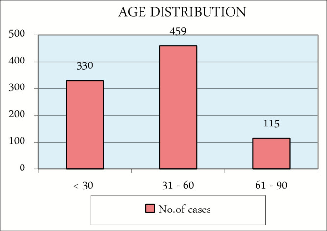
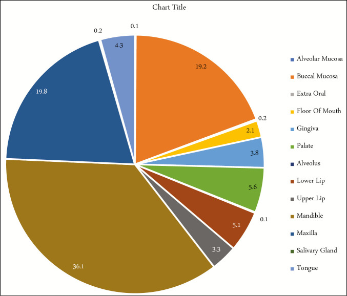
In our retrospective study, we analyzed a total of 904 histological diagnosis, with 463 (51%) derived from male patients and 441 (49%) from female patients with a ratio of 1.04. The series shows a slight male predominance. A notable association was observed between age and type of lesion, and between age and anatomical location. The age range was kept from 5 to 85 years. The highest frequency of the disease was among the age-group of 31–60 years [Figure 1]. The lesion-affected different sites are shown in Figure 2, and the most common regions are the mandible (36.1%), maxilla (19.6%), and buccal mucosa (19.2%).
Figure 1.

Distribution of cases by age
Figure 2.
Site distribution of the lesions
Most of the lesions were found due to the awareness of the presence of the lesion (70%), whereas the rest (30%) were found during clinical exploration. Most of the biopsies performed were excisional, and nearly 40 different histological diagnoses were established [Table 1]. The frequent hard tissue lesions were cystic in nature, and among these, the most common were periapical cyst (12.94%) followed by dentigerous cyst (3.3%) and residual cyst (1.4%). In addition, periapical granuloma and ameloblastoma constitute 3.5% and 5.3% of the lesions, respectively.
Table 1.
Specific diagnosis and relative frequencies
| Diagnosis | No. of cases | Percentage |
|---|---|---|
| Adenoid cystic carcinoma | 3 | 0.33 |
| Ameloblastic fibroma | 6 | 0.66 |
| Ameloblastoma | 48 | 5.31 |
| Amelogenesis imperfecta | 3 | 0.33 |
| Central giant cell granuloma | 10 | 1.11 |
| Chronic nonspecific ulcer | 7 | 0.77 |
| Dentigerous cyst | 30 | 3.32 |
| Dysplasia | 84 | 9.29 |
| Hyperkeratosis | 11 | 1.22 |
| Inflammatory hyperplasia | 25 | 2.77 |
| Intramucosal nevus | 3 | 0.33 |
| Leukoplakia | 3 | 0.33 |
| Lichen planus | 32 | 3.54 |
| Lichenoid reaction | 5 | 0.55 |
| Lipoma | 4 | 0.44 |
| Mucocele | 32 | 3.54 |
| Nasopalatine dental cyst | 5 | 0.55 |
| Odontogenic keratocyst | 22 | 2.43 |
| Odontogenic myxoma | 4 | 0.44 |
| Oral sub mucosal fibrosis | 4 | 0.44 |
| Ossifying fibroma | 10 | 1.11 |
| Osteoid osteoma | 3 | 0.33 |
| Osteomyelitis | 36 | 3.98 |
| Periapical granuloma | 32 | 3.54 |
| Periapical abscess | 5 | 0.55 |
| Periapical cyst | 117 | 12.94 |
| Pericoronitis | 16 | 1.77 |
| Pleomorphic adenoma | 6 | 0.66 |
| Pyogenic capillary hemangioma | 5 | 0.55 |
| Pyogenic granuloma | 86 | 9.51 |
| Ranula | 4 | 0.44 |
| Residual cyst | 13 | 1.44 |
| Squamous cell carcinoma | 66 | 7.3 |
| Squamous papilloma | 5 | 0.55 |
| Traumatic fibroma | 144 | 15.93 |
| Verrucous hyperplasia | 4 | 0.44 |
| Others (less than two cases) | 11 | 1.22 |
In soft tissue lesion, we recorded 15.93% traumatic fibromas, whereas the pyogenic granuloma and dysplasia represented 9.5% and 9.2%, respectively. The most prevalent malignant neoplasm occurring in soft tissue was squamous cell carcinoma, which represents 7.3% of the total lesion. The other common soft tissue lesions found were mucocele (3.54%), inflammatory hyperplasia (2.7%), and hyperkeratosis (1.2%). Adenoid cystic carcinoma and mucoepidermoid carcinoma constitute 0.3% and 0.2%, respectively.
DISCUSSION
In this study, 50.4% of the lesions were present among the age-group of patients between 31 and 60 years, whereas a study conducted by Satorres et al.[1] reported that 49% of the lesions were present among the patients in the age range 40–60 years. In this incidence, no notable differences were observed between males (51%) and females (49%).
In this study, we found that periapical cysts (12.9%) with inflammatory origin were the most common odontogenic cysts compared to dentigerous cyst (3.3%). These were more common among the people present in second and third decades of life, consistent with the findings of a study by De Souza et al.[3] and more prevalent among males.
The dentigerous cyst is a developmental cyst formed by the expansion of its follicle around the crown of an unerupted tooth. It was more common in males. The mandibular posterior region and anterior maxilla were the common sites of the lesion. According to Jones et al.,[4] the frequency of dentigerous cysts was related to the frequently impacted lower third molars and upper canines. In an epidemiological study, the frequency of periapical granulomas observed in all biopsies was 3.54%. According to Satorres et al.[1] in their 77 documented periapical alterations, 52% were root cysts and 48% were periapical granulomas.
The most common inflammatory or reactive lesion in our study was irritational fibroma (15.9%), which was comparable to that reported by Naderi et al.[5] It was more prevalent in males, and buccal mucosa was the most commonly affected. Followed by fibroma, the next common connective tissue lesion was pyogenic granuloma (9.5%). Most of the lesions occurred in anterior gingiva and with a female predilection over male in the ratio of 1:3. The etiology includes local irritants, trauma, calculus, and hormonal imbalances, which makes it more common in females, especially during pregnancy.[6]
The most common odontogenic tumor in this study was ameloblastoma, similar to other studies conducted in Asian[7] and African[8] countries. Oral squamous cell carcinoma was the most common malignant epithelial lesion in our study, which was in accordance with a study conducted by Uma[9] It showed a predilection for elderly males older than 50 years of age due to high-risk habits, such as alcohol and tobacco use, than that for females. The occurrence of oral squamous cell carcinoma is less in young patients because of decreased exposure time to risk factors.
The prevalence of lichen planus was 3.54%, which had a higher prevalence compared to that in studies by Mathew et al.[10] and Ikeda et al.[11] The prevalence of lichen lesions in this study was 0.55%, which was low compared to studies by Mathew et al.[10] (1.6%) and Ikeda et al.[11] (1.8%). In mucoceles, a high incidence (10%) of these lesions was reported by Nair et al.[12] In contrast to this, we have lower incidence of 3.5% in our study.
Demographic data, such as patient’s socioeconomic status, location, occupation, and oral habits, can help to identify risk groups to some extent. Owing to the lack of demographic details from the lab report, we were unable to evaluate risk factor and the associated lesion in the study. Such potential information helps in understanding the characteristics of oral and maxillofacial lesion among the patients reported in our institution.
CONCLUSION
Cystic lesions and traumatic fibroma were the most common findings in and around Madurai. As the causes leading to this situation were not recorded, we can point to the factors inherent to the patients and professional factors with irregular follow-up. Lastly, we would like to emphasize the importance of biopsies in the study of various pathological reasons, considering the prognosis and establishing an early diagnosis, leading in effective treatment.
Financial support and sponsorship
Nil.
Conflicts of interest
There are no conflicts of interest.
REFERENCES
- 1.Satorres M, Faura M, Bresco M, Berini L, Gay Escoda C. Prevalencia de lesiones orales biopsiadas en un Servicio de Cirugía Bucal. Med Oral Patol Oral Cir Buc. 2001;6:296–305. [Google Scholar]
- 2.Fierro-Garibay C, Almendros-Marqués N, Berini-Aytés L, Gay-Escoda C. Prevalence of biopsied oral lesions in a Department of Oral Surgery (2007-2009) J Clin Exp Dent. 2011;3:e73–7. [Google Scholar]
- 3.De Souza LB, Gordón-Núñez MA, Nonaka CF, de Medeiros MC, Torres TF, Emiliano GB. Odontogenic cysts: demographic profile in a Brazilian population over a 38-year period. Med Oral Patol Oral Cir Bucal. 2010;15:e583–90. [PubMed] [Google Scholar]
- 4.Jones AV, Craig GT, Franklin CD. Range and demographics of odontogenic cysts diagnosed in a UK population over a 30‐year period. J Oral Pathol Med. 2006;35:500–7. doi: 10.1111/j.1600-0714.2006.00455.x. [DOI] [PubMed] [Google Scholar]
- 5.Naderi NJ, Eshghyar N, Esfehanian H. Reactive lesions of the oral cavity: a retrospective study on 2068 cases. Dent Res J. 2012;9:251. [PMC free article] [PubMed] [Google Scholar]
- 6.Assadat Hashemi Pour M, Rad M, Mojtahedi A. A survey of soft tissue tumor-like lesions of oral cavity: a clinicopathological study. Iran J Pathol. 2008;3:81–7. [Google Scholar]
- 7.Luo HY, Li TJ. Odontogenic tumors: a study of 1309 cases in a Chinese population. Oral Oncol. 2009;45:706–11. doi: 10.1016/j.oraloncology.2008.11.001. [DOI] [PubMed] [Google Scholar]
- 8.Adebayo ET, Ajike SO, Adekeye EO. A review of 318 odontogenic tumors in Kaduna, Nigeria. J Oral Maxillofac Surg. 2005;63:811–9. doi: 10.1016/j.joms.2004.03.022. [DOI] [PubMed] [Google Scholar]
- 9.Uma M. Pattern of distribution of biopsied oral lesions in a Tertiary Health Care Centre – a 10 year retrospective study. IOSR J Dent Med Sci. 2017;16:77–83. [Google Scholar]
- 10.Mathew AL, Pai KM, Sholapurkar AA, Vengal M. The prevalence of oral mucosal lesions in patients visiting a dental school in Southern India. Ind J Dent Res. 2008;19:99–103. doi: 10.4103/0970-9290.40461. [DOI] [PubMed] [Google Scholar]
- 11.Ikeda N, Handa Y, Khim SP, Durward C, Axéll T, Mizuno T, et al. Prevalence study of oral mucosal lesions in a selected Cambodian population. Community Dent Oral Epidemiol. 1995;23:49–54. doi: 10.1111/j.1600-0528.1995.tb00197.x. [DOI] [PubMed] [Google Scholar]
- 12.Nair RG, Samaranayake LP, Philipsen HP, Graham RG, Itthagarun A. Prevalence of oral lesions in a selected Vietnamese population. Int Dent J. 1996;46:48–51. [PubMed] [Google Scholar]


