ABSTRACT
It is very rare (2%–6% cases) for a mandibular canine to have two root canals and the incidence of finding two roots with two root canals in a mandibular canine that too bilaterally is almost negligible. This case report discusses the presence and multidisciplinary management of such rarest configuration in both mandibular canines of a female patient. This case shows the importance of recognition of anatomical variations in successful accomplishment of root canal treatment.
KEYWORDS: Anatomic variations, endodontic treatment, mandibular canine, root canal anatomy
INTRODUCTION
Success of endodontic treatment relies heavily on the knowledge of root canal configuration and morphology. Missed canals are among the main causes of treatment failure in endodontics frequently because of failure of clinician to recognize root canal variations. This can adversely affect the outcome and long-term prognosis of the tooth. Mandibular canines are considered easy teeth for endodontic intervention because of their relatively simple canal configuration, that is, one root one canal in most cases (>94%). In rare cases, there can be two roots with two canals (2%–6%) and presence of this configuration bilaterally is even rarer. The following case shows the successful management of such a unique case.
CASE REPORT
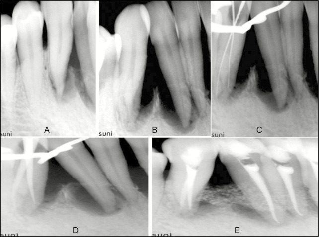
A 37-year-old female patient reported to the Department of Periodontology with complaint of mobile lower front teeth since 1 year. She had no contributory dental, medical, or family history. On intraoral examination, deep periodontal pockets and Grade II mobility were detected with respect to (w.r.t.) 22, 23, 24, 25, 26, and 27 (Universal Numbering System). On electric and thermal (cold) pulp testing, 22–27 teeth were found to be nonvital. Radiographic examination revealed vertical bone defects. On the basis of clinical and radiographical examination, diagnosis of primary periodontal lesion with secondary endodontic involvement w.r.t. 22, 23, 24, 25, 26, and 27 was made. Interdisciplinary approach involving root canal treatment of the involved teeth followed by periodontal flap surgery was planned for the patient. Intraoral periapical radiograph of 22 and 27 [Figures 1A and 2A] revealed sudden disappearance of root canal in the middle root indicating the possibility of presence of extra canal in the tooth. For confirmation, angulated radiographs [Figures 1B and 2B] were taken for both teeth and extra roots were found in each tooth.
Figure 1.
(A) Intraoral Periapical radiograph of 22. (B) Angulated radiograph of 22. (C) Working length radiograph of 22. (D) Radiograph showing master cones in 22. (E) Radiograph after 6 months
Figure 2.
(A) Intraoral periapical radiograph of 27. (B) Angulated radiograph of 27. (C) Working length radiograph of 27. (D) Radiograph showing master cones in 27. (E) Radiograph after 6 months
Informed consent for the procedure was obtained from the patient. Under L.A and rubber dam isolation access opening was carried out w.r.t. 22 and 27 using No. 2 round bur and Endo Z bur (Dentsply/Maillefer, Ballaigues, Switzerland). Root canals were negotiated with No.10 K-file and coronal flaring accomplished with orifice shaper (Protaper universal system, Dentsply Maillefer, Ballaigues, Switzerland). Working length was determined using Root ZX Apex Locator (J.Morita Corp., Kyoto, Japan) and confirmed radiographically [Figures 1C and 2C]. Two roots and two canals (buccal and lingual) were found in each tooth. Cleaning and shaping was carried out with ProTaper system up to F3 size. Canals were irrigated throughout instrumentation using 2.5% sodium hypochlorite. Calcium hydroxide (Metapex, Meta Dental Corp, Elmburt) intra-canal dressing was given for a week. After one week, obturation was done with F3 ProTaper cones [Figures 1D and 2D]. Root canal treatment was similarly performed in 23, 24, 25, and 26. Periodontal surgery was performed following root canal treatment. The periodontal defect was covered with a bone graft (Ossifi, Equinox MedicalTechnologies B.V., Amersfoort, Holland) and teeth were splinted for 3 weeks with Ribbond (Ribbond Inc. Seattle,WA). Patient was recalled for regular checkups. After 6 months, teeth were asymptomatic and healing was evident radiographically [Figures 1E and 2E].
DISCUSSION
For a successful endodontic outcome, the knowledge of root canal anatomy and its deviations is critical.[1,2] Chances of finding a single root canal in mandibular canines are very high (94%–100%) [Table 1].[3,4,5,6,7,8,9] Most variations occur in the form of single root with two canals.[3,4,5,6,7,8,9] Variations such as two roots–three canals,[10,11] three canals–two foramen,[12] and two roots–two canals[13,14,15] and bilateral two root canals in single root[16] have been reported. But findings, that is, two canals in two roots on both mandibular canines in same patient are very rare and are only reported in isolated case reports.[17,18] Different studies and case reports performed on mandibular canine are presented in Tables 1 and 2, respectively. Missed canals because of failure to gauze the variations are among the frequent causes of the endodontic treatment failure.[19]
Table 1.
Studies on root canal patterns in mandibular canines
| Authors | Type of study | Country/year | Type I | Type II | Type III | Type IV | Type V | Additional type |
|---|---|---|---|---|---|---|---|---|
| Vertucci[3] | Clearing | USA/1984 | 100 | |||||
| Pecora et al.[4] | Clearing | Brazil/1993 | 100 | |||||
| Caliskan et al.[5] | Clearing | Turkey/1995 | 93.48 | 4.35 | 2.17 | |||
| Sert and Bayirli[6] | Clearing (M) | Turkey/2004 | 91 | 3 | 4 | 2 | 1 | |
| Clearing (F) | 96 | 4 | ||||||
| Rahimi et al.[7] | Clearing | Iran/2011 | 91.6 | 6.11 | 2.29 | 0 | 0 | 0 |
| Aminsobhani et al.[8] | CBCT (M) | Iran/2013 | 36 | 5.1 | 1.4 | 6.4 | 1.3 | |
| CBCT (F) | 35.8 | 5.2 | 1.4 | 6.4 | 1.0 | |||
| Amardeep et al.[9] | CBCT | India/2014 | 81.6 | 2.8 | 11.6 | 0.8 | 2 | 1.2 |
CBCT = cone beam computed tomography
Table 2.
Case reports on different variations shown by mandibular canine
| Authors | Country/year | Two canals | Three canals |
|---|---|---|---|
| Heling et al.[10] | Israel/1995 | Two roots and three root canals | |
| Orguneser and Kartal[12] | Turkey/1998 | Three canals and two foramina | |
| D’Arcangelo et al.[13] | Italy/2001 | Two cases with two canals in each tooth | |
| Victorino et al.[17] | Brazil/2009 | Bilateral two roots––two canals | |
| Andrei et al.[14] | Romania/2011 | Two roots two canals in single tooth | |
| Moogi et al.[15] | India/2012 | Two roots two canals | |
| Fuentes and Borie[18] | Chile/2013 | Bilateral two root two canal | |
| Subha et al.[16] | India/2013 | Bilateral two root canals in single root | |
| Jadhav[11] | India/2014 | Two roots and three canals |
Diagnostic radiograph is first and most important tool at the disposal of dentist, which can guide about the identification or possibility of presence of anatomical variations. Abrupt disruption of root canal in the middle of root in radiograph in present case arose the suspicion of additional canal in each tooth.[20] Multiple radiographs with changed angulation were used to confirm the variation, as single radiographic view has 33% probability of failure to diagnose canal division.[21] There were two distinct roots with buccal and lingual roots in both teeth as was also observed by Versiani et al.[22]
Use of endodontic explorer (examination), endo-sonic tips (dentin removal), methylene blue dye, champagne bubble test, and bleeding spots observation can also aid in locating root canal openings.[19] Use of magnification, that is, dental operating microscope (DOM) is another vital aid in finding and negotiating canal orifices.[23] Cone beam computed tomography (CBCT) has high radiation exposure to the patient so should be used in extreme cases such as calcification of root canals.
Once canals were identified root canal treatment was performed using standard procedure. As it was a combined perio-endo lesion, root canal treatment was followed by flap surgery and bone grafting 1 week after the root canal treatment. To reduce the mobility and promote healing, physiologic splinting was carried out.
CONCLUSION
Success of endodontic treatment relies heavily on the knowledge of root canal configuration and morphology. Clinician must be aware of the possibility of finding rare configurations in seemingly “easy teeth” and should use every technology such as radiography, magnification, and CBCT at his/her disposal to avoid missed canals and thus failure. Interdisciplinary approach for the management of combined lesions is essential for the successful accomplishment of treatment.
Financial support and sponsorship
Nil.
Conflicts of interest
There are no conflicts of interest.
REFERENCE
- 1.Mittal N, Arora S. Endodontic treatment of mandibular second premolar with three root canals using dental operating microscope. Endodontology. 2009;2:80–3. [Google Scholar]
- 2.Mittal N, Arora S. Implications of third and fourth root in mandibular first molar. J Int College Dent. 2009;54:2. [Google Scholar]
- 3.Vertucci FJ. Root canal anatomy of the human permanent teeth. Oral Surg Oral Med Oral Pathol. 1984;58:589–99. doi: 10.1016/0030-4220(84)90085-9. [DOI] [PubMed] [Google Scholar]
- 4.Pécora JD, Sousa Neto MD, Saquy PC. Internal anatomy, direction and number of roots and size of human mandibular canines. Braz Dent J. 1993;4:53–7. [PubMed] [Google Scholar]
- 5.Calişkan MK, Pehlivan Y, Sepetçioğlu F, Türkün M, Tuncer SS. Root canal morphology of human permanent teeth in a Turkish population. J Endod. 1995;21:200–4. doi: 10.1016/S0099-2399(06)80566-2. [DOI] [PubMed] [Google Scholar]
- 6.Sert S, Bayirli GS. Evaluation of the root canal configurations of the mandibular and maxillary permanent teeth by gender in the Turkish population. J Endod. 2004;30:391–8. doi: 10.1097/00004770-200406000-00004. [DOI] [PubMed] [Google Scholar]
- 7.Rahimi S, Milani AS, Shahi S, Sergiz Y, Nezafati S, Lotfi M. Prevalence of two root canals in human mandibular anterior teeth in an Iranian population. Indian J Dent Res. 2013;24:234–6. doi: 10.4103/0970-9290.116694. [DOI] [PubMed] [Google Scholar]
- 8.Aminsobhani M, Sadegh M, Meraji N, Razmi H, Kharazifard MJ. Evaluation of the root and canal morphology of mandibular permanent anterior teeth in an Iranian population by cone-beam computed tomography. J Dent (Tehran) 2013;10:358–66. [PMC free article] [PubMed] [Google Scholar]
- 9.Somalinga Amardeep N, Raghu S, Natanasabapathy V. Root canal morphology of permanent maxillary and mandibular canines in Indian population using cone beam computed tomography. Anat Res Int. 2014;2014:731859. doi: 10.1155/2014/731859. [DOI] [PMC free article] [PubMed] [Google Scholar]
- 10.Heling I, Gottlieb-Dadon I, Chandler NP. Mandibular canine with two roots and three root canals. Endod Dent Traumatol. 1995;11:301–2. doi: 10.1111/j.1600-9657.1995.tb00509.x. [DOI] [PubMed] [Google Scholar]
- 11.Jadhav GR. Endodontic management of a two rooted, three canaled mandibular canine with a fractured instrument. J Conserv Dent. 2014;17:192–5. doi: 10.4103/0972-0707.128046. [DOI] [PMC free article] [PubMed] [Google Scholar]
- 12.Orguneser A, Kartal N. Three canals and two foramina in a mandibular canine. J Endod. 1998;24:444–5. doi: 10.1016/S0099-2399(98)80031-9. [DOI] [PubMed] [Google Scholar]
- 13.D’Arcangelo C, Varvara G, De Fazio P. Root canal treatment in mandibular canines with two roots: a report of two cases. Int Endod J. 2001;34:331–4. doi: 10.1046/j.1365-2591.2001.00376.x. [DOI] [PubMed] [Google Scholar]
- 14.Andrei OC, Mărgărit R, Gheorghiu IM. Endodontic treatment of a mandibular canine with two roots. Rom J Morphol Embryol. 2011;52:923–6. [PubMed] [Google Scholar]
- 15.Moogi PP, Hegde RS, Prasanth BR, Kumar GV, Biradar N. Endodontic treatment of mandibular canine with two roots and two canals. J Contemp Dent Pract. 2012;13:902–4. doi: 10.5005/jp-journals-10024-1250. [DOI] [PubMed] [Google Scholar]
- 16.Subha N, Prabu M, Prabhakar V, Abarajithan M. Spiral computed tomographic evaluation and endodontic management of a maxillary canine with two canals. J Conserv Dent. 2013;16:272–6. doi: 10.4103/0972-0707.111333. [DOI] [PMC free article] [PubMed] [Google Scholar]
- 17.Victorino FR, Bernardes RA, Baldi JV, Moraes IG, Bernardinelli N, Garcia RB, et al. Bilateral mandibular canines with two roots and two separate canals: case report. Braz Dent J. 2009;20:84–6. doi: 10.1590/s0103-64402009000100015. [DOI] [PubMed] [Google Scholar]
- 18.Fuentes R, Borie E. Bilateral two-rooted mandibular canines in the same individual: a case report. Int J Odontostomat. 2013;7:471–3. [Google Scholar]
- 19.Vertucci FJ. Root canal morphology and its relationship to endodontic procedures. Endod Topics. 2005;10:3–29. [Google Scholar]
- 20.Slowey RR. Radiographic aids in the detection of extra root canals. Oral Surg Oral Med Oral Pathol. 1974;37:762–72. doi: 10.1016/0030-4220(74)90142-x. [DOI] [PubMed] [Google Scholar]
- 21.Nattress BR, Martin DM. Predictability of radiographic diagnosis of variations in root canal anatomy in mandibular incisor and premolar teeth. Int Endod J. 1991;24:58–62. doi: 10.1111/j.1365-2591.1991.tb00808.x. [DOI] [PubMed] [Google Scholar]
- 22.Versiani MA, Pecora JD, Sousa-Neto MD. The anatomy of two-rooted mandibular canines determined using microcomputed tomography. Int Endod J. 2011;44:682–7. doi: 10.1111/j.1365-2591.2011.01879.x. [DOI] [PubMed] [Google Scholar]
- 23.Mittal N, Arora S. Role of microendodontics in detection of root canal orifices: a comparative study between naked eye, loupes and surgical operating microscope. J Med Sci Clin Res. 2015;3:7810–6. [Google Scholar]


