Figure 5.
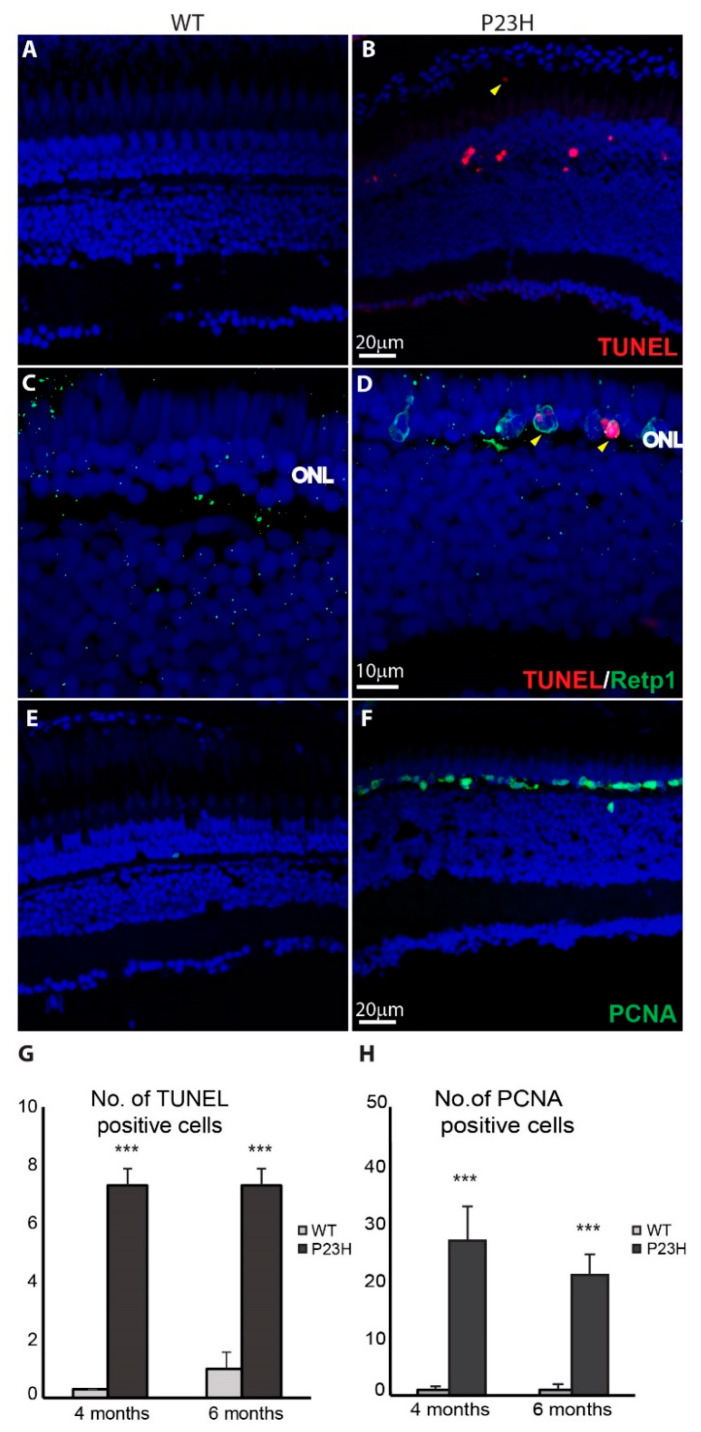
Cell death and cell proliferation in the P23H transgenic zebrafish. (A,B) Cell death detection using TUNEL staining shows TUNEL-positive dying cells (red) in the P23H zebrafish retina (4 months old). The yellow arrowhead shows a TUNEL-positive cell in the sub-retinal space. (C,D) Yellow arrowheads show the colocalization of Retp1 (green) with TUNEL labeling in the P23H transgenic fish. (E,F) PCNA immunostaining shows many PCNA-positive proliferating cells (green) in the ONL of the P23H zebrafish, but very few in WT. ONL: outer nuclear layer. (G) Numbers of TUNEL positive cells and (H) PCNA-positive cells per 210 µm image field in 4-month old and 6-month old WT and P23H retina (n = 6 fish per genotype; error bars are ± SD; *** p < 0.001).

