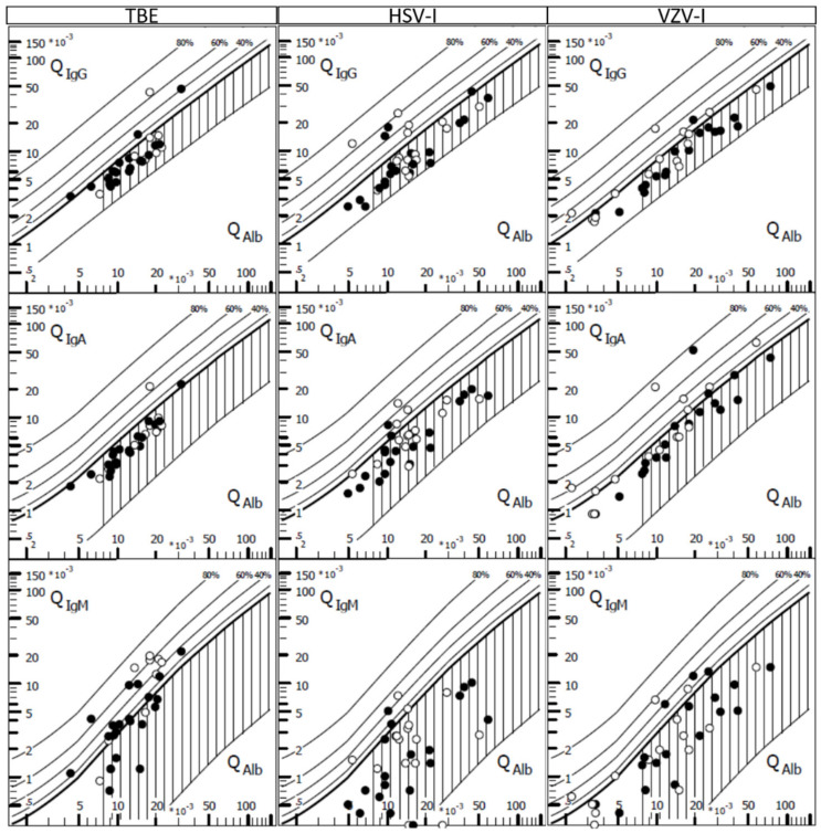
Figure 1.
Intrathecal immunoglobulins in tick-borne encephalitis (TBE) and CNS infections by herpes simplex virus (HSV-I) and varicella zoster virus (VZV-I) CSF/serum quotient diagrams for IgG, IgA and IgM with hyperbolic discrimination functions in tick-borne encephalitis (TBE), and CNS infections by herpes simplex virus (HSV-I) and varicella zoster virus (VZV-I). The upper curve of the reference range represents the discrimination line between brain-derived and blood-derived immunoglobulin fractions in the CSF. Filled figures indicate first diagnostic lumbar puncture and open figures indicate one follow-up lumbar puncture. Graphic Program by Albaum IT-Solutions was used to visualize the Reiber diagrams.

