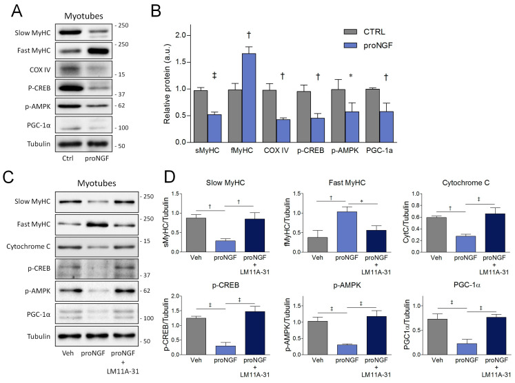
Figure 6.
Effects of proNGF/p75NTR axis on slow/oxidative markers in differentiated myotubes. (A) Western blots and (B) densitometric analysis of the slow myosin heavy chain (sMyHC), fast MyHC (fMyHC), COX IV, CREB phosphorylation (p-CREB), AMPK phosphorylation (p-AMPK) and PGC-1α in the myotubes treated with proNGF. (C) Western blots and (D) densitometric analysis of slow myosin heavy chain (sMyHC), fast MyHC (fMyHC), cytochrome C, CREB phosphorylation (p-CREB), AMPK phosphorylation (p-AMPK) and the PGC-1α of myotubes derived from terminally differentiated SCDMs treated with proNGF or proNGF + LM11A-31. SCDMs were allowed to reach terminal differentiation into myotubes for 72 h. Subsequently, mature myotubes were treated with vehicle (PBS), proNGF (100 ng/mL) and proNGF (100 ng/mL) + LM11A-31 (100 nM) for 48 h. Tubulin was employed as the loading control. Results are expressed as the mean ± SD and were obtained from three independent experiments. Statistical analysis was performed by using the Student’s t test in panel B, whereas one-way ANOVA was used to calculate statistical significance in panel D. * p < 0.05; † p < 0.01; ‡ p < 0.001.

