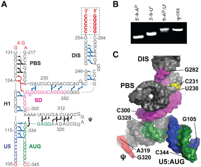
Figure 7.
NMR structure of HIV-1NL4-3 ΨCESm. (A) Fragment-annealed sample used to identify long-range adenosine-H2 detected NOEs (denoted by arrows). Non-native residues are shown in red; U5, blue; AUG, green; and SD, pink. (B) Native polyacrylamide gel electrophoresis showing non-covalent annealing of differentially labeled 5′- and 3′- fragments. (C) Representation of the tandem three-way junction NMR structure adopted by ΨCESm (colors match panel A; conformationally dynamic nucleotides, yellow; Ψ tetraloop, orange). Adapted from [41], with permission.

