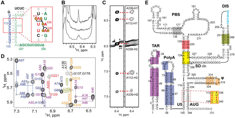
Figure 8.
NMR studies of the intact, dimeric HIV-1NL4-3 5′-leader. (A) Illustration depicting mutations in the lr-AID substitution and 1H-1H NOEs. (B) Region of 1D 1H NMR spectra showing (top) the native TAR A46-H2 signal, (middle) A46G substitution, (bottom) A338-H2 signal observed for the lr-AID substitution. (C) 2D 1H-1H NOESY spectra of the same A338-H2 signal observed an isolated U5:AUG hairpin (left) and intact dimer (right) containing the lr-AID substitution. (D) Region of the 2D 1H-1H NOESY spectrum of A2rGrCr-labeled dimeric HIV-1NL4-3 RNA; color-coding matches the elements shown in (E). (E) Secondary structure of the HIV-1NL4-3 in its DIS-exposed, dimer-promoting state. Color-shaded boxes denoted resolved and assigned 2D 1H-1H NOESY signals; yellow denotes sites that were only assignable in mutant constructs using truncated constructs or lrAID substitution. Panels (A–C) adapted from [71] and panels (D,E) from [221], with permission.

