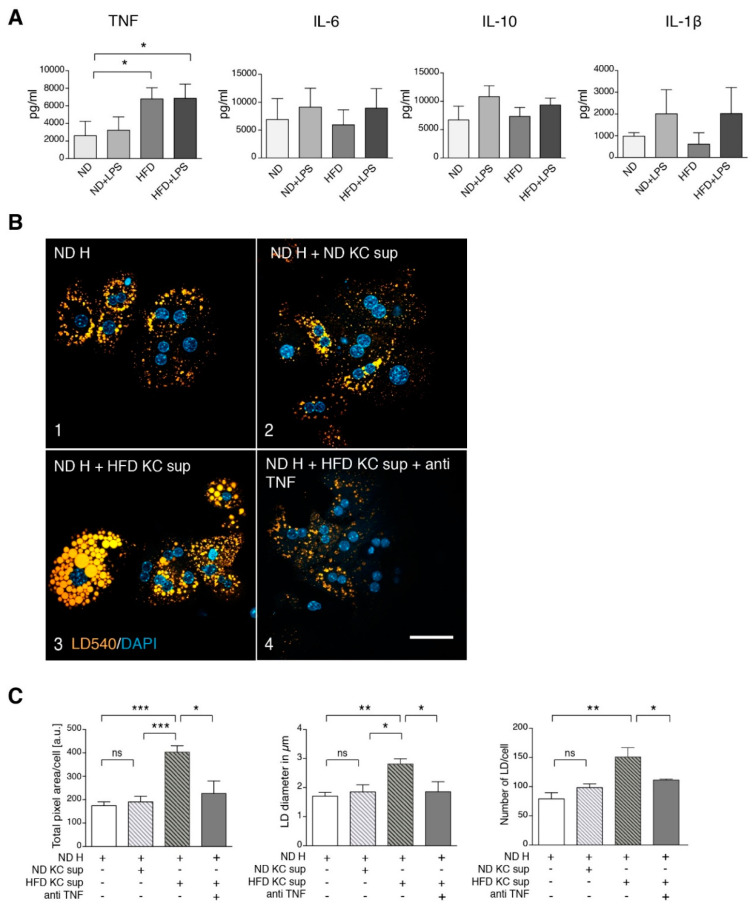
Figure 3.
Kupffer cell supernatant from HFD mice stimulates steatosis in ND hepatocytes via tumor necrosis factor (TNF). (A) Cytokine secretion by Kupffer cells from untreated and LPS-treated ND and HFD mice. Mice were treated as indicated, Kupffer cells were isolated and cultured for 16 h. Secreted cytokines were measured by ELISA (TNF n ≥ 3, IL-1β n ≥ 3, IL-6 n ≥ 4, IL-10 n ≥ 2). (B) Freshly isolated hepatocytes (H) from ND mice were cultured in the absence (frame 1) or presence of 16 h pre-conditioned supernatant of Kupffer cells (KC sup) from ND (frame 2) or HFD mice (frame 3 and 4) as indicated. In frame 4, a neutralizing TNF antibody was added. Microscopy images of cells that were stained for lipid droplets (LD540) and nuclei (DAPI) are shown. Scale bar: 50 µm. (C) Quantification of the total fluorescent LD area/cell, average LD diameter in µm and LD number/cell of the various conditions (n ≥ 3). Mean values with SEM of at least three independent experiments are shown. H: hepatocytes, KC: Kupffer cells, ND: controlled normal diet, HFD: high-fat diet, sup: supernatant. Significance was analyzed with a two-tailed Student’s t-test. Statistical significance is indicated as follows: ns, not significant, * p < 0.05, ** p < 0.01, *** p < 0.001.

