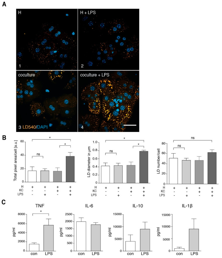
Figure 7.
Intracellular lipid droplet accumulation in hepatocytes upon in vitro LPS treatment depends on the presence of Kupffer cells. (A) Primary mouse hepatocytes (H) were incubated for 16 h in the absence (frame 1) or presence of LPS in vitro (frame 2), in co-culture with Kupffer cells (KC) (frame 3) or in co-culture with Kupffer cells and LPS in vitro (frame 4). The lipid droplet (LD) accumulation is visualized by staining with LD540 (orange) and DAPI (blue). Scale bar: 50 µm. (B) Quantification of total fluorescent LD area und LD number per cell as well as LD size (n = 3). (C) Kupffer cells were isolated, followed by 16 h of cell culture in the presence of 100 ng/mL LPS. Secreted TNF, IL-6, IL-10 and IL-1β was measured in the Kupffer cell culture supernatant by ELISA. TNF n = 4; IL-6 n = 6; IL-10 n = 3; IL-1β n = 3. All data are presented as mean value with SEM. Significance was analyzed with a two-tailed Student’s t-test. Statistical significance is indicated as follows: ns, not significant, * p < 0.05.

