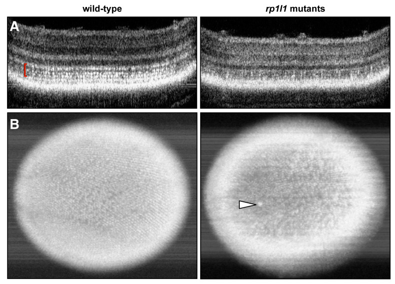
Figure 5.
rp1l1 mutant zebrafish have fewer hyper-reflective inner segment punctae and gaps in the photoreceptor mosaic. Optical coherence tomography (OCT) of wild-type and rp1l1 mutant zebrafish retinas at 12 months. (A) OCT B-scans through zebrafish retinas. In the wild-type animals, the photoreceptor inner segments (seen as hyper-reflective dots, red brackets) are abundant and uniformly distributed. In mutant animals, this part of the retina is very patchy and appears to have fewer punctae. (B) En face projections through the retina. In the mutant animals, the mosaic appears patchy and disrupted. There is also a large hyper-reflective speck in the retina of the mutant animal (arrowhead), which typically indicates an immune cell response.

