Abstract
Simple Summary
Acute myeloid leukemia (AML) is a haematologic disease in which oncogenic mutations in the receptor tyrosine kinase FLT3 frequently lead to leukaemic development. Potent treatment of AML patients is still hampered by inefficient targeting of leukemic stem cells expressing constitutive active FLT3 mutants. This review summarizes the current knowledge about the regulation of FLT3 activity at cellular level and discusses therapeutical options to affect the tumor cells and the microenvironment to impair the haematological aberrations.
Abstract
Fms-like tyrosine kinase 3 (FLT3) is a member of the class III receptor tyrosine kinases (RTK) and is involved in cell survival, proliferation, and differentiation of haematopoietic progenitors of lymphoid and myeloid lineages. Oncogenic mutations in the FLT3 gene resulting in constitutively active FLT3 variants are frequently found in acute myeloid leukaemia (AML) patients and correlate with patient’s poor survival. Targeting FLT3 mutant leukaemic stem cells (LSC) is a key to efficient treatment of patients with relapsed/refractory AML. It is therefore essential to understand how LSC escape current therapies in order to develop novel therapeutic strategies. Here, we summarize the current knowledge on mechanisms of FLT3 activity regulation and its cellular consequences. Furthermore, we discuss how aberrant FLT3 signalling cooperates with other oncogenic lesions and the microenvironment to drive haematopoietic malignancies and how this can be harnessed for therapeutical purposes.
Keywords: acute myeloid leukaemia (AML), FMS-like tyrosine kinase 3 (FLT3), oncogenic signaling, re-sistance development, cancer cell vulnerability, haematopoietic niche
1. Introduction
Fms-like tyrosine kinase 3 (FLT3) is a member of the PDGFR (class III) receptor-tyrosine kinase (RTK) family and is expressed in human CD34+ haematopoietic stem cells (HSC), lymphoid progenitors, and progenitors cells of the granulocyte/macrophage lineage, including the common myeloid progenitor and the granulocyte/macrophage progenitors. As such, FLT3 participates in the maintenance of pluripotent HSC and contributes to proliferation and differentiation of B-cell progenitors, myelomonocytic and dendritic cells [1,2,3].
1.1. Classes of Activating FLT3 Mutations
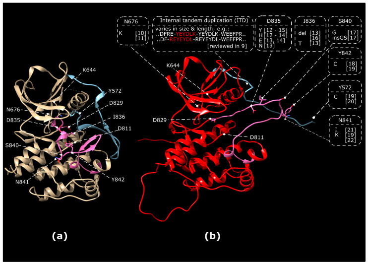
Overexpression of wild type (WT) or oncogenic forms of FLT3 have been implicated in several haematopoietic malignancies [4,5] and inflammatory disorders [6]. Mutations in the FLT3 gene are present in approximately 30% of newly diagnosed AML [7]. Internal tandem duplication (ITD) mutations and tyrosine kinase domain (TKD) mutations are the most common classes of FLT3 abberations in AML. The overall incidence is approximately 23% for FLT3 ITD mutations and 7% for FLT3 TKD mutations [8]. The majority of ITD mutations occurs in the juxtamembrane region of FLT3, where it disrupts its autoinhibitory function (Figure 1). ITD mutations can also be located in the first kinase domain [8]. The ITD insertion site, which can vary in sequence and length is associated with resistance to chemotherapy and inferior outcome. Its integration site in the kinase domain was identified as an unfavorable prognostic factor for achievement of a complete remission, relapse-free survival, and overall survival of AML patients [9].
Figure 1.
Activating ITD in the juxtamembrane domain and point mutations in the tyrosine kinase domain (TKD) of FLT3. (a) Inactive, autoinhibited conformation of FLT3 (1RJB.pdb). Residues involved in ATP-binding and catalysis (K644, D829) and residues subjected to mutation in leukemia are indicated. (b) Model of active FLT3 kinase domain demonstrating the region for ITD insertions and known frequent TKD point mutations resulting in activation of FLT3. Homology modelling of FLT3 kinase domain was based the active conformation of the CSF-1 receptor (3LCD.pdb) using MODELLER within the UCSF Chimera 1.14 software package. The activation segment of the kinase domain is marked in pink, the juxtamembrane domain is marked in blue. N676K [10,11], ITD reviewed in [9]. References: D835Y [12,13,14,15], D835H [12,13,14], D835E [13,14], D835N [13], D835V [13], I836 del. [13,16], I836T [13], S840G [17], S840insGS [17], Y842C [18,19], Y572C [19,20], N841I [21], N841K [19,22].
TKD mutations are predominantly found in the activation segment of the kinase domain and stabilise an active conformation of the activation segment (Figure 1). TKD mutations are most frequently observed at D835 and I836 with the substitution D835Y being the most frequently occurring TKD mutation. However, other substitutions within the activation segment were also reported. These substitutions stabilise an “open”, active conformation in which the activation segment is flipped out (Figure 1b), enabling access of ATP to the aspartate residue at position 829 that serves as a catalytic base [23]. Importantly, FLT3 TKD mutations display differential sensitivity towards TKI [24].
Upon TKI treatment, secondary mutations can occur, conferring TKI-resistance via alteration of the ATP/inhibitor-binding pocket. Additionally, these secondary mutations can, on their own, confer ligand-independent kinase activation. This has been in particular shown for the N676K substitution (Figure 1), which was first identified as TKI resistance-conferring mutation [12] and later established as an oncogenic driver mutation [25,26].
1.2. Biogenesis, Signalling and Regulation of the FLT3 Receptor Tyrosine Kinase
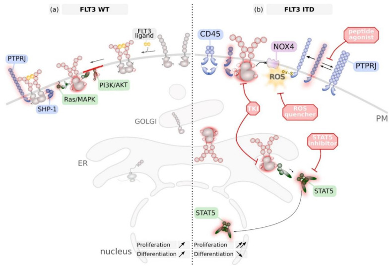
The FLT3 protein is co-translationally translocated into the endoplasmic reticulum (ER). Here, the luminal-faced N-terminus of the receptor undergoes multistep glycosylation and folding, as mediated by the ER luminal enzyme machinery [1]. The ER quality control system ensures that only properly folded FLT3 molecules egress the ER, get further glycosylated at the Golgi system to finally traffic to the plasma membrane (Figure 2a) [27,28].
Figure 2.
Biosynthesis and major signalling pathways activated by FLT3 wildtype (WT) (a) and FLT3 with internal tandem duplication (ITD) mutation (b). (a) Wildtype (WT) FLT3 is synthesized and processed in the endoplasmic reticulum (ER) and the GOLGI and reaches the plasma membrane (PM) as inactive monomer. Binding of the FLT3 ligand (FL) induces dimerization, autophosphorylation and induction of downstream signalling. This comprises activation of the Ras/mitogen activated protein kinase (MAPK) pathway and the phosphoinositol-3 kinase (PI3K)/AKT pathway from the PM and to some extent STAT5 phosphorylation from endosomes (not shown). Consequently, FLT3 WT signalling induces proliferation and differentiation of haematopoietic progenitor cells. FLT3 WT is inactivated via dephosphorylation by protein tyrosine phosphatases (PTPs) such as the transmembrane PTPRJ but also cytoplasmic PTPs such as SHP-1. (b) FLT3 with internal tandem duplication (ITD) mutations in the juxtamembrane region is constitutively and ligand-independently active at the PM and preferentially at endomembranes such as ER and endosomes (not shown). In particular, STAT5 is aberrantly activated at endomembranes. FLT3 ITD induces reactive oxygen species (ROS) production via activation of NADPH oxidase (NOX) 4. As a consequence, PTPRJ gets inactivated via oxidation of catalytic active cysteine residues. Dimerization of PTPRJ also reduces its catalytic activity. FLT3 ITD signalling can be blunted by the use of specific tyrosine kinase inhibitors (TKI) that are in clinical use. However, secondary mutations in FLT3 can cause inhibitor-resistance. Potential novel therapeutics comprise inhibitors of STAT5, ROS quencher and peptides that prevent dimerization of PTPRJ, thereby enhancing its activity.
There is an increasing body of evidence that activating mutations in receptor tyrosine kinases (RTK) cause aberrant intracellular localisation of these proteins. The reader is referred to a recent review [27]; which summarises molecular mechanisms and consequences of RTK mislocalization. Activating mutations in FLT3 were also shown to result in prolonged association of mutant FLT3 within the ER quality control and, therefore, resulting in a predominant ER localisation of FLT3 [28]. Aberrant activation of FLT3 at endomembranes results in altered downstream signalling quality [29,30]. In particular, the ER-resident pool of mutant FLT3 is responsible for phosphorylation and activation of STAT5 and was demonstrated to contribute to oncogenic transformation (Figure 2b) [29].
Constitutive phosphorylation of the receptor is a key determinant for the intracellular retention. An inactivating K644A point mutation of FLT3 ITD, treatment with FLT3 kinase inhibitors or overexpression of protein-tyrosine phosphatases promoted FLT3 surface localization [28,31]. Thus, in an “intracellular active kinase load” model Chan suggested that recruitment of phosphotyrosine-binding domain-containing proteins causes the retardation [32], but the molecular mechanism of FLT3 ITD retention in intracellular compartments is currently still not known.
The dormant FLT3 resides in the cell membrane as an auto-inhibited monomer. Binding of its cognate ligand FL (FLT3 ligand) invokes conformational changes in the receptor ectodomain to establish a dimeric receptor assembly (Figure 2a). As a consequence of dimerization, the adjacent intracellular TKDs are trans-activated by auto-phosphorylation [33]. Stimulation of FLT3 mediates activation of several signal transduction pathways, including the mitogen-activated protein kinases ERK1/2 and the phosphoinositide-3-kinase/Akt signaling cascades (Figure 2a) [34,35].
Ligand-mediated phosphorylation of intracellular tyrosines induces FLT3 receptor endocytosis similar to that which has been observed for other RTKs such as the EGFR [36] or c-Kit [37]. FL-mediated internalization of both FLT3 WT and FLT3 ITD follows mainly a clathrin-dependent mode [38], and is associated with the induction of FLT3 degradation. Despite activity-independent internalization, ligand-induced receptor degradation depends on FLT3 kinase activity [38]. FLT3 WT and surface-bound FLT3 ITD exhibit similar ligand-induced internalization and degradation characteristics.
Protein tyrosine phosphatases (PTP) were shown to predominantly antagonize FLT3 phosphorylation. Overexpression of ER-resident PTP1B [39] or perinuclear SHP-1 [40] resulted in dephoshorylation of oncogenic FLT3 ITD and stimulated its maturation [28]. Conversely, FLT3 interacting PTP SHP-2 was found to positively influence FLT3 signalling pathways [41,42]. Depletion or pharmacologic inactivation of SHP2 positively contributed to FLT3 ITD-induced haematopoietic progenitor hyper-proliferation and leukaemogenesis [43,44]. Expression of dual-specificity phosphatase (DUSP) 6 is up-regulated in FLT3 ITD positive cells and AML blasts, and was found to contribute to FLT3 ITD-mediated cell transformation [45]. Depletion of the membrane-localized receptor-type tyrosine-protein phosphatase eta (PTPRJ, Dep-1), and similarly receptor-type tyrosine-protein phosphatase C (PTPRC, CD45), resulted in enhanced phosphorylation and receptor-mediated downstream signalling activity of the FLT3 WT protein in myeloid cell lines. Direct interaction of PTPRJ and FLT3 was demonstrated by co-immunoprecipitation and in situ proximity ligation [46,47]. PTPRJ depletion stimulated colony formation of FLT3 ITD-expressing myeloid cells [48]. The absence of an antagonistic role of PTPRJ on FLT3 ITD was explained by oxidation of PTPRJ catalytic cysteines due to high FLT3 ITD-induced ROS levels [48]. Quenching of FLT3 ITD-mediated ROS-formation resulted in restoration of PTPRJ activity and subsequent inhibition of cell transformation [48]. Interference with NOX4-mediated overproduction of ROS resulted in PTPRJ re-activation in FLT3 ITD-expressing cells [37,38]. Breeding of FLT3 ITD knock-in mice [49] to Ptprj- or Ptprc-deficient mice, respectively, demonstrated that FLT3 ITD-signalling is controlled by PTPRJ and PTPRC in vivo (Figure 2b) [50,51]. Importantly, low level expression of both receptor PTPs correlated with a poor prognosis of FLT3 ITD positive AML patients [40,41].
Beside its effect on PTP activity, elevated ROS levels can also directly alter kinase function via cysteine oxidation as it was demonstrated for PDGFR [52], EGFR [53] and recently also for FLT3 [54]. Consistently, treatment of cells expressing FLT3 ITD with ROS-quenching agents attenuated signal transduction. Comprehensive analysis of cysteine-to-serine mutant FLT3 ITD proteins revealed critical roles of several cysteine residues for kinase activity and transforming signalling, further supporting cysteine modification as potential mechanism of activity regulation [54].
1.3. FLT3 Regulates HSC Self-Renewal and Aging
All haematopoietic lineages propagate from a pluripotent HSC that exclusively possess the capacity for self-renewal to maintain life-long blood lineage replenishment. Ligand-mediated activation of FLT3 is one of the regulators of HSC self-renewal and differentiation [55,56]. In humans, FLT3 expression is detected on reconstituting short term (ST)-HSC, the Lin negative Sca-1+ c-Kit+ FLT3+ compartment [57], while there is no definitive evidence for its expression in human long-term (LT)-HSC [56]. In contrast in mice, Flt3 transcripts and FLT3 protein were detected in the LT-HSC compartment suggesting a previously unrecognized role of FLT3 in LT-HSC homeostasis and establishes an intrinsic link between normal stem cell quiescence/homeostasis and development of myeloproliferative neoplasm [58].
While young LT-HSC have a quiescent cell cycle state and an unbiased differentiation capacity, their ageing results in enhanced proliferation and skewing of lineage commitment towards myeloid cells [59]. Loss of immune function and an increased incidence of myeloid leukaemia are two of the most clinically significant consequences of ageing of the haematopoietic system [60]. The NAD-dependent deacetylase sirtuin 7 (SIRT7) was shown to act as checkpoint for HSC maintenance. High SIRT7 level maintains HSC in a proper balance of quiescence, proliferation and lineage commitment. Aged HSC are specified by reduced SIRT7 [59,61]. Down regulation of SIRT7 in FLT3 ITD-expressing cells was recently established as relevant pathomechanism in AML [62]. Pharmacologic inhibition of FLT3 ITD or positive treatment response of patients restored SIRT7 expression, suggesting that FLT3 ITD regulates HSC ageing and differentiation via SIRT7 [62]. The detailed molecular mechanism, how SIRT7 affects HSC differentiation and transformation in FLT3 mutant AML still remains elusive [63]. In contrast, overexpression of SIRT1 by a c-MYC-related network has been shown to contribute to the LSC maintenance in FLT3 ITD-positive AML [64].
2. FLT3 in Leukaemia
2.1. Initiating Events in Leukemogenesis
AML is characterized by clonal evolution and genetic heterogeneity of poorly differentiated cell clones derived from the haematopoietic system [65,66]. Several studies identified pre-cancerous signatures in exome data of healthy individuals [67,68,69]. More than 2% of healthy individuals (5–6% of people older than 70 years) contain mutations that may represent pre-malignant events linked to clonal haematopoietic expansion [69]. In particular, DNMT3a, ASXL1 and TET2 were significantly enriched for protein disruptive mutations. Strikingly, none of the samples analysed contained mutations in the proto-oncogene FLT3. This observation indicates that mutations in the FLT3 gene are late events in the clonal expansion of haematopoietic stem and progenitor cells.
2.2. Involvement of FLT3 in Other Haematological Diseases
Mutations in the FLT3 gene are not restricted to AML. Albeit uncommon in myelodysplastic syndromes (MDS), increased frequencies of FLT3 mutations are associated with MDS progressing to secondary AML [70,71]. FLT3 mutations are in general rare in acute lymphoblastic leukemia (ALL). Mutant FLT3 and its overexpression were observed in Early T cell precursor T-lineage ALL [72] and Philadelphia chromosome-like ALL [73]. Adolescents and young adults with ALL also showed higher frequencies of FLT3 mutations [74]. Zhang and co-worker even suggested the FLT3 pathway as potential therapeutic target for polycomb repressive complex 2 (PRC2)-mutated T-cell ALL [75]. To decipher the molecular mechanisms involved in the transition from the chronic phase to blast crisis in chronic myelogenous leukemia (CML), gene expression profiles at diagnosis in patients at the chronic phase and in blast crisis showed that increased abundance of FLT3-expressing cells attenuated imatinib-induced apoptosis [76].
2.3. Cell-Intrinsic Oncogenes Cooperating With Mutant FLT3
FLT3, DNMT3a and NPM1 are the most frequently mutated genes in cytogenetically normal AML [69,77]. Both, transgenic [49] and knock-in [78] mouse models expressing FLT3 ITD in the haematopoietic compartment revealed that FLT3 ITD mutations enhance survival and proliferation of lymphoid and myeloid progenitor cells and induced in particular a myeloproliferative syndrome [79], resembling chronic myelomonocytic leukaemia [78]. Thus, FLT3 ITD itself is not sufficient to induce AML in rodent and zebra fish models [80], but rather needs an initiating oncogenic event to fully drive leukemogenesis. Accordingly, in mouse models, FLT3 ITD was shown to cooperate with other oncogenic mutations found in patients to induce human AML-like syndromes. The earliest indication resulted from mice co-expressing FLT3 ITD with either partial tandem duplication of the gene encoding the histone methyltransferase mixed-lineage leukaemia (MLL) [81] or a MLL-AF9 fusion protein [82]. After a period of latency, these mice develop AML with short life span and extra-medullary involvement. Fusion of the nucleopore protein NUP98 to the homeobox protein HOXD13 occurs in human myelodysplastic syndrome [80]. Transgenic expression of NUP98-HOXD13 was shown in a mouse model to cooperate with FLT3 ITD to induce AML with short latency and 100% penetrance [83].
Mutations in the gene encoding the multifunctional nucleolar protein nucleophosmin (NPM) 1 occur frequently in myeloid neoplasia, disrupt its nuclear localisation and were shown in mouse models to develop late on-set leukaemia [80]. In mouse models, FLT3 ITD cooperates with NPM1 mutations to result in rapid leukaemogenesis resembling human AML [84,85,86].
DNMT3A is a de novo DNA methyltransferase that is involved in CpG methylation and hence inactivation of genes, including maternal and paternal imprinting. Mutations in DNMT3A are preleukaemic lesions frequently mutated in apparently healthy persons. Thus, these mutations may represent characteristic early events in the development of haematologic cancers [67,68,87]. Genetic deletion of murine Dnmt3A along with subsequent expression of mutant FLT3 ITD in adult mice resulted in the development of acute myeloid and lymphocytic leukaemias [87]. Taken together, mutations of FLT3 follow leukaemia-initiating events during the course of AML development.
2.4. Interaction of FLT3 Mutant Blasts With the Haematopoietic Niche
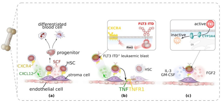
The physiologic HSC niche in the adult is located in the bone marrow and provides juxta- and paracrine signals to ensure maintenance and proliferation of HSC and to stimulate their differentiation into less potent, lineage-restricted progenitor cells. In particular, perivascular stromal cells play a crucial role for homing and residence of HSC by providing soluble and membrane-bound chemokine (C-X-C) ligand (CXCL) 12 (Figure 3a) and promote proliferation of HSC by the secretion of stem cell factor (SCF, encoded by KITLG). For further details on the cellular and molecular organisation of the haematopoietic niche, the reader is referred to recent excellent reviews [88,89].
Figure 3.
Interaction of haematopoietic stem cells (HSC) and FLT3 ITD-positive blast cells with the haematopoietic bone marrow niche. (a) Under physiological conditions, HSC are recruited and immobilised via the chemokine CXCL12, produced by bone marrow stromal cells in the perivascular niche. On HSC CXCL12 binds to and activates the G-protein coupled chemokine receptor CXCR4. HSC give rise to more restricted progenitor cells that fuel differentiated peripheral blood cells. (b) FLT3 ITD+ leukaemic blast cells shape the bone marrow haematopoietic niche. Through activation of the cytoplasmic serine/threonine kinase Pim1, FLT3 ITD enhances CXCR4 signalling and mediates leukaemic blast recruitment to the perivascular niche. Through the induction of tumour necrosis factor (TNF)-expression in endothelial cells, leukaemic blast cells drive TNF-mediated decay of stroma cells and in consequence, impaired normal haematopoiesis. (c) The perivascular niche, in particular stromal cells contribute to tyrosine kinase inhibitor (TKI)-resistance through (I) secretion of cytokines and growth factors that enable bypassing of inhibited FLT3 ITD and (II) CYP3A4-mediated catabolism of TKI in stromal cells.
In haematopoietic malignancies, the interaction of leukaemic cells with the haematopoietic niche is altered which contributes to pathology and therapy resistance. Patients suffering from haematopoietic malignancies including AML display loss of normal blood cell replenishment, which is associated with altered composition of the HSC compartment [90,91]. FLT3 ITD-induced myeloproliferation reduced the number of normal HSC in FLT3 ITD-transgenic mice and transplantation experiments. This effect was linked to enhanced expression of tumour necrosis factor (TNF) in endothelial cells and consequently in a reduction of mesenchymal stromal cells and endothelial cells (Figure 3b) [92]. Administration of etanercept, a recombinant dimerised version of TNFR2 ectodomain [93], which binds TNF and hence blocks TNF signalling, restored normal haematopoiesis in the mouse model [92]. Furthermore, there is strong evidence that FLT3 ITD-signalling enhances CXCR4 signalling in leukaemic cells [94,95]. FLT3 ITD-induced Pim1 kinase activity results in phosphorylation of CXCR4 intracellular domain and enhanced signalling of the chemokine receptor (Figure 3b) [96] and, therefore, enhances migration of FLT3 ITD+ leukaemic cells towards CXCL12 via enhanced Rho-associated kinase (ROCK) [97]. As a result, homing and residence of leukaemic cells to the HSC niche is enhanced.
The stromal cells of the haematopoietic stem cell niche were demonstrated to confer therapy resistance by different means. Recently, it was shown that bone marrow stromal cells express several cytochrom P450 isoforms, among them also CYP3A4 which was previously shown to be responsible for hepatic metabolism of TKI [98]. CYP3A4 in bone marrow stromal cells also contributed to resistance against three clinically used TKI (Figure 3c) [99]. Furthermore, stromal cells provide FL, which stimulates FLT3 expressed from the WT allele. Signalling of FLT3 WT on leukaemic blasts that also express a mutant FLT3 allele was shown to confer resistance to TKI [100].
In addition, bone marrow stromal cells were shown to confer TKI resistance [101] via provision of paracrine signals. IL-3 and GM-CSF were shown to rescue FLT3 ITD+ cells from tyrosine kinase inhibition (Figure 3c) [102] in vitro, which was at least in part via upregulation of the RTK Axl (Figure 3c and Figure 4d) [103]. However, FLT3 ligand (FL) and fibroblast growth factor (FGF) 2 were identified to be most potent in conferring TKI resistance and patients relapsing under TKI treatment demonstrated increased FGF2 expression in bone marrow stromal cells [104].
Figure 4.
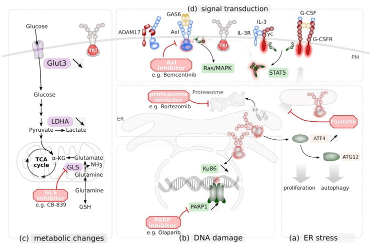
Targeting novel vulnerabilities in FLT3 mutant cells as novel therapeutic strategy. (a) FLT3 ITD induces expression of activating transcription factor (ATF) 4 that enhances the unfolded protein response (UPR) and triggers autophagy via autophagy related protein (ATG) 12. Export from the ER and degradation of misfolded proteins via the proteasome is part of the UPR. FLT3 ITD+ blasts are susceptible to proteasome inhibition via e.g. the clinically approved Bortezomib. Inhibtion of ER-to-Golgi trafficking via Flustatin further enhances UPR and UPR-mediated cell death in these cells. (b) FLT3 ITD signalling reduces Ku86 expression and consequently DNA damage repair by non-homologues end-joining. As a consequence, FLT3 ITD+ blasts rely on poly (ADP-ribose) polymerase (PARP) 1-mediated DNA damage response, making them vulnerable for PARP inhibitors such as Olaparib. (c) TKI-mediated inhibition of FLT3 ITD reduces expression of Glucose transporter Glut3 and lactate dehydrogenase (LDHA). As a consequence, ITD+ blasts rely on Glutamine as carbone source for the tricarboxylic acid cycle (TCA) cycle, making them vulnerable for glutaminase (GLS)-inhibitors such as the drug candidate CB-839 (d) Tyrosine kinase inhibitor (TKI)-mediated inhibition of FLT3 ITD signalling in leukaemic blast cells can be bypassed by expression and cytokine-mediated activation of the receptor tyrosine kinases Axl (by its ligand GAS6) or granulocyte colony stimulating factor receptor G-CSFR, or by activation of the interleukin (IL)-3 receptor complex. Signalling via Axl is regulated via ADAM17-mediated limited proteolysis. Simultaneous inhibition of FLT3 and Axl enhances therapeutic response. PM, plasma membrane; ER endoplasmatic reticulum.
3. FLT3 as Therapeutic Target in Leukaemia
3.1. Small Molecule Inhibition of FLT3
Several small-molecule TKI targeting FLT3 have been developed and tested. (Recently reviewed in [105,106].) First-generation multi-kinase inhibitors (sorafenib, midostaurin, lestaurtinib) are characterized by a broad-spectrum of drug targets, whereas second generation inhibitors (quizartinib, crenolanib, gilteritinib) show more potent and specific FLT3 inhibition, and are thereby accompanied by less toxic effects. In 2017, midostaurin (PKC412) was approved by the FDA as first TKI to treat adult patients with newly diagnosed AML who are positive for oncogenic FLT3, in combination with chemotherapy [107]. The addition of midostaurin to standard chemotherapy significantly prolonged overall and event-free survival among patients with AML and a FLT3 mutation [108]. While Midostaurin acts rather unspecific on kinases in general, Quizartinib (AC220) inhibits selectively tyrosine kinases [109]. Phase I and II clinical trials have shown its survival benefit over conventional chemotherapy in patients with FLT3 ITD-positive relapsed/refractory AML [110]. Due to poly-pharmacology commonly observed for TKI, target space of approved or tested drugs were recently analysed for their target spectrum on human kinases. Here, cabozantinib, a TKI used to treat medullary thyroid cancer and renal cell carcinoma by targeting tyrosine kinases c-Met, VEGFR2, and also AXL and RET, was repurposed to abrogate oncogenic FLT3 ITD activity in AML patients [111].
3.2. Novel Alternative Approaches to Affect Oncogenic FLT3 Activity
Receptor dimerization is the prerequisite for FLT3 autophosphorylation. Thus, small-molecule inhibitiors of dimerization has become an attractive tool to interfere with protein–protein complex formation and subsequent down-stream signalling [112,113,114] that might open a new avenue the target FLT3.
Enhancing enzymatic activity of counteracting PTPs is a potential rational approach to restrain oncogenic FLT3 kinase activity. As a proof-of-principle, PTPRJ activity was shown to be enhanced by Thrombospondin (TSP1) and to enhance dephosphorylation of its substrates [115,116]. Alternatively, interference with RPTP dimerization was identified to increase enzymatic phosphatase activity. Allosteric acting peptides preventing PTPRJ dimerization were recently demonstrated to reduce phosphorylation of EGFR and to antagonize EGFR-driven cell phenotypes [117] and might therefore represent a novel approach to regulate FLT3 activity (Figure 2b). Furthermore, chemical quenching of high ROS levels in FLT3 ITD positive cells were suggested to prevent reversible oxidation and consequently reactivate PTPRJ (Figure 2b) [118,119].
Statins were demonstrated to prevent complex FLT3 glycosylation resulting in loss of receptor surface localization and induction of cell death, as well as mitigation of TKI resistance [120]. Fluvastatin (Lescol), a generic drug approved in the USA in 2012, was shown to overcome resistance against the TKI sorafenib or the activation of the IL-3 compensatory pathway (Figure 4a). Important fluvastatin treatment in vivo reduced engraftment of BaF3 FLT3 ITD cells in Balb/c mice [120]. Combined inhibition of receptor glycosylation by fluvastatin or tunicamycin with TKI AC220 caused synergistic cell killing, which was highly selective for cell lines and primary AML cells expressing FLT3 ITD [121]. Synergistic effects observed in response to the combinatory application of pharmacologic inhibitors targeting FLT3 ITD downstream mediators are the basis for novel treatment strategies to deplete leukaemic cells. For instance, STAT5 inhibition synergistically increased cytotoxic effects of the JAK1/2 inhibitor Ruxolitinib or the p300/pCAF inhibitor Garcinol in leukaemic cells [122].
3.3. Exploiting Novel Vulnerabilities in FLT3 Mutant Blasts
As indicated above, current therapies of FLT3 mutant haematopoietic malignancies involve the directed use of TKI. However, resistance development through either secondary FLT3 mutations or through the protection of FLT3 mutant cell clones by the bone marrow niche is a major obstacle for successful treatment. Therefore, novel therapeutic approaches are highly warranted. One attractive approach is to exploit novel vulnerabilities that are induced by mutant FLT3.
Activating mutations of FLT3 induce an unstable kinase conformation, resulting in the dependency on chaperones such as the heat shock protein (HSP) 90. Targeting HSP90 by geldanamycin and analogues has therefore been considered as TKI co-treatment that is also able to prevent TKI-resistance formation [123,124,125,126]. Novel therapeutic opportunities are also created through kinase-dependent impaired receptor maturation (see above), which has been recently reviewed elsewhere [27].
Expression of the protein arginine methyltransferase (PRMT) 1 is increased in AML blasts and it was demonstrated that in FLT3 ITD+ AML blasts, FLT3 is the major target of PRMT1. Genetic ablation of PRMT1 increased apoptosis of FLT3 ITD+ blasts and prolonged survival of FLT3 ITD-expressing mice [127]. Consequently, pharmacological inhibition of PRMT1 decreased survival of FLT3 ITD+ blasts [127].
Recently, it was demonstrated that epidermal growth factor (EGF) receptor (EGFR) signalling enhances DNA damage repair (DDR) capacity in hepatocytes [128]. Apparently, RTK signalling is linked to DDR by a so far unknown mechanism. FLT3 ITD-signalling was also shown to be linked to DDR. In particular, expression of breast cancer-associated (BRCA) 1, which is part of a protein complex mediating homology-directed repair (HR) of DNA double strand breaks is enhanced in FLT3 ITD-expressing cells [129]. In contrast, efficiency of DNA double strand break repair by non-homologues end-joining (NHEJ) was decreased in cells expressing FLT3 ITD via down-regulation of Ku86 and up-regulation of the error-prone PARP1-dependent NHEJ pathway [130]. TKI-mediated FLT3 ITD inhibition results in a significant decrease of both, HR and NHEJ-mediated repair of DNA double strand breaks [129]. This seems to be conferred at least in part via Pim1 kinase activation [131]. Due to the resulting enhanced genotoxic stress, these cells were sensitized to PARP1 inhibition (Figure 4b) [129], similar as BRCA1-deficient mammary carcinoma cells [132].
Most likely due to its retention in intracellular compartments, FLT3 ITD was shown to be sensitive to the clinical approved proteasome inhibitor bortezomib [133], known to induce ER stress. The transcription factor ATF4 is present in ER stress and autophagy pathways (Figure 4b). It was demonstrated that FLT3 ITD induces autophagy and upon inhibition of autophagy, proliferation of leukaemic cells was impaired in vitro and in vivo [134].
A genome-wide CRISPR/Cas9 screen revealed that FLT3-ITD-expressing cells display alterations in their metabolic dependencies. Genetic deficiency in GLS, encoding for glutaminase, the first enzyme in glutamine catabolism, demonstrated to be synthetic lethal together with TKIs, indicating that FLT3-ITD-expressing cells maintain mitochondrial function and redox functions through glutamine catabolism (Figure 4c) [135]. Hence, the identification of novel synthetic lethalities, in particular, dependency on metabolic pathways and targeting key enzymes in these pathways represents a novel therapeutic approach that might synergize with tyrosine kinase inhibition.
Increased expression of the RTK Axl seems to be a current theme in therapy-resistant tumours [136,137,138,139], and was initially identified in EGFR inhibitor-resistant non-small cell lung carcinoma [139]. As already outlined above, FLT3-ITD-expressing leukaemic cells develop TKI-resistance via enhanced expression of Axl [140,141]. Up-regulation of Axl can also occur via reduced ectodomain shedding mediated by ADAM proteases [142]. Activation of Axl most likely occurs via autocrine secretion of its ligand Gas6 (Figure 4d) [140]. Resistance against FLT3 inhibitors was substantially reduced by co-inhibition of Axl [140,141]. Therefore, dual targeting of FLT3 and Axl seems to be a promising strategy to prevent TKI resistance and enhance therapeutic response.
CAR-T cell technology has been developed to target malignant cell populations by direct killing of cells that expose a particular antigen. The prerequisite for successful targeted immunotherapies is the identification of suitable target antigens, which are restricted to malignant cells including malignant stem cells. For an excellent review on the state-of-the art of preclinical and clinical studies on suitable target antigens for CAR-T cell therapy in AML patients, see Hoffmann et al., [143]. Activated receptor tyrosine kinases are promising targets to defeat FLT3 ITD-positive AML blast cells. Since FLT3 ITD is retained in its biogenesis route (see above), TKI mediated receptor inactivation to promote FLT3 ITD surface localisation, is a prerequisite to target FLT3 ITD-positive cell clones efficiently. Combination of small molecule TKI crenolanib with CAR-T cells targeting FLT3 has been demonstrated as proof-of-concept that efficiency of CAR T-cell immunotherapy can be enhanced by the use of small molecule inhibitors. However, despite the curative potential of FLT3-CAR T-cell-based immunotherapies based on the graft-versus-leukaemia effect, severe side-effects have to be considered. Importantly, since FLT3-CAR T-cells recognize normal HSC, and consequently disrupt normal haematopoiesis, it has to be considered that adoptive therapy with FLT3-CAR T-cells will require subsequent CAR T-cell depletion and allogeneic HSC transplantation to reconstitute the haematopoietic system [144]. Furthermore, FLT3-CAR T-cell-based therapies have the danger of life-threatening graft-versus-host disease. Enhancing the graft-versus-leukaemia effect, but preventing graft-versus-host disease is an essential challenge for future therapy improvement [143].
3.4. Preventing Protective Signals from the Bone Marrow Niche
Bone marrow stromal cells were shown to protect FLT3 ITD AML cells against TKI treatment by induction of enhanced STAT5 phosphorylation and enhanced AXL expression [103]. Thus, a further alternative therapeutic approach would be to target the protective bone marrow stromal niche and the interaction with the bone marrow niche. On one hand, inhibition of CYP3A4 has been shown to prevent metabolic TKI degradation. On the other hand, TKI resistance is also conferred by paracrine secretion of cytokines and growth factors such as IL-3, G-CSF or FGF-2, bypassing FLT3 inhibition by STAT5 and Ras/MAPK activation (Figure 4d). Some of these factors are synthesized as membrane-bound forms and released via limited proteolysis mediated by a disintegrin and metalloproteases (ADAM). Albeit, experimental evidence is still lacking, it is conceivable that inhibition of ADAM proteases could reduce TKI resistance formation. In contrast, membrane-bound proteins might also contribute to TKI resistance, as was previously suggested [145]. However, addition of the hypomethylating agent azacytidine was able to overcome TKI resistance in leukaemic blasts conferred by stromal cell contact [145].
4. Conclusions
Taken together, there are growing experimental data on how haematopoietic malignant cells exploit cellular components, signalling pathways and metabolic enzymes, resulting in relapse or refraction of targeted therapies. Combination of TKI-based therapy with targeting cooperating molecules or cells would result in synergistic effects to target resistance development and uncontrolled clonal development of leukaemic cells. Therefore, adopted clinical approaches synergistically attacking aberrant signaling events of leukaemic cells is the way to enhance therapeutic efficiency.
Author Contributions
J.P.M. and D.S.-A. wrote and revised the manuscript. All authors have read and agreed to the published version of the manuscript.
Funding
This paper was supported by the Deutsche Forschungsgemeinschaft, grant Mu955/14-1 and Mu955/15-1 to JPM and grant SFB841, project C1 to DSA.
Conflicts of Interest
The other authors declare that they have no conflict of interest.
References
- 1.Maroc N., Rottapel R., Rosnet O., Marchetto S., Lavezzi C., Mannoni P., Birnbaum D., Dubreuil P. Biochemical characterization and analysis of the transforming potential of the FLT3/FLK2 receptor tyrosine kinase. Oncogene. 1993;8:909–918. [PubMed] [Google Scholar]
- 2.Kikushige Y., Yoshimoto G., Miyamoto T., Iino T., Mori Y., Iwasaki H., Niiro H., Takenaka K., Nagafuji K., Harada M., et al. Human Flt3 is expressed at the hematopoietic stem cell and the granulocyte/macrophage progenitor stages to maintain cell survival. J. Immunol. 2008;180:7358–7367. doi: 10.4049/jimmunol.180.11.7358. [DOI] [PubMed] [Google Scholar]
- 3.Toffalini F., Demoulin J.B. New insights into the mechanisms of hematopoietic cell transformation by activated receptor tyrosine kinases. Blood. 2010;116:2429–2437. doi: 10.1182/blood-2010-04-279752. [DOI] [PubMed] [Google Scholar]
- 4.Stirewalt D.L., Radich J.P. The role of FLT3 in haematopoietic malignancies. Nat. Rev. Cancer. 2003;3:650–665. doi: 10.1038/nrc1169. [DOI] [PubMed] [Google Scholar]
- 5.Sanz M., Burnett A., Lo-Coco F., Lowenberg B. FLT3 inhibition as a targeted therapy for acute myeloid leukemia. Curr. Opin. Oncol. 2009;21:594–600. doi: 10.1097/CCO.0b013e32833118fd. [DOI] [PubMed] [Google Scholar]
- 6.Dehlin M., Bokarewa M., Rottapel R., Foster S.J., Magnusson M., Dahlberg L.E., Tarkowski A. Intra-articular fms-like tyrosine kinase 3 ligand expression is a driving force in induction and progression of arthritis. PLoS ONE. 2008;3:e3633. doi: 10.1371/journal.pone.0003633. [DOI] [PMC free article] [PubMed] [Google Scholar]
- 7.Hogan F.L., Williams V., Knapper S. FLT3 Inhibition in Acute Myeloid Leukaemia—Current Knowledge and Future Prospects. Curr. Cancer Drug Targets. 2020;20:513–531. doi: 10.2174/1570163817666200518075820. [DOI] [PubMed] [Google Scholar]
- 8.Levis M., Perl A.E. Gilteritinib: Potent targeting of FLT3 mutations in AML. Blood Adv. 2020;4:1178–1191. doi: 10.1182/bloodadvances.2019000174. [DOI] [PMC free article] [PubMed] [Google Scholar]
- 9.Kayser S., Schlenk R.F., Londono M.C., Breitenbuecher F., Wittke K., Du J., Groner S., Spath D., Krauter J., Ganser A., et al. Insertion of FLT3 internal tandem duplication in the tyrosine kinase domain-1 is associated with resistance to chemotherapy and inferior outcome. Blood. 2009;114:2386–2392. doi: 10.1182/blood-2009-03-209999. [DOI] [PubMed] [Google Scholar]
- 10.Huang K., Yang M., Pan Z., Heidel F.H., Scherr M., Eder M., Fischer T., Busche G., Welte K., von Neuhoff N., et al. Leukemogenic potency of the novel FLT3-N676K mutant. Ann. Hematol. 2016;95:783–791. doi: 10.1007/s00277-016-2616-z. [DOI] [PubMed] [Google Scholar]
- 11.Heidel F., Solem F.K., Breitenbuecher F., Lipka D.B., Kasper S., Thiede M.H., Brandts C., Serve H., Roesel J., Giles F., et al. Clinical resistance to the kinase inhibitor PKC412 in acute myeloid leukemia by mutation of Asn-676 in the FLT3 tyrosine kinase domain. Blood. 2006;107:293–300. doi: 10.1182/blood-2005-06-2469. [DOI] [PubMed] [Google Scholar]
- 12.Abu-Duhier F.M., Goodeve A.C., Wilson G.A., Care R.S., Peake I.R., Reilly J.T. Identification of novel FLT-3 Asp835 mutations in adult acute myeloid leukaemia. Br. J. Haematol. 2001;113:983–988. doi: 10.1046/j.1365-2141.2001.02850.x. [DOI] [PubMed] [Google Scholar]
- 13.Yamamoto Y., Kiyoi H., Nakano Y., Suzuki R., Kodera Y., Miyawaki S., Asou N., Kuriyama K., Yagasaki F., Shimazaki C., et al. Activating mutation of D835 within the activation loop of FLT3 in human hematologic malignancies. Blood. 2001;97:2434–2439. doi: 10.1182/blood.V97.8.2434. [DOI] [PubMed] [Google Scholar]
- 14.Taketani T., Taki T., Sugita K., Furuichi Y., Ishii E., Hanada R., Tsuchida M., Sugita K., Ida K., Hayashi Y. FLT3 mutations in the activation loop of tyrosine kinase domain are frequently found in infant ALL with MLL rearrangements and pediatric ALL with hyperdiploidy. Blood. 2004;103:1085–1088. doi: 10.1182/blood-2003-02-0418. [DOI] [PubMed] [Google Scholar]
- 15.Frohling S., Schlenk R.F., Breitruck J., Benner A., Kreitmeier S., Tobis K., Dohner H., Dohner K., leukemia AMLSGUAm Prognostic significance of activating FLT3 mutations in younger adults (16 to 60 years) with acute myeloid leukemia and normal cytogenetics: A study of the AML Study Group Ulm. Blood. 2002;100:4372–4380. doi: 10.1182/blood-2002-05-1440. [DOI] [PubMed] [Google Scholar]
- 16.Thiede C., Steudel C., Mohr B., Schaich M., Schakel U., Platzbecker U., Wermke M., Bornhauser M., Ritter M., Neubauer A., et al. Analysis of FLT3-activating mutations in 979 patients with acute myelogenous leukemia: Association with FAB subtypes and identification of subgroups with poor prognosis. Blood. 2002;99:4326–4335. doi: 10.1182/blood.V99.12.4326. [DOI] [PubMed] [Google Scholar]
- 17.Ghosh K., Swaminathan S., Madkaikar M., Gupta M., Kerketta L., Vundinti B. FLT3 and NPM1 mutations in a cohort of AML patients and detection of a novel mutation in tyrosine kinase domain of FLT3 gene from Western India. Ann. Hematol. 2012;91:1703–1712. doi: 10.1007/s00277-012-1509-z. [DOI] [PubMed] [Google Scholar]
- 18.Kindler T., Breitenbuecher F., Kasper S., Estey E., Giles F., Feldman E., Ehninger G., Schiller G., Klimek V., Nimer S.D., et al. Identification of a novel activating mutation (Y842C) within the activation loop of FLT3 in patients with acute myeloid leukemia (AML) Blood. 2005;105:335–340. doi: 10.1182/blood-2004-02-0660. [DOI] [PubMed] [Google Scholar]
- 19.Zhang H., Savage S., Schultz A.R., Bottomly D., White L., Segerdell E., Wilmot B., McWeeney S.K., Eide C.A., Nechiporuk T., et al. Clinical resistance to crenolanib in acute myeloid leukemia due to diverse molecular mechanisms. Nat. Commun. 2019;10:244. doi: 10.1038/s41467-018-08263-x. [DOI] [PMC free article] [PubMed] [Google Scholar]
- 20.Frohling S., Scholl C., Levine R.L., Loriaux M., Boggon T.J., Bernard O.A., Berger R., Dohner H., Dohner K., Ebert B.L., et al. Identification of driver and passenger mutations of FLT3 by high-throughput DNA sequence analysis and functional assessment of candidate alleles. Cancer Cell. 2007;12:501–513. doi: 10.1016/j.ccr.2007.11.005. [DOI] [PubMed] [Google Scholar]
- 21.Jiang J., Paez J.G., Lee J.C., Bo R., Stone R.M., DeAngelo D.J., Galinsky I., Wolpin B.M., Jonasova A., Herman P., et al. Identifying and characterizing a novel activating mutation of the FLT3 tyrosine kinase in AML. Blood. 2004;104:1855–1858. doi: 10.1182/blood-2004-02-0712. [DOI] [PubMed] [Google Scholar]
- 22.Matsuno N., Nanri T., Kawakita T., Mitsuya H., Asou N. A novel FLT3 activation loop mutation N841K in acute myeloblastic leukemia. Leukemia. 2005;19:480–481. doi: 10.1038/sj.leu.2403630. [DOI] [PubMed] [Google Scholar]
- 23.Griffith J., Black J., Faerman C., Swenson L., Wynn M., Lu F., Lippke J., Saxena K. The structural basis for autoinhibition of FLT3 by the juxtamembrane domain. Mol. Cell. 2004;13:169–178. doi: 10.1016/S1097-2765(03)00505-7. [DOI] [PubMed] [Google Scholar]
- 24.Nguyen B., Williams A.B., Young D.J., Ma H., Li L., Levis M., Brown P., Small D. FLT3 activating mutations display differential sensitivity to multiple tyrosine kinase inhibitors. Oncotarget. 2017;8:10931–10944. doi: 10.18632/oncotarget.14539. [DOI] [PMC free article] [PubMed] [Google Scholar]
- 25.Opatz S., Polzer H., Herold T., Konstandin N.P., Ksienzyk B., Zellmeier E., Vosberg S., Graf A., Krebs S., Blum H., et al. Exome sequencing identifies recurring FLT3 N676K mutations in core-binding factor leukemia. Blood. 2013;122:1761–1769. doi: 10.1182/blood-2013-01-476473. [DOI] [PubMed] [Google Scholar]
- 26.Hyrenius-Wittsten A., Pilheden M., Falques-Costa A., Eriksson M., Sturesson H., Schneider P., Wander P., Garcia-Ruiz C., Liu J., Agerstam H., et al. FLT3(N676K) drives acute myeloid leukemia in a xenograft model of KMT2A-MLLT3 leukemogenesis. Leukemia. 2019;33:2310–2314. doi: 10.1038/s41375-019-0465-1. [DOI] [PMC free article] [PubMed] [Google Scholar]
- 27.Schmidt-Arras D., Bohmer F.D. Mislocalisation of Activated Receptor Tyrosine Kinases—Challenges for Cancer Therapy. Trends Mol. Med. 2020;26:833–847. doi: 10.1016/j.molmed.2020.06.002. [DOI] [PubMed] [Google Scholar]
- 28.Schmidt-Arras D.E., Böhmer A., Markova B., Choudhary C., Serve H., Böhmer F.D. Tyrosine phosphorylation regulates maturation of receptor tyrosine kinases. Mol. Cell. Biol. 2005;25:3690–3703. doi: 10.1128/MCB.25.9.3690-3703.2005. [DOI] [PMC free article] [PubMed] [Google Scholar]
- 29.Schmidt-Arras D., Böhmer S.A., Koch S., Müller J.P., Blei L., Cornils H., Bauer R., Korasikha S., Thiede C., Böhmer F.D. Anchoring of FLT3 in the endoplasmic reticulum alters signaling quality. Blood. 2009;113:3568–3576. doi: 10.1182/blood-2007-10-121426. [DOI] [PubMed] [Google Scholar]
- 30.Choudhary C., Olsen J.V., Brandts C., Cox J., Reddy P.N., Böhmer F.D., Gerke V., Schmidt-Arras D.E., Berdel W.E., Müller-Tidow C., et al. Mislocalized activation of oncogenic RTKs switches downstream signaling outcomes. Mol. Cell. 2009;36:326–339. doi: 10.1016/j.molcel.2009.09.019. [DOI] [PubMed] [Google Scholar]
- 31.Reiter K., Polzer H., Krupka C., Maiser A., Vick B., Rothenberg-Thurley M., Metzeler K.H., Dorfel D., Salih H.R., Jung G., et al. Tyrosine kinase inhibition increases the cell surface localization of FLT3-ITD and enhances FLT3-directed immunotherapy of acute myeloid leukemia. Leukemia. 2018;32:313–322. doi: 10.1038/leu.2017.257. [DOI] [PMC free article] [PubMed] [Google Scholar]
- 32.Chan P.M. Differential signaling of Flt3 activating mutations in acute myeloid leukemia: A working model. Protein Cell. 2011;2:108–115. doi: 10.1007/s13238-011-1020-7. [DOI] [PMC free article] [PubMed] [Google Scholar]
- 33.Hubbard G.P., Wolffram S., Lovegrove J.A., Gibbins J.M. Ingestion of quercetin inhibits platelet aggregation and essential components of the collagen-stimulated platelet activation pathway in humans. J. Thromb. Haemost. 2004;2:2138–2145. doi: 10.1111/j.1538-7836.2004.01067.x. [DOI] [PubMed] [Google Scholar]
- 34.Schmidt-Arras D., Schwable J., Böhmer F.D., Serve H. Flt3 receptor tyrosine kinase as a drug target in leukemia. Curr. Pharm. Des. 2004;10:1867–1883. doi: 10.2174/1381612043384394. [DOI] [PubMed] [Google Scholar]
- 35.Kazi J.U., Ronnstrand L. FMS-like Tyrosine Kinase 3/FLT3, From Basic Science to Clinical Implications. Physiol. Rev. 2019;99:1433–1466. doi: 10.1152/physrev.00029.2018. [DOI] [PubMed] [Google Scholar]
- 36.Lamaze C., Schmid S.L. Recruitment of epidermal growth factor receptors into coated pits requires their activated tyrosine kinase. J. Cell Biol. 1995;129:47–54. doi: 10.1083/jcb.129.1.47. [DOI] [PMC free article] [PubMed] [Google Scholar]
- 37.Jahn T., Seipel P., Coutinho S., Urschel S., Schwarz K., Miething C., Serve H., Peschel C., Duyster J. Analysing c-kit internalization using a functional c-kit-EGFP chimera containing the fluorochrome within the extracellular domain. Oncogene. 2002;21:4508–4520. doi: 10.1038/sj.onc.1205559. [DOI] [PubMed] [Google Scholar]
- 38.Kellner F., Keil A., Schindler K., Tschongov T., Hunninger K., Loercher H., Rhein P., Bohmer S.A., Bohmer F.D., Muller J.P. Wild-type FLT3 and FLT3 ITD exhibit similar ligand-induced internalization characteristics. J. Cell Mol. Med. 2020 doi: 10.1111/jcmm.15132. [DOI] [PMC free article] [PubMed] [Google Scholar]
- 39.Frangioni J.V., Beahm P.H., Shifrin V., Jost C.A., Neel B.G. The nontransmembrane tyrosine phosphatase PTP-1B localizes to the endoplasmic reticulum via its 35 amino acid C-terminal sequence. Cell. 1992;68:545–560. doi: 10.1016/0092-8674(92)90190-N. [DOI] [PubMed] [Google Scholar]
- 40.Tenev T., Bohmer S.A., Kaufmann R., Frese S., Bittorf T., Beckers T., Bohmer F.D. Perinuclear localization of the protein-tyrosine phosphatase SHP-1 and inhibition of epidermal growth factor-stimulated STAT1/3 activation in A431 cells. Eur. J. Cell Biol. 2000;79:261–271. doi: 10.1078/S0171-9335(04)70029-1. [DOI] [PubMed] [Google Scholar]
- 41.Müller J.P., Schönherr C., Markova B., Bauer R., Stocking C., Böhmer F.D. Role of SHP2 for FLT3-dependent proliferation and transformation in 32D cells. Leukemia. 2008;22:1945–1948. doi: 10.1038/leu.2008.73. [DOI] [PubMed] [Google Scholar]
- 42.Zhang S., Broxmeyer H.E. Flt3 ligand induces tyrosine phosphorylation of gab1 and gab2 and their association with shp-2, grb2, and PI3 kinase. Biochem. Biophys. Res. Commun. 2000;277:195–199. doi: 10.1006/bbrc.2000.3662. [DOI] [PubMed] [Google Scholar]
- 43.Nabinger S.C., Li X.J., Ramdas B., He Y., Zhang X., Zeng L., Richine B., Bowling J.D., Fukuda S., Goenka S., et al. The protein tyrosine phosphatase, Shp2, positively contributes to FLT3-ITD-induced hematopoietic progenitor hyperproliferation and malignant disease in vivo. Leukemia. 2013;27:398–408. doi: 10.1038/leu.2012.308. [DOI] [PMC free article] [PubMed] [Google Scholar]
- 44.Pandey R., Ramdas B., Wan C., Sandusky G., Mohseni M., Zhang C., Kapur R. SHP2 inhibition reduces leukemogenesis in models of combined genetic and epigenetic mutations. J. Clin. Investig. 2019;129:5468–5473. doi: 10.1172/JCI130520. [DOI] [PMC free article] [PubMed] [Google Scholar]
- 45.Arora D., Kothe S., van den Eijnden M., Hooft van Huijsduijnen R., Heidel F., Fischer T., Scholl S., Tolle B., Bohmer S.A., Lennartsson J., et al. Expression of protein-tyrosine phosphatases in Acute Myeloid Leukemia cells: FLT3 ITD sustains high levels of DUSP6 expression. Cell Commun. Signal. 2012;10:19. doi: 10.1186/1478-811X-10-19. [DOI] [PMC free article] [PubMed] [Google Scholar]
- 46.Arora D., Stopp S., Bohmer S.A., Schons J., Godfrey R., Masson K., Razumovskaya E., Ronnstrand L., Tanzer S., Bauer R., et al. Protein-tyrosine phosphatase DEP-1 controls receptor tyrosine kinase FLT3 signaling. J. Biol. Chem. 2011;286:10918–10929. doi: 10.1074/jbc.M110.205021. [DOI] [PMC free article] [PubMed] [Google Scholar]
- 47.Bohmer S.A., Weibrecht I., Soderberg O., Bohmer F.D. Association of the protein-tyrosine phosphatase DEP-1 with its substrate FLT3 visualized by in situ proximity ligation assay. PLoS ONE. 2013;8:e62871. doi: 10.1371/journal.pone.0062871. [DOI] [PMC free article] [PubMed] [Google Scholar]
- 48.Godfrey R., Arora D., Bauer R., Stopp S., Muller J.P., Heinrich T., Bohmer S.A., Dagnell M., Schnetzke U., Scholl S., et al. Cell transformation by FLT3 ITD in acute myeloid leukemia involves oxidative inactivation of the tumor suppressor protein-tyrosine phosphatase DEP-1/ PTPRJ. Blood. 2012;119:4499–4511. doi: 10.1182/blood-2011-02-336446. [DOI] [PubMed] [Google Scholar]
- 49.Lee B.H., Williams I.R., Anastasiadou E., Boulton C.L., Joseph S.W., Amaral S.M., Curley D.P., Duclos N., Huntly B.J., Fabbro D., et al. FLT3 internal tandem duplication mutations induce myeloproliferative or lymphoid disease in a transgenic mouse model. Oncogene. 2005;24:7882–7892. doi: 10.1038/sj.onc.1208933. [DOI] [PubMed] [Google Scholar]
- 50.Kresinsky A., Bauer R., Schnoder T.M., Berg T., Meyer D., Ast V., Konig R., Serve H., Heidel F.H., Bohmer F.D., et al. Loss of DEP-1 (Ptprj) promotes myeloproliferative disease in FLT3-ITD acute myeloid leukemia. Haematologica. 2018;103:e505–e509. doi: 10.3324/haematol.2017.185306. [DOI] [PMC free article] [PubMed] [Google Scholar]
- 51.Kresinsky A., Schnoder T.M., Jacobsen I.D., Rauner M., Hofbauer L.C., Ast V., Konig R., Hoffmann B., Svensson C.M., Figge M.T., et al. Lack of CD45 in FLT3-ITD mice results in a myeloproliferative phenotype, cortical porosity, and ectopic bone formation. Oncogene. 2019;38:4773–4787. doi: 10.1038/s41388-019-0757-y. [DOI] [PubMed] [Google Scholar]
- 52.Sundaresan M., Yu Z.X., Ferrans V.J., Irani K., Finkel T. Requirement for generation of H2O2 for platelet-derived growth factor signal transduction. Science. 1995;270:296–299. doi: 10.1126/science.270.5234.296. [DOI] [PubMed] [Google Scholar]
- 53.Rhee C.M., Kalantar-Zadeh K., Streja E., Carrero J.J., Ma J.Z., Lu J.L., Kovesdy C.P. The relationship between thyroid function and estimated glomerular filtration rate in patients with chronic kidney disease. Nephrol. Dial. Transpl. 2015;30:282–287. doi: 10.1093/ndt/gfu303. [DOI] [PMC free article] [PubMed] [Google Scholar]
- 54.Bohmer A., Barz S., Schwab K., Kolbe U., Gabel A., Kirkpatrick J., Ohlenschlager O., Gorlach M., Bohmer F.D. Modulation of FLT3 signal transduction through cytoplasmic cysteine residues indicates the potential for redox regulation. Redox Biol. 2020;28:101325. doi: 10.1016/j.redox.2019.101325. [DOI] [PMC free article] [PubMed] [Google Scholar]
- 55.Gilliland D.G., Griffin J.D. The roles of FLT3 in hematopoiesis and leukemia. Blood. 2002;100:1532–1542. doi: 10.1182/blood-2002-02-0492. [DOI] [PubMed] [Google Scholar]
- 56.Parcells B.W., Ikeda A.K., Simms-Waldrip T., Moore T.B., Sakamoto K.M. FMS-like tyrosine kinase 3 in normal hematopoiesis and acute myeloid leukemia. Stem Cells. 2006;24:1174–1184. doi: 10.1634/stemcells.2005-0519. [DOI] [PubMed] [Google Scholar]
- 57.Adolfsson J., Mansson R., Buza-Vidas N., Hultquist A., Liuba K., Jensen C.T., Bryder D., Yang L., Borge O.J., Thoren L.A., et al. Identification of Flt3+ lympho-myeloid stem cells lacking erythro-megakaryocytic potential a revised road map for adult blood lineage commitment. Cell. 2005;121:295–306. doi: 10.1016/j.cell.2005.02.013. [DOI] [PubMed] [Google Scholar]
- 58.Chu S.H., Heiser D., Li L., Kaplan I., Collector M., Huso D., Sharkis S.J., Civin C., Small D. FLT3-ITD knockin impairs hematopoietic stem cell quiescence/homeostasis, leading to myeloproliferative neoplasm. Cell Stem Cell. 2012;11:346–358. doi: 10.1016/j.stem.2012.05.027. [DOI] [PMC free article] [PubMed] [Google Scholar]
- 59.Ocampo A., Izpisua Belmonte J.C. Stem cells: Holding your breath for longevity. Science. 2015;347:1319–1320. doi: 10.1126/science.aaa9608. [DOI] [PMC free article] [PubMed] [Google Scholar]
- 60.Rossi D.J., Bryder D., Zahn J.M., Ahlenius H., Sonu R., Wagers A.J., Weissman I.L. Cell intrinsic alterations underlie hematopoietic stem cell aging. Proc. Natl. Acad. Sci. USA. 2005;102:9194–9199. doi: 10.1073/pnas.0503280102. [DOI] [PMC free article] [PubMed] [Google Scholar]
- 61.Mohrin M., Shin J., Liu Y., Brown K., Luo H., Xi Y., Haynes C.M., Chen D. Stem cell aging. A mitochondrial UPR-mediated metabolic checkpoint regulates hematopoietic stem cell aging. Science. 2015;347:1374–1377. doi: 10.1126/science.aaa2361. [DOI] [PMC free article] [PubMed] [Google Scholar]
- 62.Kaiser A., Schmidt M., Huber O., Frietsch J.J., Scholl S., Heidel F.H., Hochhaus A., Muller J.P., Ernst T. SIRT7, an influence factor in healthy aging and the development of age-dependent myeloid stem-cell disorders. Leukemia. 2020;34:2206–2216. doi: 10.1038/s41375-020-0803-3. [DOI] [PMC free article] [PubMed] [Google Scholar]
- 63.Wei W., Jing Z.X., Ke Z., Yi P. Sirtuin 7 plays an oncogenic role in human osteosarcoma via downregulating CDC4 expression. Am. J. Cancer Res. 2017;7:1788–1803. [PMC free article] [PubMed] [Google Scholar]
- 64.Li L., Osdal T., Ho Y., Chun S., McDonald T., Agarwal P., Lin A., Chu S., Qi J., Li L., et al. SIRT1 activation by a c-MYC oncogenic network promotes the maintenance and drug resistance of human FLT3-ITD acute myeloid leukemia stem cells. Cell Stem Cell. 2014;15:431–446. doi: 10.1016/j.stem.2014.08.001. [DOI] [PMC free article] [PubMed] [Google Scholar]
- 65.Ding L., Ley T.J., Larson D.E., Miller C.A., Koboldt D.C., Welch J.S., Ritchey J.K., Young M.A., Lamprecht T., McLellan M.D., et al. Clonal evolution in relapsed acute myeloid leukaemia revealed by whole-genome sequencing. Nature. 2012;481:506–510. doi: 10.1038/nature10738. [DOI] [PMC free article] [PubMed] [Google Scholar]
- 66.Dohner H., Weisdorf D.J., Bloomfield C.D. Acute Myeloid Leukemia. New Engl. J. Med. 2015;373:1136–1152. doi: 10.1056/NEJMra1406184. [DOI] [PubMed] [Google Scholar]
- 67.Genovese G., Kahler A.K., Handsaker R.E., Lindberg J., Rose S.A., Bakhoum S.F., Chambert K., Mick E., Neale B.M., Fromer M., et al. Clonal hematopoiesis and blood-cancer risk inferred from blood DNA sequence. N. Engl. J. Med. 2014;371:2477–2487. doi: 10.1056/NEJMoa1409405. [DOI] [PMC free article] [PubMed] [Google Scholar]
- 68.Jaiswal S., Fontanillas P., Flannick J., Manning A., Grauman P.V., Mar B.G., Lindsley R.C., Mermel C.H., Burtt N., Chavez A., et al. Age-related clonal hematopoiesis associated with adverse outcomes. N. Engl. J. Med. 2014;371:2488–2498. doi: 10.1056/NEJMoa1408617. [DOI] [PMC free article] [PubMed] [Google Scholar]
- 69.Xie M., Lu C., Wang J., McLellan M.D., Johnson K.J., Wendl M.C., McMichael J.F., Schmidt H.K., Yellapantula V., Miller C.A., et al. Age-related mutations associated with clonal hematopoietic expansion and malignancies. Nat. Med. 2014;20:1472–1478. doi: 10.1038/nm.3733. [DOI] [PMC free article] [PubMed] [Google Scholar]
- 70.Ganguly B.B., Kadam N.N. Mutations of myelodysplastic syndromes (MDS): An update. Mutat. Res. 2016;769:47–62. doi: 10.1016/j.mrrev.2016.04.009. [DOI] [PubMed] [Google Scholar]
- 71.Martin-Izquierdo M., Abaigar M., Hernandez-Sanchez J.M., Tamborero D., Lopez-Cadenas F., Ramos F., Lumbreras E., Madinaveitia-Ochoa A., Megido M., Labrador J., et al. Co-occurrence of cohesin complex and Ras signaling mutations during progression from myelodysplastic syndromes to secondary acute myeloid leukemia. Haematologica. 2020 doi: 10.3324/haematol.2020.248807. [DOI] [PMC free article] [PubMed] [Google Scholar]
- 72.Liu Y., Easton J., Shao Y., Maciaszek J., Wang Z., Wilkinson M.R., McCastlain K., Edmonson M., Pounds S.B., Shi L., et al. The genomic landscape of pediatric and young adult T-lineage acute lymphoblastic leukemia. Nat. Genet. 2017;49:1211–1218. doi: 10.1038/ng.3909. [DOI] [PMC free article] [PubMed] [Google Scholar]
- 73.Roberts K.G., Li Y., Payne-Turner D., Harvey R.C., Yang Y.L., Pei D., McCastlain K., Ding L., Lu C., Song G., et al. Targetable kinase-activating lesions in Ph-like acute lymphoblastic leukemia. New Engl. J. Med. 2014;371:1005–1015. doi: 10.1056/NEJMoa1403088. [DOI] [PMC free article] [PubMed] [Google Scholar]
- 74.Tomizawa D. Acute leukemia in adolescents and young adults. Rinsho Ketsueki. 2017;58:2160–2167. doi: 10.11406/rinketsu.58.2160. [DOI] [PubMed] [Google Scholar]
- 75.Zhang J., Zhang Y., Zhang M., Liu C., Liu X., Yin J., Wu P., Chen X., Yang W., Zhang L., et al. FLT3 pathway is a potential therapeutic target for PRC2-mutated T-cell acute lymphoblastic leukemia. Blood. 2018;132:2520–2524. doi: 10.1182/blood-2018-04-845628. [DOI] [PubMed] [Google Scholar]
- 76.Kim K.I., Park J., Ahn K.S., Won N.H., Kim B.K., Shin W.G., Yoon S.S., Oh J.M. Molecular characterization and prognostic significance of FLT3 in CML progression. Leukemia Res. 2010;34:995–1001. doi: 10.1016/j.leukres.2009.11.008. [DOI] [PubMed] [Google Scholar]
- 77.Garg S., Reyes-Palomares A., He L., Bergeron A., Lavallee V.P., Lemieux S., Gendron P., Rohde C., Xia J., Jagdhane P., et al. Hepatic leukemia factor is a novel leukemic stem cell regulator in DNMT3A, NPM1, and FLT3-ITD triple-mutated AML. Blood. 2019;134:263–276. doi: 10.1182/blood.2018862383. [DOI] [PubMed] [Google Scholar]
- 78.Lee B.H., Tothova Z., Levine R.L., Anderson K., Buza-Vidas N., Cullen D.E., McDowell E.P., Adelsperger J., Frohling S., Huntly B.J., et al. FLT3 mutations confer enhanced proliferation and survival properties to multipotent progenitors in a murine model of chronic myelomonocytic leukemia. Cancer Cell. 2007;12:367–380. doi: 10.1016/j.ccr.2007.08.031. [DOI] [PMC free article] [PubMed] [Google Scholar]
- 79.Li L., Piloto O., Nguyen H.B., Greenberg K., Takamiya K., Racke F., Huso D., Small D. Knock-in of an internal tandem duplication mutation into murine FLT3 confers myeloproliferative disease in a mouse model. Blood. 2008;111:3849–3858. doi: 10.1182/blood-2007-08-109942. [DOI] [PMC free article] [PubMed] [Google Scholar]
- 80.Skayneh H., Jishi B., Hleihel R., Hamieh M., Darwiche N., Bazarbachi A., El Sabban M., El Hajj H. A Critical Review of Animal Models Used in Acute Myeloid Leukemia Pathophysiology. Genes (Basel) 2019;10:614. doi: 10.3390/genes10080614. [DOI] [PMC free article] [PubMed] [Google Scholar]
- 81.Zorko N.A., Bernot K.M., Whitman S.P., Siebenaler R.F., Ahmed E.H., Marcucci G.G., Yanes D.A., McConnell K.K., Mao C., Kalu C., et al. Mll partial tandem duplication and Flt3 internal tandem duplication in a double knock-in mouse recapitulates features of counterpart human acute myeloid leukemias. Blood. 2012;120:1130–1136. doi: 10.1182/blood-2012-03-415067. [DOI] [PMC free article] [PubMed] [Google Scholar]
- 82.Stubbs M.C., Kim Y.M., Krivtsov A.V., Wright R.D., Feng Z., Agarwal J., Kung A.L., Armstrong S.A. MLL-AF9 and FLT3 cooperation in acute myelogenous leukemia: Development of a model for rapid therapeutic assessment. Leukemia. 2008;22:66–77. doi: 10.1038/sj.leu.2404951. [DOI] [PMC free article] [PubMed] [Google Scholar]
- 83.Greenblatt S., Li L., Slape C., Nguyen B., Novak R., Duffield A., Huso D., Desiderio S., Borowitz M.J., Aplan P., et al. Knock-in of a FLT3/ITD mutation cooperates with a NUP98-HOXD13 fusion to generate acute myeloid leukemia in a mouse model. Blood. 2012;119:2883–2894. doi: 10.1182/blood-2011-10-382283. [DOI] [PMC free article] [PubMed] [Google Scholar]
- 84.Mupo A., Celani L., Dovey O., Cooper J.L., Grove C., Rad R., Sportoletti P., Falini B., Bradley A., Vassiliou G.S. A powerful molecular synergy between mutant Nucleophosmin and Flt3-ITD drives acute myeloid leukemia in mice. Leukemia. 2013;27:1917–1920. doi: 10.1038/leu.2013.77. [DOI] [PMC free article] [PubMed] [Google Scholar]
- 85.Mallardo M., Caronno A., Pruneri G., Raviele P.R., Viale A., Pelicci P.G., Colombo E. NPMc+ and FLT3_ITD mutations cooperate in inducing acute leukaemia in a novel mouse model. Leukemia. 2013;27:2248–2251. doi: 10.1038/leu.2013.114. [DOI] [PubMed] [Google Scholar]
- 86.Rau R., Magoon D., Greenblatt S., Li L., Annesley C., Duffield A.S., Huso D., McIntyre E., Clohessy J.G., Reschke M., et al. NPMc+ cooperates with Flt3/ITD mutations to cause acute leukemia recapitulating human disease. Exp. Hematol. 2014;42:101–113.e5. doi: 10.1016/j.exphem.2013.10.005. [DOI] [PMC free article] [PubMed] [Google Scholar]
- 87.Poitras J.L., Heiser D., Li L., Nguyen B., Nagai K., Duffield A.S., Gamper C., Small D. Dnmt3a deletion cooperates with the Flt3/ITD mutation to drive leukemogenesis in a murine model. Oncotarget. 2016;7:69124–69135. doi: 10.18632/oncotarget.11986. [DOI] [PMC free article] [PubMed] [Google Scholar]
- 88.Mendelson A., Frenette P.S. Hematopoietic stem cell niche maintenance during homeostasis and regeneration. Nat. Med. 2014;20:833–846. doi: 10.1038/nm.3647. [DOI] [PMC free article] [PubMed] [Google Scholar]
- 89.Crane G.M., Jeffery E., Morrison S.J. Adult haematopoietic stem cell niches. Nat. Rev. Immunol. 2017;17:573–590. doi: 10.1038/nri.2017.53. [DOI] [PubMed] [Google Scholar]
- 90.Agarwal P., Bhatia R. Influence of Bone Marrow Microenvironment on Leukemic Stem Cells: Breaking Up an Intimate Relationship. Adv. Cancer Res. 2015;127:227–252. doi: 10.1016/bs.acr.2015.04.007. [DOI] [PubMed] [Google Scholar]
- 91.Krause D.S., Scadden D.T. A hostel for the hostile: The bone marrow niche in hematologic neoplasms. Haematologica. 2015;100:1376–1387. doi: 10.3324/haematol.2014.113852. [DOI] [PMC free article] [PubMed] [Google Scholar]
- 92.Mead A.J., Neo W.H., Barkas N., Matsuoka S., Giustacchini A., Facchini R., Thongjuea S., Jamieson L., Booth C.A.G., Fordham N., et al. Niche-mediated depletion of the normal hematopoietic stem cell reservoir by Flt3-ITD-induced myeloproliferation. J. Exp. Med. 2017;214:2005–2021. doi: 10.1084/jem.20161418. [DOI] [PMC free article] [PubMed] [Google Scholar]
- 93.Noack M., Miossec P. Selected cytokine pathways in rheumatoid arthritis. Semin. Immunopathol. 2017;39:365–383. doi: 10.1007/s00281-017-0619-z. [DOI] [PubMed] [Google Scholar]
- 94.Fukuda S., Broxmeyer H.E., Pelus L.M. Flt3 ligand and the Flt3 receptor regulate hematopoietic cell migration by modulating the SDF-1alpha(CXCL12)/CXCR4 axis. Blood. 2005;105:3117–3126. doi: 10.1182/blood-2004-04-1440. [DOI] [PubMed] [Google Scholar]
- 95.Voermans C., van Heese W.P., de Jong I., Gerritsen W.R., van Der Schoot C.E. Migratory behavior of leukemic cells from acute myeloid leukemia patients. Leukemia. 2002;16:650–657. doi: 10.1038/sj.leu.2402431. [DOI] [PubMed] [Google Scholar]
- 96.Grundler R., Brault L., Gasser C., Bullock A.N., Dechow T., Woetzel S., Pogacic V., Villa A., Ehret S., Berridge G., et al. Dissection of PIM serine/threonine kinases in FLT3-ITD-induced leukemogenesis reveals PIM1 as regulator of CXCL12-CXCR4-mediated homing and migration. J. Exp. Med. 2009;206:1957–1970. doi: 10.1084/jem.20082074. [DOI] [PMC free article] [PubMed] [Google Scholar]
- 97.Onishi C., Mori-Kimachi S., Hirade T., Abe M., Taketani T., Suzumiya J., Sugimoto T., Yamaguchi S., Kapur R., Fukuda S. Internal tandem duplication mutations in FLT3 gene augment chemotaxis to Cxcl12 protein by blocking the down-regulation of the Rho-associated kinase via the Cxcl12/Cxcr4 signaling axis. J. Biol. Chem. 2014;289:31053–31065. doi: 10.1074/jbc.M114.568287. [DOI] [PMC free article] [PubMed] [Google Scholar]
- 98.Alonso S., Su M., Jones J.W., Ganguly S., Kane M.A., Jones R.J., Ghiaur G. Human bone marrow niche chemoprotection mediated by cytochrome P450 enzymes. Oncotarget. 2015;6:14905–14912. doi: 10.18632/oncotarget.3614. [DOI] [PMC free article] [PubMed] [Google Scholar]
- 99.Chang Y.T., Hernandez D., Alonso S., Gao M., Su M., Ghiaur G., Levis M.J., Jones R.J. Role of CYP3A4 in bone marrow microenvironment-mediated protection of FLT3/ITD AML from tyrosine kinase inhibitors. Blood Adv. 2019;3:908–916. doi: 10.1182/bloodadvances.2018022921. [DOI] [PMC free article] [PubMed] [Google Scholar]
- 100.Chen F., Ishikawa Y., Akashi A., Naoe T., Kiyoi H. Co-expression of wild-type FLT3 attenuates the inhibitory effect of FLT3 inhibitor on FLT3 mutated leukemia cells. Oncotarget. 2016;7:47018–47032. doi: 10.18632/oncotarget.10147. [DOI] [PMC free article] [PubMed] [Google Scholar]
- 101.Parmar A., Marz S., Rushton S., Holzwarth C., Lind K., Kayser S., Dohner K., Peschel C., Oostendorp R.A., Gotze K.S. Stromal niche cells protect early leukemic FLT3-ITD+ progenitor cells against first-generation FLT3 tyrosine kinase inhibitors. Cancer Res. 2011;71:4696–4706. doi: 10.1158/0008-5472.CAN-10-4136. [DOI] [PubMed] [Google Scholar]
- 102.Sung P.J., Sugita M., Koblish H., Perl A.E., Carroll M. Hematopoietic cytokines mediate resistance to targeted therapy in FLT3-ITD acute myeloid leukemia. Blood Adv. 2019;3:1061–1072. doi: 10.1182/bloodadvances.2018029850. [DOI] [PMC free article] [PubMed] [Google Scholar]
- 103.Dumas P.Y., Naudin C., Martin-Lanneree S., Izac B., Casetti L., Mansier O., Rousseau B., Artus A., Dufossee M., Giese A., et al. Hematopoietic niche drives FLT3-ITD acute myeloid leukemia resistance to quizartinib via STAT5-and hypoxia-dependent upregulation of AXL. Haematologica. 2019;104:2017–2027. doi: 10.3324/haematol.2018.205385. [DOI] [PMC free article] [PubMed] [Google Scholar]
- 104.Traer E., Martinez J., Javidi-Sharifi N., Agarwal A., Dunlap J., English I., Kovacsovics T., Tyner J.W., Wong M., Druker B.J. FGF2 from Marrow Microenvironment Promotes Resistance to FLT3 Inhibitors in Acute Myeloid Leukemia. Cancer Res. 2016;76:6471–6482. doi: 10.1158/0008-5472.CAN-15-3569. [DOI] [PMC free article] [PubMed] [Google Scholar]
- 105.Antar A.I., Otrock Z.K., Jabbour E., Mohty M., Bazarbachi A. FLT3 inhibitors in acute myeloid leukemia: Ten frequently asked questions. Leukemia. 2020;34:682–696. doi: 10.1038/s41375-019-0694-3. [DOI] [PubMed] [Google Scholar]
- 106.Mosquera Orgueira A., Bao Perez L., Mosquera Torre A., Peleteiro Raindo A., Cid Lopez M., Diaz Arias J.A., Ferreiro Ferro R., Antelo Rodriguez B., Gonzalez Perez M.S., Albors Ferreiro M., et al. FLT3 inhibitors in the treatment of Acute Myeloid Leukemia: Current status and future perspectives. Minerva Med. 2020 doi: 10.23736/S0026-4806.20.06989-X. [DOI] [PubMed] [Google Scholar]
- 107.Levis M. Midostaurin approved for FLT3-mutated AML. Blood. 2017;129:3403–3406. doi: 10.1182/blood-2017-05-782292. [DOI] [PubMed] [Google Scholar]
- 108.Stone R.M., Mandrekar S.J., Sanford B.L., Laumann K., Geyer S., Bloomfield C.D., Thiede C., Prior T.W., Dohner K., Marcucci G., et al. Midostaurin plus Chemotherapy for Acute Myeloid Leukemia with a FLT3 Mutation. N. Engl. J. Med. 2017;377:454–464. doi: 10.1056/NEJMoa1614359. [DOI] [PMC free article] [PubMed] [Google Scholar]
- 109.Davis M.I., Hunt J.P., Herrgard S., Ciceri P., Wodicka L.M., Pallares G., Hocker M., Treiber D.K., Zarrinkar P.P. Comprehensive analysis of kinase inhibitor selectivity. Nat. Biotechnol. 2011;29:1046–1051. doi: 10.1038/nbt.1990. [DOI] [PubMed] [Google Scholar]
- 110.Naqvi K., Ravandi F. FLT3 inhibitor quizartinib (AC220) Leuk. Lymphoma. 2019;60:1866–1876. doi: 10.1080/10428194.2019.1602263. [DOI] [PubMed] [Google Scholar]
- 111.Klaeger S., Heinzlmeir S., Wilhelm M., Polzer H., Vick B., Koenig P.A., Reinecke M., Ruprecht B., Petzoldt S., Meng C., et al. The target landscape of clinical kinase drugs. Science. 2017;358 doi: 10.1126/science.aan4368. [DOI] [PMC free article] [PubMed] [Google Scholar]
- 112.Qi J., Dong Z., Liu J., Peery R.C., Zhang S., Liu J.Y., Zhang J.T. Effective Targeting of the Survivin Dimerization Interface with Small-Molecule Inhibitors. Cancer Res. 2016;76:453–462. doi: 10.1158/0008-5472.CAN-15-1874. [DOI] [PubMed] [Google Scholar]
- 113.Herrero A., Pinto A., Colon-Bolea P., Casar B., Jones M., Agudo-Ibanez L., Vidal R., Tenbaum S.P., Nuciforo P., Valdizan E.M., et al. Small Molecule Inhibition of ERK Dimerization Prevents Tumorigenesis by RAS-ERK Pathway Oncogenes. Cancer Cell. 2015;28:170–182. doi: 10.1016/j.ccell.2015.07.001. [DOI] [PubMed] [Google Scholar]
- 114.Poli G., Gelain A., Porta F., Asai A., Martinelli A., Tuccinardi T. Identification of a new STAT3 dimerization inhibitor through a pharmacophore-based virtual screening approach. J. Enzym. Inhib. Med. Chem. 2016;31:1011–1017. doi: 10.3109/14756366.2015.1079184. [DOI] [PubMed] [Google Scholar]
- 115.Takahashi K., Mernaugh R.L., Friedman D.B., Weller R., Tsuboi N., Yamashita H., Quaranta V., Takahashi T. Thrombospondin-1 acts as a ligand for CD148 tyrosine phosphatase. Proc. Natl. Acad. Sci. USA. 2012;109:1985–1990. doi: 10.1073/pnas.1106171109. [DOI] [PMC free article] [PubMed] [Google Scholar]
- 116.Takahashi K., Sumarriva K., Kim R., Jiang R., Brantley-Sieders D.M., Chen J., Mernaugh R.L., Takahashi T. Determination of the CD148-Interacting Region in Thrombospondin-1. PLoS ONE. 2016;11:e0154916. doi: 10.1371/journal.pone.0154916. [DOI] [PMC free article] [PubMed] [Google Scholar]
- 117.Bloch E., Sikorski E.L., Pontoriero D., Day E.K., Berger B.W., Lazzara M.J., Thevenin D. Disrupting the transmembrane domain-mediated oligomerization of protein tyrosine phosphatase receptor J inhibits EGFR-driven cancer cell phenotypes. J. Biol. Chem. 2019;294:18796–18806. doi: 10.1074/jbc.RA119.010229. [DOI] [PMC free article] [PubMed] [Google Scholar]
- 118.Jayavelu A.K., Muller J.P., Bauer R., Bohmer S.A., Lassig J., Cerny-Reiterer S., Sperr W.R., Valent P., Maurer B., Moriggl R., et al. NOX4-driven ROS formation mediates PTP inactivation and cell transformation in FLT3ITD-positive AML cells. Leukemia. 2016;30:473–483. doi: 10.1038/leu.2015.234. [DOI] [PubMed] [Google Scholar]
- 119.Jayavelu A.K., Moloney J.N., Bohmer F.D., Cotter T.G. NOX-driven ROS formation in cell transformation of FLT3-ITD-positive AML. Exp. Hematol. 2016;44:1113–1122. doi: 10.1016/j.exphem.2016.08.008. [DOI] [PubMed] [Google Scholar]
- 120.Williams A.B., Li L., Nguyen B., Brown P., Levis M., Small D. Fluvastatin inhibits FLT3 glycosylation in human and murine cells and prolongs survival of mice with FLT3/ITD leukemia. Blood. 2012;120:3069–3079. doi: 10.1182/blood-2012-01-403493. [DOI] [PMC free article] [PubMed] [Google Scholar]
- 121.Tsitsipatis D., Jayavelu A.K., Muller J.P., Bauer R., Schmidt-Arras D., Mahboobi S., Schnoder T.M., Heidel F., Bohmer F.D. Synergistic killing of FLT3ITD-positive AML cells by combined inhibition of tyrosine-kinase activity and N-glycosylation. Oncotarget. 2017;8:26613–26624. doi: 10.18632/oncotarget.15772. [DOI] [PMC free article] [PubMed] [Google Scholar]
- 122.Wingelhofer B., Maurer B., Heyes E.C., Cumaraswamy A.A., Berger-Becvar A., de Araujo E.D., Orlova A., Freund P., Ruge F., Park J., et al. Pharmacologic inhibition of STAT5 in acute myeloid leukemia. Leukemia. 2018;32:1135–1146. doi: 10.1038/s41375-017-0005-9. [DOI] [PMC free article] [PubMed] [Google Scholar]
- 123.Minami Y., Kiyoi H., Yamamoto Y., Yamamoto K., Ueda R., Saito H., Naoe T. Selective apoptosis of tandemly duplicated FLT3-transformed leukemia cells by Hsp90 inhibitors. Leukemia. 2002;16:1535–1540. doi: 10.1038/sj.leu.2402558. [DOI] [PubMed] [Google Scholar]
- 124.Yao Q., Nishiuchi R., Li Q., Kumar A.R., Hudson W.A., Kersey J.H. FLT3 expressing leukemias are selectively sensitive to inhibitors of the molecular chaperone heat shock protein 90 through destabilization of signal transduction-associated kinases. Clin. Cancer Res. 2003;9:4483–4493. [PubMed] [Google Scholar]
- 125.George P., Bali P., Cohen P., Tao J., Guo F., Sigua C., Vishvanath A., Fiskus W., Scuto A., Annavarapu S., et al. Cotreatment with 17-allylamino-demethoxygeldanamycin and FLT-3 kinase inhibitor PKC412 is highly effective against human acute myelogenous leukemia cells with mutant FLT-3. Cancer Res. 2004;64:3645–3652. doi: 10.1158/0008-5472.CAN-04-0006. [DOI] [PubMed] [Google Scholar]
- 126.Katayama K., Noguchi K., Sugimoto Y. Heat shock protein 90 inhibitors overcome the resistance to Fms-like tyrosine kinase 3 inhibitors in acute myeloid leukemia. Oncotarget. 2018;9:34240–34258. doi: 10.18632/oncotarget.26045. [DOI] [PMC free article] [PubMed] [Google Scholar]
- 127.He X., Zhu Y., Lin Y.C., Li M., Du J., Dong H., Sun J., Zhu L., Wang H., Ding Z., et al. PRMT1-mediated FLT3 arginine methylation promotes maintenance of FLT3-ITD(+) acute myeloid leukemia. Blood. 2019;134:548–560. doi: 10.1182/blood.2019001282. [DOI] [PMC free article] [PubMed] [Google Scholar]
- 128.Guedj A., Volman Y., Geiger-Maor A., Bolik J., Schumacher N., Kunzel S., Baines J.F., Nevo Y., Elgavish S., Galun E., et al. Gut microbiota shape ‘inflamm-ageing’ cytokines and account for age-dependent decline in DNA damage repair. Gut. 2020;69:1064–1075. doi: 10.1136/gutjnl-2019-318491. [DOI] [PubMed] [Google Scholar]
- 129.Maifrede S., Nieborowska-Skorska M., Sullivan-Reed K., Dasgupta Y., Podszywalow-Bartnicka P., Le B.V., Solecka M., Lian Z., Belyaeva E.A., Nersesyan A., et al. Tyrosine kinase inhibitor-induced defects in DNA repair sensitize FLT3(ITD)-positive leukemia cells to PARP1 inhibitors. Blood. 2018;132:67–77. doi: 10.1182/blood-2018-02-834895. [DOI] [PMC free article] [PubMed] [Google Scholar]
- 130.Li L., Zhang L., Fan J., Greenberg K., Desiderio S., Rassool F.V., Small D. Defective nonhomologous end joining blocks B-cell development in FLT3/ITD mice. Blood. 2011;117:3131–3139. doi: 10.1182/blood-2010-05-286070. [DOI] [PMC free article] [PubMed] [Google Scholar]
- 131.Doshi K.A., Trotta R., Natarajan K., Rassool F.V., Tron A.E., Huszar D., Perrotti D., Baer M.R. Pim kinase inhibition sensitizes FLT3-ITD acute myeloid leukemia cells to topoisomerase 2 inhibitors through increased DNA damage and oxidative stress. Oncotarget. 2016;7:48280–48295. doi: 10.18632/oncotarget.10209. [DOI] [PMC free article] [PubMed] [Google Scholar]
- 132.McCann K.E. Advances in the use of PARP inhibitors for BRCA1/2-associated breast cancer: Talazoparib. Future Oncol. 2019;15:1707–1715. doi: 10.2217/fon-2018-0751. [DOI] [PubMed] [Google Scholar]
- 133.Larrue C., Saland E., Boutzen H., Vergez F., David M., Joffre C., Hospital M.A., Tamburini J., Delabesse E., Manenti S., et al. Proteasome inhibitors induce FLT3-ITD degradation through autophagy in AML cells. Blood. 2016;127:882–892. doi: 10.1182/blood-2015-05-646497. [DOI] [PubMed] [Google Scholar]
- 134.Heydt Q., Larrue C., Saland E., Bertoli S., Sarry J.E., Besson A., Manenti S., Joffre C., Mansat-De Mas V. Oncogenic FLT3-ITD supports autophagy via ATF4 in acute myeloid leukemia. Oncogene. 2018;37:787–797. doi: 10.1038/onc.2017.376. [DOI] [PMC free article] [PubMed] [Google Scholar]
- 135.Gallipoli P., Giotopoulos G., Tzelepis K., Costa A.S.H., Vohra S., Medina-Perez P., Basheer F., Marando L., Di Lisio L., Dias J.M.L., et al. Glutaminolysis is a metabolic dependency in FLT3(ITD) acute myeloid leukemia unmasked by FLT3 tyrosine kinase inhibition. Blood. 2018;131:1639–1653. doi: 10.1182/blood-2017-12-820035. [DOI] [PMC free article] [PubMed] [Google Scholar]
- 136.Shu S., Wu H.J., Ge J.Y., Zeid R., Harris I.S., Jovanovic B., Murphy K., Wang B., Qiu X., Endress J.E., et al. Synthetic Lethal and Resistance Interactions with BET Bromodomain Inhibitors in Triple-Negative Breast Cancer. Mol. Cell. 2020;78:1096–1113.e8. doi: 10.1016/j.molcel.2020.04.027. [DOI] [PMC free article] [PubMed] [Google Scholar]
- 137.McDaniel N.K., Iida M., Nickel K.P., Longhurst C.A., Fischbach S.R., Rodems T.S., Kranjac C.A., Bo A.Y., Luo Q., Gallagher M.M., et al. AXL Mediates Cetuximab and Radiation Resistance Through Tyrosine 821 and the c-ABL Kinase Pathway in Head and Neck Cancer. Clin. Cancer Res. 2020;26:4349–4359. doi: 10.1158/1078-0432.CCR-19-3142. [DOI] [PMC free article] [PubMed] [Google Scholar]
- 138.Brand T.M., Iida M., Stein A.P., Corrigan K.L., Braverman C.M., Luthar N., Toulany M., Gill P.S., Salgia R., Kimple R.J., et al. AXL mediates resistance to cetuximab therapy. Cancer Res. 2014;74:5152–5164. doi: 10.1158/0008-5472.CAN-14-0294. [DOI] [PMC free article] [PubMed] [Google Scholar] [Research Misconduct Found]
- 139.Zhang Z., Lee J.C., Lin L., Olivas V., Au V., LaFramboise T., Abdel-Rahman M., Wang X., Levine A.D., Rho J.K., et al. Activation of the AXL kinase causes resistance to EGFR-targeted therapy in lung cancer. Nat. Genet. 2012;44:852–860. doi: 10.1038/ng.2330. [DOI] [PMC free article] [PubMed] [Google Scholar]
- 140.Park I.K., Trotta R., Yu J., Caligiuri M.A. Axl/Gas6 pathway positively regulates FLT3 activation in human natural killer cell development. Eur. J. Immunol. 2013;43:2750–2755. doi: 10.1002/eji.201243116. [DOI] [PMC free article] [PubMed] [Google Scholar]
- 141.Park I.K., Mundy-Bosse B., Whitman S.P., Zhang X., Warner S.L., Bearss D.J., Blum W., Marcucci G., Caligiuri M.A. Receptor tyrosine kinase Axl is required for resistance of leukemic cells to FLT3-targeted therapy in acute myeloid leukemia. Leukemia. 2015;29:2382–2389. doi: 10.1038/leu.2015.147. [DOI] [PMC free article] [PubMed] [Google Scholar]
- 142.Miller M.A., Oudin M.J., Sullivan R.J., Wang S.J., Meyer A.S., Im H., Frederick D.T., Tadros J., Griffith L.G., Lee H., et al. Reduced Proteolytic Shedding of Receptor Tyrosine Kinases Is a Post-Translational Mechanism of Kinase Inhibitor Resistance. Cancer Discov. 2016;6:382–399. doi: 10.1158/2159-8290.CD-15-0933. [DOI] [PMC free article] [PubMed] [Google Scholar]
- 143.Hofmann S., Schubert M.L., Wang L., He B., Neuber B., Dreger P., Muller-Tidow C., Schmitt M. Chimeric Antigen Receptor (CAR) T Cell Therapy in Acute Myeloid Leukemia (AML) J. Clin. Med. 2019;8:200. doi: 10.3390/jcm8020200. [DOI] [PMC free article] [PubMed] [Google Scholar]
- 144.Jetani H., Garcia-Cadenas I., Nerreter T., Thomas S., Rydzek J., Meijide J.B., Bonig H., Herr W., Sierra J., Einsele H., et al. CAR T-cells targeting FLT3 have potent activity against FLT3(-)ITD(+) AML and act synergistically with the FLT3-inhibitor crenolanib. Leukemia. 2018;32:1168–1179. doi: 10.1038/s41375-018-0009-0. [DOI] [PubMed] [Google Scholar]
- 145.Garz A.K., Wolf S., Grath S., Gaidzik V., Habringer S., Vick B., Rudelius M., Ziegenhain C., Herold S., Weickert M.T., et al. Azacitidine combined with the selective FLT3 kinase inhibitor crenolanib disrupts stromal protection and inhibits expansion of residual leukemia-initiating cells in FLT3-ITD AML with concurrent epigenetic mutations. Oncotarget. 2017;8:108738–108759. doi: 10.18632/oncotarget.21877. [DOI] [PMC free article] [PubMed] [Google Scholar]


