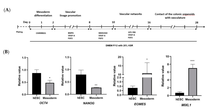
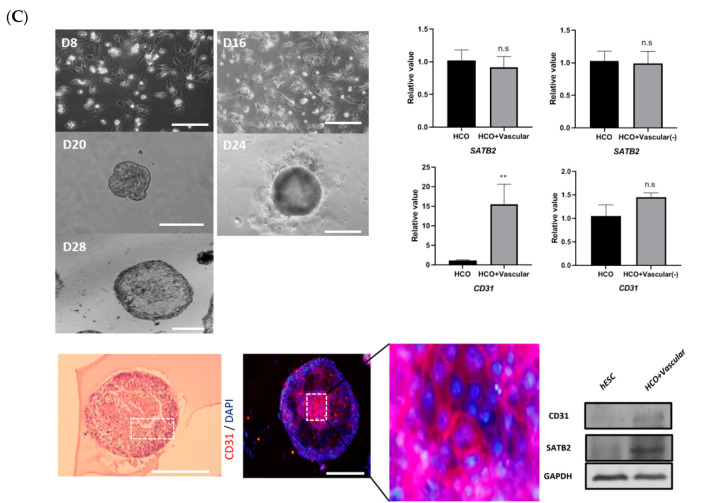
Figure 4.
Development of HCOs containing blood vessels. (A) Timeline for development of blood vessel-containing HCOs. (B) Expression of pluripotency markers (OCT4 and NANOG) and mesoderm differentiation markers (EOMES and MIXL1) as detected via qRT-PCR. (C) The morphology of blood vessel structures at days 8, 16, 20, 24, and 28. Immunofluorescent staining of blood vessel endothelial cell marker (CD31). Detection of SATB2 and CD31 by Western blotting, with GAPDH used as the loading control. H&E staining of HCO containing blood vessels, and immunofluorescent staining of CD31. Expression of colon-specific marker (SATB2) and blood vessel endothelial cell marker (CD31) as detected via qRT-PCR. n.s. indicates non significance. * p ≤ 0.1, ** p ≤ 0.01, and *** p ≤ 0.001. All the scale bars indicate 200 µm.


