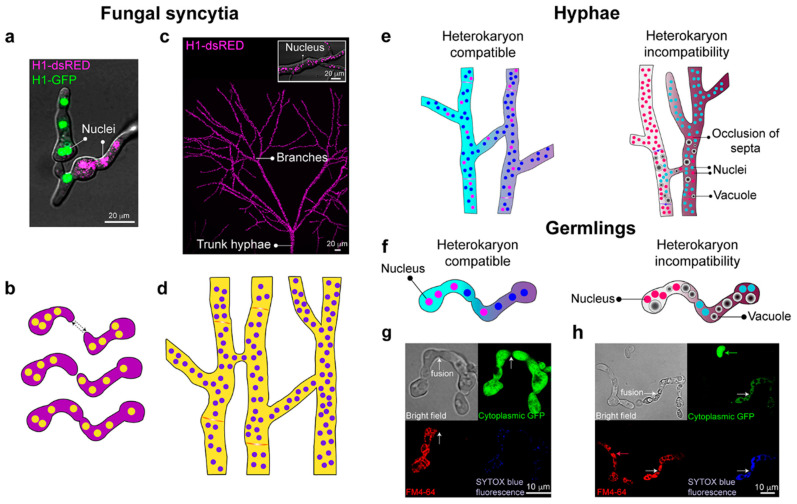
Figure 2.
Formation of interconnected fungal syncytia. (a) The fusion of compatible strains of Neurospora crassa, whose nuclei are labeled with either histone H1-GFP or H1-dsRED (green and magenta, respectively). (b) Genetically identical germs grow to each other (dashed arrows), fuse, and give rise to interconnected multinucleate syncytia. (c) Heterogeneity of the mycelial network and syncytia formation. Magenta dots are nuclei marked with histone H1-DsRed. The box shows a close-up of a hypha, showing the marked nuclei. (d) Hyphal fusion within a colony contributes to an interconnected syncytium. (e) In hyphae, heterokaryon formation can occur when there are no differences at het (heterokaryon) or vic (vegetative incompatibility) loci. In contrast, genetic differences at these loci result in heterokaryon incompatibility, which triggers compartmentalization of the fusion compartment due to occlusion of the septum, vacuolization of the hyphae, and eventual cell death. (f) In germlings, heterokaryon formation can occur when there are no differences at the rcd-1 (regulator of cell death-1) and plp-1/sec-9 (patatin-like phospholipase-1) loci. In contrast, differences at these loci result in heterokaryon incompatibility, rapidly triggering a cell death reaction that is a similar process in hyphae [38,39]. (g) Micrographs show the fusion of compatible germlings. One of the germlings is marked with cytoplasmic Green Fluorescent Protein (GFP) (green) and has undergone cell fusion with a compatible germling stained with FM4-64 fluorescent dye (red). Fusion is evident by the fact that GFP fluorescence can be observed in both germlings due to cytoplasmic mixing, and cell death does not occur, as indicated with the absence of SYTOX Blue fluorescence (death cell stain). (h) Cell fusion between germlings with genetic differences at the plp/sec-9 loci results in rapid cellular vacuolization and death, as demonstrated with the staining of SYTOX Blue fluorescence. White arrows indicate fusion events. Micrographs also show two germlings that have not undergone cell fusion and are healthy (green; GFP and red arrows; FM4-64). Micrographs (g,h) courtesy of Dr. Jens Heller (UCB Glass Laboratory).

