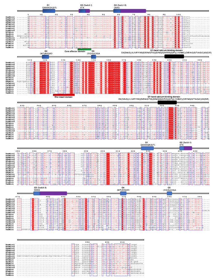
Figure 11.
Sequence alignment of the Miro subfamily. The amino acid sequences of Miro GTPases were aligned using ClustalX, the secondary structures predicted using ESPript. Regions of the Miro proteins are indicated above the alignment; blue are the G domains (G1, G2, G3, G4, and G5) domains; purple represent the switch domains (I and II) and black the EF hand calcium binding domains with their respected motifs. Blue frames are indicators of the conserved residues; white letters in red boxes represent identity, red letters in white boxes represent similarity.

