Abstract
The bacterial communities that are established during natural meat fermentation depend on the processing conditions and the type of meat substrate used. Six pork samples of variable quality (reflected in pH values) and six less conventional meats (beef, horse, hare, wild deer, wild duck, and wild boar) were naturally fermented under controlled conditions in model systems. The development of lactic acid bacteria (LAB), coagulase-negative staphylococci (CNS), and enterobacteria was followed using culture-dependent techniques and (GTG)5-PCR fingerprinting of genomic DNA from the isolates obtained. Taken together, Latilactobacillus sakei was the most abundant LAB species, although Latilactobacillus curvatus was more manifest in high-pH pork. Within staphylococci, common species were encountered (i.e., Staphylococcus equorum, Staphylococcus saprophyticus, and Staphylococcus xylosus), although some atypical ones (i.e., Staphylococcus succinus) were also recovered. Within enterobacteria, Serratia spp. prevailed in more acidic pork batches and in beef, whereas Hafnia spp. prevailed in game meat fermentations. Enterobacterial counts were particularly high in fermentations with low acidity, namely for some pork batches, hare, wild duck, and wild boar. These findings should be considered when naturally fermented meat products are manufactured, as the use of game meat or meat with high pH can give rise to safety concerns.
Keywords: meat fermentation, game meat, enterobacteria, lactic acid bacteria, staphylococcus, pH
1. Introduction
Fermented meats, such as fermented sausages, occupy an important place in European food markets [1,2]. Fermented sausages are made from raw meat mixed with fat, curing salt, and spices. They should be considered as the endpoint of a series of complex events, whereby the ingredients are stuffed into casings, fermented, and matured [3,4,5]. In Europe, the use of pork meat is common, followed by beef or horse meat [6,7,8]. In some cases, albeit more rarely, fermented sausages are prepared from game, such as wild boar and deer [9,10,11,12,13,14]. From a microbiological standpoint, this variety presents ample opportunity for investigating the diversity of microbial consortia that can be potentially established during different types of meat fermentation.
In general, microbial communities are variable and difficult to predict, which is especially the case in natural fermentations [15]. Nonetheless, the consortia always consist of lactic acid bacteria (LAB) which drive acidification and coagulase-negative staphylococci (CNS) which contribute to colour and flavour formation [16,17,18,19,20]. Within the group of LAB, Latilactobacillus sakei (formerly known as Lactobacillus sakei) and, to a lesser degree, Latilactobacillus curvatus (formerly known as Lactobacillus curvatus) and Leuconostoc spp. are most commonly encountered [16,17,18]. Whereas there are usually no specific quality or safety concerns associated with Latl. sakei prevalence, leuconostocs and Latl. curvatus can sometimes produce problematic levels of biogenic amines [19,20]. As far as the CNS communities are concerned, a rather marked species diversity can be encountered, although Staphylococcus xylosus, Staphylococcus saprophyticus, and Staphylococcus equorum are usually the most prevalent species in most natural fermentation processes. Many other CNS species have also been observed, such as Staphylococcus warneri, Staphylococcus epidermidis, Staphylococcus sciuri, and Staphylococcus succinus [17,21]. Some CNS species are possibly of concern due to their opportunistic pathogenic nature whereas truly pathogenic coagulase-positive staphylococci may also occur [22]. In a natural fermentation, however, there is no absolute control over which microorganisms are present during fermentation so that food safety risks need to be monitored. Furthermore, it is possible that enterobacteria may be found. Enterobacteria are generally undesired in food fermentations as some species can be pathogenic and/or linked with the presence of biogenic amines [21,23,24].
To mediate unfavourable microbiological consortia, the use of high-quality meat, inoculated with desirable microorganisms under controlled conditions is advisable in view of a safe meat fermentation [16]. A current trend for artisan-type foods and home fermentation is nonetheless discernible, with a renewed interest in natural fermentation [25]. This may lead to the use of less conventional meats, such as game meat. Although this offers an interesting option for product diversification, it could be argued that if the traditional and empirical knowledge that is needed to safely execute such practices is less prevalent, potential risk is created [26]. There has only been a limited amount of studies into the microbiota of game meat, such as wild boar and deer [8,9,12,13,14]. Typically, LAB and CNS communities behave as expected and initial spoilage bacteria tend to decline over time, although more information is needed. Even when using conventional pork mince, variability in the product characteristics may cause fluctuations in product safety and quality [8]. The use of low-quality, dark-firm-dry (DFD) pork meat may cause such fluctuations, which is likely due to its high pH [27]. High pH values are indeed a major modulator of bacterial dynamics during fermentation [15].
Given this complex and interlinked fermentation process, there is a need for more integrated and systematic studies of the relationship between meat quality, pH, and microbiota. Therefore, the aim of this study was to chart the bacterial communities that develop during the fermentation of beef, horse, and game meats (i.e., hare, wild deer, wild boar, and wild duck), compared to what occurs during the fermentation of pork mince of variable quality (as reflected in pH) in model systems. The focus was on the major bacterial groups (i.e., LAB, CNS, and enterobacteria), not on the presence of specific pathogens. The research was limited to the effects of the early fermentation stage, being the most critical, and does not account for further potential changes during drying.
2. Materials and Methods
2.1. Sample Acquisition
To mimic the fermentation stage of fermented sausage production (excluding the drying stage), a previously developed fermented meat model system was used [15]. To evaluate the impact of the initial pH, a total of six different pork meat batches of different quality were purchased from a local butcher (Brussels, Belgium), according to their colour and exudation level and reflected in their pH value. They were referred to as pork batters 1 to 6. Two batches of raw beef, horse, hare, wild boar, duck, and deer meat were purchased from local supermarkets (Brussels, Belgium). Each meat batter consisted of fresh mince (2 kg, 14% (m/m) fat fraction), sodium nitrate at 150 mg/kg (VWR International, Darmstadt, Germany), ascorbic acid at 500 mg/kg (Sigma-Aldrich, St. Louis, MO, USA), and sodium chloride at 3.0% (m/m; VWR International).
Each mixture was divided into a total of four plastic 60-mL containers per batch, containing about 60 g per container (closed lid) to fill the volume and permit fermentation in the absence of air. The containers were deposited into an incubator at 23°C. Samples were taken for analysis at the start and after 7 days of fermentation. Previous analysis has indicated that the bacterial diversity in the fermented meat models mentioned above stabilizes at around 3 days of fermentation, which is the usual industrial practice, and does not differ much from the situation after one week. Nonetheless, in the present study, a 7-day sampling point was chosen to maximize the effects of selection pressure and because natural, artisan-type fermentation processes often take several more days [15].
At each time point, two containers were analysed. For the bacterial enumerations, one sample per container was taken (duplicate data). Subsequently, for pH and water activity (aw) measurements, three measurements were performed (see below).
2.2. Enumeration and Isolation of Microorganisms
Colony enumeration and isolation were performed as described previously [23]. Some 10–15 g of meat sample was aseptically transferred into a stomacher bag (Seward, Worthing, West 99 Sussex, UK), with the addition of recovery diluents in ratio 1:10 (depending on the weight of the meat sample) (sterile solution of 0.85% (m/v) NaCl (VWR International) and 0.1% (m/v) bacteriological peptone (Oxoid, Basingstoke, Hampshire, UK)). This mixture was homogenized at maximum speed for 2 min in a Laboratory Blender Stomacher 400 (Seward) and appropriate decimal dilutions in saline (0.85% (m/v) NaCl) were prepared and spread on three selective agar media. Mannitol salt-phenol red-agar (MSA; VWR International), de Man-Rogosa-Sharpe (MRS) agar (Oxoid), and RAPID’Enterobacteriaceae agar (RAPID’Entero agar; Bio-Rad laboratories, Hercules, CA, USA) were used for the enumeration of presumptive catalase-positive cocci (in particular CNS), LAB, and enterobacteria, respectively. The MSA and MRS agar media were incubated at 30 °C for 72 h and the RAPID’Entero agar medium at 30 °C for 24 h. MSA, MRS agar, and RAPID’Entero agar media containing 30 to 300 colonies were retained to pick colonies (20–30%) to follow the bacterial community dynamics. These colonies were randomly selected and transferred into brain heart infusion (BHI; Oxoid). The cultures were incubated overnight at 30 °C and used for DNA extraction as well as for storage at −80 °C in cryovials containing 25% (v/v) of glycerol.
2.3. pH and aw Measurements
The pH of the meat batter was measured with a DY-P10 pH meter (Sartorius, Göttingen, Germany) equipped with an insertion pH probe (VWR International, Darmstadt, Germany). The aw was measured at 25 °C with a Hydropalm 23 aw meter (Rotronic, New York, NY, USA). Three independent measurements were performed per sample.
2.4. Classification and Identification of Bacterial Isolates through (GTG)5-PCR Fingerprinting of Genomic DNA
Genomic DNA extraction from cell pellets obtained by microcentrifugation at 13,000 rpm of 1.5 mL of an overnight culture of the isolates mentioned above was performed with a Nucleospin 96 tissue kit (Macherey Nagel, Düren, Germany), according to the manufacturer’s instructions. Prior to extraction, all cell pellets were washed with Tris-ethylene diaminetetraacetic acid (EDTA)-sucrose buffer (TES buffer; 50 mM Tris base (Calbiochem, Darmstadt, Germany), 1 mM EDTA (Sigma-Aldrich), and 6.7% (m/v) sucrose (VWR International); pH 8.0). Subsequently, (GTG)5-PCR fingerprints of the genomic DNA were obtained that were subjected to image analysis, as described previously [28]. Numerical cluster analysis of the fingerprints obtained was performed with the BioNumerics 5.1 software (Applied Maths, Sint-Martens-Latem, Belgium). The clustering of the fingerprints was calculated based on the Pearson product-moment correlation coefficient. Dendrograms were then generated with the unweighted pair group method with arithmetic average (UPGMA) clustering algorithm. To confirm the species identity assigned to each cluster, random isolates were selected and identified by sequencing the 16S rRNA gene and/or rpoA, rpoB, and tuf genes, as described previously [29,30]. For the molecular identification of Latl. sakei and Latl. curvatus, the reverse primers Ls (5′-ATG AAA CTA TTA AAT TGG TAC-3′) and Lc (5′-TTG GTA CTA TTT AAT TCT TAG-3′), coupled with the forward primer 16S (5′-GCT GGA TCA CCT CCT TTC-3′), were used [31]. The amplification conditions were as described by these authors.
2.5. Statistics
Intrasample diversity (alpha-diversity) was assessed by calculating the Simpson (diversity) and Pielou (evenness) indexes. To identify discrete bacterial communities between the samples and to quantify the effect of the different meat types on the bacterial communities within these fermentation processes, a permutational analysis of variance (PERMANOVA) based on the Bray–Curtis dissimilarity matrix was performed. This analysis was followed by a similarity percentage analysis (SIMPER) to assess the bacterial differences between meat types. Further, Pearson correlation coefficients were calculated to test the effect of pH on the bacterial counts and relative abundances of the species found using the Hmisc package (version 4.4-0) [32]. The packages vegan, (version 2.5-5) [33], and RVAideMemoire, (version 0.9-73) [34], were implemented for both intrasample and intersample variability assessment. All samples were also subjected to a principal component analysis (PCA) to explore the isolate identification data. PCA was performed using vegan and visualized by using the package ggplot2 (version 3.1.1) [35]. Subsequently, one-way analysis of variance (ANOVA) was conducted for the determination of differences in bacterial enumerations and of pH between all fermentation samples, followed by a series of post-hoc pairwise comparisons with Tukey’s test. A threshold value of 0.05 was considered to be significant for all statistical procedures applied. All statistical analyses and tests performed were executed through the RStudio software (version 3.5.2) [36].
3. Results
3.1. The Effect of Meat Quality as Reflected in Initial pH Values on the Bacterial Community Dynamics during Fermentation of Pork Mince
The various raw pork minces were analysed with respect to their pH and aw values. The initial pH of the sausage pork batter varied from 6.02 ± 0.01 to 5.62 ± 0.01 and decreased after 7 days of fermentation to values from varied from 5.90 ± 0.02 to 5.21 ± 0.01, respectively (Table 1). The variability of pH was reflected in significance difference between the samples, both at day 0 and 7 (Table 1). Moreover, the drop in pH of the pork meat samples with the highest initial acidity (pork batters 5 and 6) was significantly higher than for the rest of the pork meat samples (p < 0.05). The initial aw was on average 0.982 ± 0.003 but decreased as fermentation proceeded over time (Table S1).
Table 1.
Community dynamics of the microbiota of naturally fermented pork mince and the resulting pH course after 0 and 7 days of fermentation and standard deviation (SD). The bacterial counts on de Man-Rogosa-Sharpe (MRS) agar, mannitol salt-phenol red-agar (MSA), and RAPID’Enterobacteriaceae agar (RAPID′Entero agar) are expressed as log (cfu/g). Data sets with superscripts were significantly heterogeneous according to ANOVA; within each sampling time, means sharing the same superscript were not significantly different from each other using post-hoc tests.
| Time (days) | Sample | pH | SD | MRS Agar Counts (log(cfu/g)) | SD | MSA Counts (log(cfu/g)) | SD | RAPID’Entero Agar Counts (log(cfu/g)) | SD |
|---|---|---|---|---|---|---|---|---|---|
| 0 | 1 | 6.02 a | 0.01 | 5.76 | 0.60 | 3.70 | 0.30 | 3.93 | 0.26 |
| 2 | 5.99 a | 0.02 | 5.12 | 0.36 | 3.90 | 0.10 | 4.02 | 0.10 | |
| 3 | 5.85 a,b | 0.01 | 4.19 | 0.50 | 4.32 | 0.28 | 3.86 | 0.18 | |
| 4 | 5.80 a,b | 0.01 | 4.27 | 0.45 | 3.89 | 0.15 | 3.75 | 0.50 | |
| 5 | 5.72 b | 0.01 | 4.6 | 0.30 | 3.81 | 0.11 | 3.00 | 0.30 | |
| 6 | 5.62 b | 0.01 | 4.97 | 0.45 | 4.57 | 0.50 | 2.77 | 0.15 | |
| 7 | 1 | 5.90 a | 0.01 | 8.27 | 0.25 | 5.40 | 0.22 | 5.80 a | 0.41 |
| 2 | 5.78 a | 0.01 | 8.33 | 0.18 | 5.85 | 0.25 | 5.33 a | 0.22 | |
| 3 | 5.65 a | 0.01 | 8.47 | 0.22 | 6.37 | 0.46 | 3.79 b | 0.42 | |
| 4 | 5.56 a | 0.01 | 8.12 | 0.15 | 5.43 | 0.20 | 3.90 b | 0.21 | |
| 5 | 5.24 b | 0.01 | 7.91 | 0.20 | 5.68 | 0.32 | 3.10 b | 0.45 | |
| 6 | 5.21 b | 0.01 | 8.84 | 0.71 | 5.80 | 0.64 | 2.60 b | 0.33 |
Presumptive LAB populations, as measured on MRS agar media, ranged from 4.19 ± 0.50 log (cfu/g) to 5.76 ± 0.60 log (cfu/g) in the initial pork meat batters and increased to levels between 7.91 ± 0.20 log (cfu/g) and 8.84 ± 0.40 log (cfu/g) after 7 days of fermentation (Table 1). Staphylococcal counts, as measured on MSA media, were initially situated between 3.70 ± 0.30 log (cfu/g) and 4.57 ± 0.50 log (cfu/g). After 7 days of fermentation, they increased in numbers that varied from 5.40 ± 1.00 log (cfu/g) to 6.37 ± 0.80 log (cfu/g). Lastly, presumable enterobacterial populations, derived from the counts on RAPID’Entero agar media, equalled 3.75 ± 0.50 log (cfu/g) to 4.02 ± 0.10 log (cfu/g) initially (Table 1). At low pH values (samples of pork batters 5 and 6), the counts of presumable enterobacterial populations remained stable during fermentation. At high pH values, however, presumable enterobacterial populations increased over time. The counts on RAPID’Entero agar media of meat batters with the highest end-pH values (samples of pork batters 1 and 2) were significantly higher than those in samples of pork batters 3–6 by the end of the experiment (p < 0.05). This was not the case for the counts on MRS agar (p = 0.92) and MSA media (p = 0.84).
As mentioned above, biodiversity was assessed by using (GTG)5-PCR fingerprinting. The accession numbers of the representative isolates identified, as well as their percentages of sequence identity, are listed in Table 2.
Table 2.
Identities of the 16S rRNA, and/or rpoB, tuf, and rpoA genes sequenced from genomic DNA of representative bacterial isolates picked from MRS agar, MSA, and RAPID’Entero agar, and the accession numbers of the entries with the highest identity.
| Bacterial Species | Gene | Identity (%) | Accession Number |
|---|---|---|---|
| Carnobacterium spp. | 16S rRNA | 99 | NR_113798.1, NR_042093.1 |
| Latilactobacillus curvatus | 16S rRNA | 100 | NR_114915.1 |
| Latilactobacillus sakei | 16S rRNA | 100 | NR_113821.1, NR_115172.1 |
| Lactiplantibacillus plantarum | 16S rRNA | 100 | NR_104573.1 |
| Lactococcus lactis | 16S rRNA | 100 | NR_113925.1 |
| Leuconostoc carnosum | 16S rRNA | 100 | NR_040811.1 |
| Leuconostoc mesenteroides | 16S rRNA | 99 | NR_157602.1 |
| Weissella fabalis | 16S rRNA | 98 | NR_108858.1 |
| Brochothrix thermosphacta | 16S rRNA | 100 | NR_113587.1 |
| Macrococcus caseolyticus | 16S rRNA, tuf | 98 | NR_119262.1, AP009484.1 |
| Corynebacterium variabilis | 16S rRNA | 100 | NR_025314.1 |
| Enterococcus faecium | rpoB | 100 | CP021885.1, NR_115764.1 |
| Enterococcus hirae | rpoB, tuf | 100 | CP003504.1, CP023011.2 |
| Staphylococcus xylosus | rpoB, tuf | 99 | CP008724.1, CP031275.1 |
| Staphylococcus equorum | rpoB | 100 | CP013980.1 |
| Staphylococcus saprophyticus | rpoB | 100 | CP022093.2, CP014113.2 |
| Staphylococcus epidermidis | rpoB | 99 | CP009046.1 |
| Staphylococcus vitulinus | rpoB, tuf | 100 | HM352960.1, KY011914.1 |
| Staphylococcus capitis | rpoB | 99 | CP007601.1 |
| Staphylococcus haemolyticus | rpoB | 99 | CP013911.1 |
| Staphylococcus succinus | rpoB | 100 | CP018199.1 |
| Staphylococcus pasteuri | tuf | 99 | CP017463.1 |
| Staphylococcus hyicus | rpoB | 99 | CP008747.1 |
| Staphylococcus aureus | rpoB | 99 | AP017922.1 |
| Bacillus safensis | rpoB | 100 | CP018197.1 |
| Serratia proteamaculans | rpoA | 100 | CP000826.1 |
| Serratia liquefaciens | rpoA | 100 | CP033893.1, CP014017.2 |
| Hafnia alvei | rpoA | 100 | CP015379.1 |
| Hafnia paralvei | rpoA | 99 | CP014031.2 |
| Rahnella aquatilis | rpoA | 99 | CP003244.1 |
| Lelliotia amnigena | rpoA | 99 | CP015774.2 |
| Citrobacter sp. | rpoA | 99 | CP022049.2 |
| Proteus bulgari | rpoA | 100 | CP033736.1 |
| Enterobacter sp. | rpoA | 100 | CP041062.1 |
| Klebsiella sp. | rpoA | 99 | CP011077.1 |
| Pantoea agglomerans | rpoA | 99 | CP016889.1 |
| Pseudomonas sp. | 16S rRNA | 99 | NR_148763.1 |
| Kurthia sp. | 16S rRNA | 100 | NR_118296.1 |
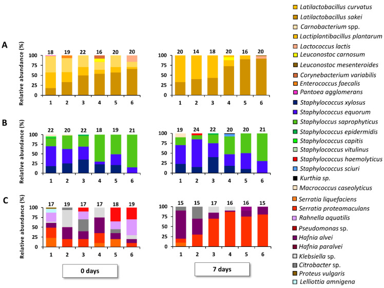
Regarding LAB, the following species were isolated during the pork meat fermentations: Latl. sakei, Latl. curvatus, Lactococcus lactis, Carnobacterium spp., Leuconostoc carnosum, and Enterococcus sp. Initially, LAB communities consisted mainly of Latl. sakei, Latl. curvatus, and carnobacteria. However, carnobacteria vanished by the end of the fermentation processes (Figure 1, Table S2). Additionally, a consistent prevalence shift from Latl. sakei to Latl. curvatus was seen over the pH spectrum of the various samples, both at the start and after 7 days of fermentation. A strong correlation between Latl. sakei and the acidification level was found (r = −0.94, p < 0.05).
Figure 1.
Course of bacterial species isolated from MRS agar (A), MSA (B), and RAPID′Entero agar (C) of pork batters 1, 2, 3, 4, 5, and 6, fermented at 23 °C for 7 days. Τhe number of isolates for identification is mentioned above each bar. The bacterial species diversity is displayed as relative abundances, calculated based on the number of isolates (N) obtained per time point (0 and 7 days of meat fermentation).
Within the species diversity recovered from MSA media, a coprevalence of S. xylosus, S. equorum, and S. saprophyticus was found (Figure 1; Table S3). In addition, S. epidermidis, S. vitulinus, Staphylococcus capitis, S. pasteuri, Staphylococcus haemolyticus, Kurthia sp., and Macrococcus caseolyticus were present as minor species. The most striking trend was that S. saprophyticus became increasingly manifest as pH values decreased across the samples.
With respect to the RAPID’Entero agar isolates, a high species diversity was seen at the start of the fermentation processes (Figure 1; Table S4). The initial communities were composed of Serratia proteamaculans, Serratia liquefaciens, Hafnia alvei, Hafnia paralvei, Rahnella aquatilis, Klebsiella sp., Citrobacter sp., and Proteus vulgaris. After 7 days of fermentation, however, Serratia spp. took over at the low pH values (Figure 1; Table S4). A Pearson correlation analysis revealed that Serratia spp. and pH were significantly correlated (r = −0.72, p < 0.05).
3.2. Alpha- and Beta-Diversity of the Pork Mince Fermentation Processes
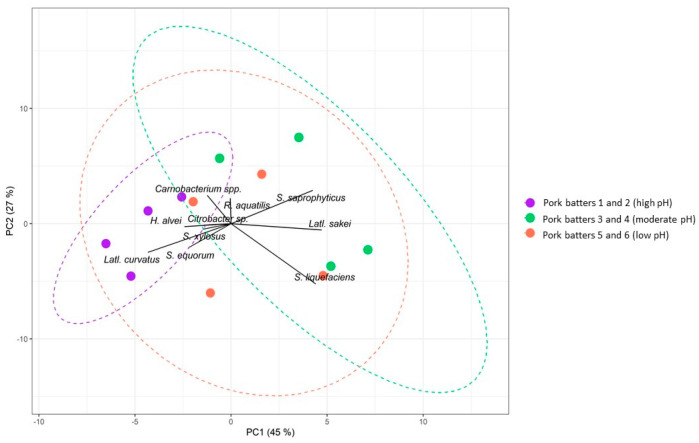
In general, the bacterial diversity of all pork-based meat fermentation processes was high (Table S5). When differentiating samples according to high (samples of pork batters 1 and 2), moderate (samples of pork batters 3 and 4), and low pH (samples of pork batters 5 and 6), a significant impact was seen on the bacterial species diversity (p < 0.05), based on a PERMANOVA. These differences in community compositions of the different samples were supported by PCA (Figure 2). This also underpinned the distinction between high-pH and low-pH samples based on their bacterial community structures. Bacterial profiles in samples from the meat batters with high initial pH were significantly different (p < 0.05) from those at moderate and low initial pH. SIMPER analysis confirmed that the bacterial differentiation between the fermentation processes of high-, moderate-, and low-pH pork meat could be attributed to differences in the following bacterial species present: Latl. sakei, Latl. curvatus, carnobacteria, S. saprophyticus, S. equorum, S. liquefaciens, and H. alvei.
Figure 2.
Centred principal component analysis (PCA) biplot based on Bray–Curtis dissimilarity scores of the bacterial community structures, with factor loadings, of pork fermentation processes. Only factor loadings with an absolute value above 1 were considered. Colours indicate different sample groups characterized by high pH (purple), moderate pH (orange), or low pH (green). The proportion of variance for every PC is indicated between brackets. The dotted lines represent 90% normal confidence ellipses for the sample groups.
3.3. The Effect of Less Conventional Meat Types on the Bacterial Community Dynamics during Fermentation
The initial aw of the meat samples did not vary much between the different meat types and was on average 0.981 ± 0.003 initially and 0.966 ± 0.001 after 7 days of fermentation (Table S6). In contrast, a correlation between pH and the meat type was found (p < 0.05), and variations in pH were found according to the meat type, albeit not always significant (Table 3). Fermentations of hare, wild boar, and wild duck were characterized by higher pH values than was the case for beef, horse, and wild deer, although not significant (p = 0.15).
Table 3.
Community dynamics of the microbiota of various naturally fermented, less conventional meat types and the resulting pH course after 0 and 7 days of fermentation, displayed as average of two biological replicates carried out for each meat type, and standard deviation (SD). The bacterial counts on MRS agar, MSA, and RAPID′Entero agar are expressed as log (cfu/g).
| Time (days) | Sample | pH | SD | MRS Agar Counts (log(cfu/g)) | SD | MSA Counts (log(cfu/g)) | SD | RAPID’Entero Agar Counts | SD |
|---|---|---|---|---|---|---|---|---|---|
| 0 | Hare | 5.94 | 0.15 | 5.28 | 0.59 | 3.26 | 0.33 | 5.55 | 0.80 |
| Wild duck | 5.84 | 0.12 | 7.56 | 0.75 | 3.78 | 0.37 | 7.67 | 1.43 | |
| Wild boar | 5.82 | 0.02 | 6.64 | 0.07 | 3.84 | 0.50 | 6.72 | 1.74 | |
| Beef | 5.67 | 0.06 | 4.28 | 0.08 | 3.97 | 0.04 | 4.08 | 0.01 | |
| Horse | 5.54 | 0.03 | 4.65 | 0.98 | 4.40 | 0.39 | 4.33 | 0.29 | |
| Wild deer | 5.51 | 0.03 | 5.59 | 0.35 | 3.86 | 0.37 | 4.03 | 0.13 | |
| 7 | Hare | 5.52 | 0.08 | 8.79 | 0.06 | 1.76 | 2.53 | 5.41 | 0.55 |
| Wild duck | 5.96 | 0.26 | 9.07 | 0.24 | 4.78 | 1.57 | 7.12 | 2.84 | |
| Wild boar | 5.95 | 0.75 | 8.68 | 0.12 | 5.66 | 0.04 | 6.36 | 3.21 | |
| Beef | 5.27 | 0.17 | 7.72 | 0.55 | 6.03 | 0.91 | 2.68 | 0.46 | |
| Horse | 5.05 | 0.04 | 8.20 | 0.10 | 4.89 | 0.06 | 3.01 | 0.02 | |
| Wild deer | 5.10 | 0.01 | 7.88 | 0.54 | 4.90 | 0.08 | 2.73 | 0.42 |
Initial counts on MRS agar for hare, wild boar, and wild duck fermentation processes were higher (6.49 ± 0.46 log (cfu/g), on average) than for beef, horse, and wild deer (4.84 ± 0.55 log (cfu/g), on average), although not significant (p = 0.86) (Table 3). These differences were not encountered at the end of the fermentation processes, as the final counts on MRS agar for the duplicate fermentations for each meat type converged, averaging 8.39 ± 0.49 log (cfu/g) overall. The counts on MSA at the onset of the fermentation processes ranged from 3.26 ± 0.30 to 4.40 ± 0.55 log (cfu/g), and from 4.78 ± 0.08 to 6.03 ± 0.20 log (cfu/g) after 7 days of fermentation (Table 3). Notably, counts on MSA for hare fermentation samples strongly decreased during the fermentation process. In contrast, counts on RAPID’Entero agar differed between fermentation samples. For hare, wild boar, and wild duck fermentation processes, counts averaged 6.65 ± 1.06 log (cfu/g) at the beginning and 6.30 ± 0.86 log (cfu/g) after 7 days of fermentation. For the beef, horse, and wild deer fermentation processes, RAPID’Entero agar counts were generally lower, averaging 4.15 ± 0.16 log (cfu/g) at the onset of fermentation and 2.81 ± 0.18 log (cfu/g) after 7 days of fermentation (Table 3).
The LAB community profiles of all fermentation samples were similar (Table S7). They were characterized by a high prevalence of Latl. sakei, whereas carnobacteria and Latl. curvatus were retrieved at the beginning of the fermentation processes but vanished over time. Other bacterial species that were only occasionally isolated were Leuconostoc spp., Carnobacterium variabilis, Enterococcus faecium, Klebsiella sp., and H. alvei.
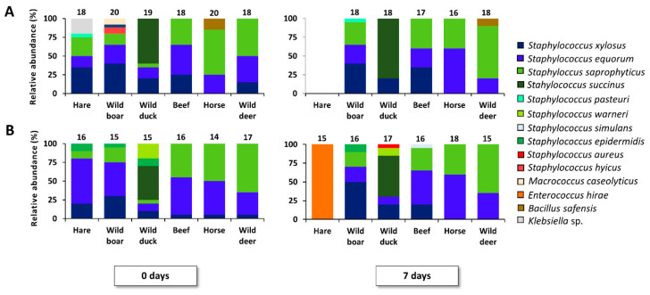
Within the MSA media isolates, more species diversity was obtained (Figure 3, Table S8). A coprevalence of S. saprophyticus, S. xylosus, and S. equorum was found in all cases, except for the hare and duck fermentation processes. An increasing relative abundance of S. saprophyticus was found in the fermentation samples that were more acidic (i.e., the beef, horse, and wild deer fermentation processes), as its presence also correlated with the pH of the meat batter (r = −0.47, p < 0.05). One replicate of the hare fermentation processes did not allow for recovery of MSA media isolates, whereas the other replicate resulted in the isolation of Enterococcus hirae at day 7. In the fermented duck samples, the presence of S. succinus stood out. Lastly, the pathogens Staphylococcus aureus and Staphylococcus hyicus were found in one replicate of the wild boar and wild duck fermentations, in very low abundance (Table S8).
Figure 3.
Course of bacterial species isolated from MSA during fermentation of less conventional meat types, including two replicates (A,B) at 23 °C for 7 days. Τhe number of isolates for identification is mentioned above each bar. The bacterial species diversity is displayed as relative abundances, calculated based on the number of isolates (N) obtained per time point (0 and 7 days of meat fermentation).
The enterobacterial profiles were variable. The main species isolated belonged to the Hafnia and Serratia genera, followed by R. aquatilis, Klebsiella sp., and Citrobacter sp. The species R. aquatilis was mainly present at the onset of the fermentation processes (Table S9). In general, a high prevalence of Hafnia spp. was found after 7 days of fermentation for all meat types, except for the beef fermentation processes, in which Serratia spp. prevailed.
3.4. Alpha- and Beta-Diversity of the Less Conventional Meat Fermentation Processes
The bacterial diversity of all meat samples investigated was relatively high, except for the hare fermentation processes that were characterized by a more narrow species diversity (Table S10). As mentioned above, this was related to the absence of isolates in one replicate and the high prevalence of E. hirae in the other.
The meat fermentation samples were statistically different (p < 0.05), indicating differences in their bacterial community structures. Pairwise comparison revealed that the bacterial profiles in the beef fermentation samples were different from those in the wild boar, wild duck, horse, and wild deer ones (p < 0.05). In turn, horse fermentation samples differed from those of wild boar and wild duck fermentation processes (p < 0.05) and wild boar fermentation samples were different from those of wild duck and deer ones (p < 0.05). Wild deer fermentation samples were different from those of hare and wild duck fermentation processes (p < 0.05) and hare fermentation samples from those of wild duck ones (p < 0.05).
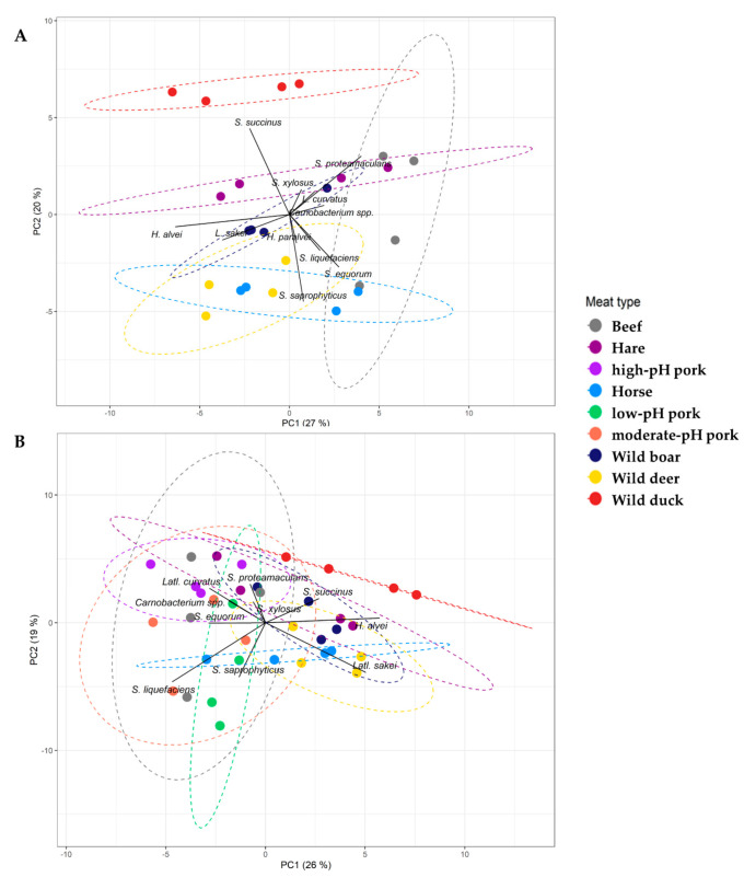
To explore differences in beta-diversity, a PCA was performed (Figure 4A). Fermentation samples from different meat types clustered closely, because of similarities in their bacterial community structures. However, certain differences in the course of the bacterial communities were obtained. SIMPER analysis showed that these differences could be attributed to the characteristic prevalence of S. succinus in duck fermentation processes, abundance of S. saprophyticus in those samples with low pH values (i.e., beef, horse, and wild deer), and prevalence of Serratia spp. in beef fermentation samples (in contrast to Hafnia spp. in the fermentation samples of the other meat types).
Figure 4.
Centred principal component analysis (PCA) biplot based on Bray–Curtis dissimilarity scores of the bacterial community structures obtained during fermentation of less conventional meat types (A) and extended with the fermented pork samples to the entire dataset (B), with factor loadings. Only factor loadings with an absolute value above 1 were considered. Colours indicate different sample groups, beef (gray), hare (dark purple), high-pH pork (light purple), horse (light blue), low-pH pork (green), moderate-pH pork (orange), wild boar (dark blue), wild deer (yellow), and wild duck (red). The proportion of variance for every PC is indicated between brackets. The dotted lines represent 90% normal confidence ellipses for the sample groups.
3.5. Beta-Diversity of the Entire Dataset of Meat Types
When analysing all the above-mentioned meat type fermentation samples within one dataset, thus including both the pork and less-conventional meats, it was uncovered that the meat type had a significant impact on the RAPID’Entero agar counts (p < 0.05) but not on the MRS agar (p = 0.08) and MSA counts (p = 0.07). The pH values and RAPID’Entero agar counts were correlated significantly (r = 0.72, p < 0.05) but this was, once more, not the case for the counts on MRS agar (p = 0.21) and MSA (p = 0.12). The meat type also had a significant impact on the bacterial species diversity (p < 0.05). A negative correlation was found between pH and Latl. sakei (r = −0.5, p <0.05), whereas a positive correlation was found between pH and Latl. curvatus (r = 0.37, p < 0.05). The pH and S. saprophyticus were negatively correlated (r = −0.41, p < 0.05). Pairwise comparisons revealed that the bacterial profiles in the wild duck fermentation samples were different from all the others and additionally, that the fermentation samples of low-pH pork differed from those of high-pH pork and hare ones (p < 0.05) (Table 4). Finally, to visualize these differences in beta-diversity of the isolate identification data, a PCA was performed (Figure 4B), in which samples with correlative bacterial community structures clustered closely.
Table 4.
P values based on pairwise comparisons between all meat fermentation samples using PERMANOVA on a distance matrix.
| Meat Type | 1 | 2 | 3 | 4 | 5 | 6 | 7 | 8 | 9 |
|---|---|---|---|---|---|---|---|---|---|
| 1. Beef | |||||||||
| 2. Hare | 0.079 | ||||||||
| 3. High-pH pork | 0.047 | 0.079 | |||||||
| 4. Horse | 0.047 | 0.105 | 0.047 | ||||||
| 5. Low-pH pork | 0.183 | 0.047 | 0.047 | 0.238 | |||||
| 6. Moderate-pH pork | 0.130 | 0.079 | 0.105 | 0.047 | 0.310 | ||||
| 7. Wild boar | 0.047 | 0.183 | 0.047 | 0.047 | 0.047 | 0.047 | |||
| 8. Wild deer | 0.047 | 0.047 | 0.047 | 0.183 | 0.047 | 0.047 | 0.047 | ||
| 9. Wild duck | 0.047 | 0.047 | 0.047 | 0.047 | 0.047 | 0.047 | 0.047 | 0.047 |
4. Discussion
Although meat fermentation normally narrows the initial microbiota down to a desirable consortium, this can be unsuccessful at times due to contaminated raw material or improper processing conditions, such as poor acidification [11,37]. In some cases, traditional fermented meat products are still obtained by natural fermentation. This may pose certain risks, especially when deviating from conventional practice and traditional knowledge [38]. As outlined further below, the present study revealed that natural meat fermentation processes may indeed lead to the development of potentially worrisome bacteria, especially when the raw meat has a high initial pH. Acidification seems to be crucial for a safe fermentation. Enterobacteria are relatively acid-sensitive and are quickly outcompeted during food fermentations that have an acidifying profile [39]. In addition, in the case of game meat, the microbiological safety can be compromised because of a higher chance of the presence of undesirable bacteria in the raw meat [40,41]. This may be due to contamination during hunting or to the fact that game meat is often characterized by high pH values [12,14].
In all meat fermentation processes studied, LAB were the prevalent species and the major drivers of fermentation. High abundance of Latl. sakei usually serves as a reassuring finding, as prevalence of this benign species is the usual state of affairs in well-performed meat fermentation processes [17,18,42,43,44]. In the present study, Latl. sakei was the most frequently isolated LAB species when using pork, as long as the pH was low enough. In the less acidified pork fermentation samples, however, Latl. curvatus became more persistent, as this species is known to prefer a higher pH than Latl. sakei [18,27]. This can be problematic since Latl. curvatus can be a major producer of biogenic amines [45]. High concentrations of biogenic amines pose a health risk, since they are neurotoxic, and they have been linked with food poisoning [46]. Higher abundance of Latl. curvatus in low-acidified pork batters indicates that, even in habitual natural pork fermentation processes, potentially undesirable microorganisms can prevail if the raw material or processing conditions deviate in certain key characteristics, such as acidification levels.
This trend was further evidenced in the CNS communities. A prevalence of S. xylosus, S. saprophyticus, and S. equorum was found in most meat fermentation processes, as is common in natural meat fermentation processes [42,47,48,49]. Yet, low acidity allowed the proliferation of several other CNS species as subdominant ones, among which S. epidermidis. The latter species is an opportunistic pathogen and a causal agent in (skin) infections [15]. It was present in low abundance in the meat batters with pH values above 5.8, as it is less competitive in acidic environments [50]. In one poorly acidified pork fermentation sample, the opportunistic pathogen Staphylococcus haemolyticus was encountered. This species is indeed able to appear during meat fermentation processes [51], although it thrives poorly under proper acidification conditions [52].
The present study showed that the use of nonconventional meat types needs to be carefully thought through, as several potentially pathogenic bacteria emerged during meat fermentation, likely due to the concomitant high-pH levels (even if this was not the focus of the study). In wild boar and duck fermentation processes, characterized by a relatively high initial pH, the pathogens S. hyicus and S. aureus were isolated, albeit at low levels, which were in accordance with previous findings [53,54,55], and do not inevitably pose a threat for enterotoxin production [56]. This nonetheless leads to potential risk and deserves attention [57]. In hare fermentation processes, no CNS species were retrieved, but E. hirae was isolated from MSA. This species has not only been associated with rabbit, wild rabbit, and hare faeces before, but its presence is also linked with urinary infections in humans [58,59,60]. In wild duck fermentation processes, S. succinus was the main species isolated. To our knowledge, this is the first time S. succinus has been reported in duck meat fermentation processes, although it has been found in other fermented meat products before [42,61]. This is not necessarily a matter of concern, but some strains of S. succinus exhibit proteolytic, lipolytic, and urease activities and can also be hemolytic and toxigenic [62,63]. Further, one should be mindful of the shortcomings and limitations of culture-based methods as they cannot identify nonculturable cells [64].
Enterobacterial counts generally showed an increase during meat fermentation processes characterized by high pH values, as such conditions favour their growth [56,65]. At the end of the fermentation processes with beef or low-pH pork, Serratia spp. prevailed within the enterobacterial group. This species has indeed a high degree of adaptation to more acidic meat environments [66]. Hafnia spp. prevailed in the nonconventional meat fermentation processes. Both Hafnia spp. and Serratia spp. are known putrescine and cadaverine producers and their presence is associated with the quality defect of green discolouration in meat too [67,68].
5. Conclusions
The present study evaluated the bacterial diversity of a variety of conventional and game meat fermentation processes, which contributes to a deeper understanding of how the meat type and acidification level influence the bacterial composition. A high starting pH in pork and other meat types used for natural fermentation processes allowed the proliferation of problematic bacteria. Hence, the use of good quality meat is of importance and particularly so for game meat fermentation processes, as such fermentation processes have not been all that well characterized yet with respect to their microbiology. Further care should be taken with natural fermentation of high-pH pork meat or with hare, wild boar, and wild duck meat. Proper acidification of the meat batter is crucial and allows the raw meat to be transformed into a safe end product. The findings obtained in the current study can be further explored by investigating the microbial diversity during actual dry-fermented sausage production on pilot scale. In time, knowledge gathered from such studies would strengthen our understanding of the impact of initial pH and different meat types on microbial ecology.
Acknowledgments
The authors would like to thank Jack James Robson for his help in revising the manuscript.
Supplementary Materials
The following are available online at https://www.mdpi.com/2304-8158/9/10/1386/s1, Table S1. Water activity (aw) values of the pork mince fermentation samples of pork batters 1, 2, 3, 4, 5, and 6, incubated at 23 °C for 0 and 7 days, Table S2. Relative abundance of identified bacterial isolates picked from MRS agar of the pork mince fermentation processes (samples of pork batters 1, 2, 3, 4, 5, and 6) at days 0 and 7, encompassing Latilactobacillus sakei, Latilactobacillus curvatus, Lactiplantibacillus plantarum (formely known as Lactobacillus plantarum), Lactococcus lactis, Carnobacterium spp., Leuconostoc carnosum, Leuconostoc mesenteroides, Corynebacterium variabilis, Enterococcus faecium, and Pantoea agglomerans, Table S3. Relative abundance of identified bacterial isolates picked from MSA of the pork mince fermentation processes (samples of pork batters 1, 2, 3, 4, 5, and 6) at days 0 and 7, encompassing Staphylococcus xylosus, Staphylococcus equorum, Staphylococcus saprophyticus, Staphylococcus epidermidis, Staphylococcus vitulinus, Staphylococcus sciuri, Staphylococcus capitis, Staphylococcus haemolyticus, Macrococcus caseolyticus, and Kurthia sp., Table S4: Relative abundance of identified bacterial isolates picked from RAPID’Entero agar of the pork mince fermentation processes (samples of pork batters 1, 2, 3, 4, 5, and 6) at days 0 and 7, encompassing Serratia proteamaculans, Serratia liquefaciens, Hafnia alvei, Hafnia paralvei, Rahnella aquatilis, Klebsiella spp., Citrobacter sp., Lelliottia amnigena, Proteus vulgaris, and Pseudomonas sp., Table S5. Alpha-diversity metrics based on the relative abundances of bacterial species found during pork mince fermentation processes (samples of pork batters 1, 2, 3, 4, 5, and 6), through (GTG)5-PCR fingerprinting of genomic DNA. The Simpson (D) and Pielou (Je) indexes were calculated for all samples to measure their diversity and evenness, respectively, Table S6. Water activity (aw) values obtained for the fermentation processes of less conventional meat types, incubated at 23°C for 0 and 7 days, Table S7. Relative abundance of identified bacterial isolates picked from MRS agar of less conventional meat fermentation processes (replicates 1 and 2) at days 0 and 7, encompassing Latilactobacillus sakei, Latilactobacillus curvatus, Lactiplantibacillus plantarum, Lactococcus lactis, Carnobacterium spp., Leuconostoc carnosum, Corynebacterium variabilis, Weissella sp., Enterococcus faecium, Hafnia alvei, and Klebsiella sp., Table S8. Relative abundance of identified bacterial isolates picked from MSA of less conventional meat fermentation processes (replicates 1 and 2) at days 0 and 7, encompassing Staphylococcus xylosus, Staphylococcus equorum, Staphylococcus saprophyticus, Staphylococcus succinus, Staphylococcus pasteuri, Staphylococcus epidermidis, Staphylococcus warneri, Staphylococcus simulans, Staphylococcus hyicus, Staphylococcus aureus, Bacillus safensis, Macrococcus caseolyticus, Enterococcus hirae, and Klebsiella sp., Table S9. Relative abundance of identified bacterial isolates picked from RAPID’Entero agar of less conventional meat fermentation processes (replicates 1 and 2) at days 0 and 7, encompassing Serratia proteamaculans, Serratia liquefaciens, Hafnia alvei, Hafnia paralvei, Rahnella aquatilis, Klebsiella sp., Lelliottia amnigena, Citrobacter sp., Proteus vulgaris, Enterobacter sp., and Pseudomonas sp., Table S10. Alpha-diversity metrics based on the relative abundances of bacterial species found during less conventional meat fermentation processes (replicates 1 and 2) at days 0 and 7, through (GTG)5-PCR fingerprinting of genomic DNA. The Simpson (D) and Pielou (Je) indexes were calculated for all samples to measure their diversity and evenness, respectively.
Author Contributions
Conceptualization, C.C. and F.L.; Methodology, C.C. and F.L.; Software, C.C., E.V.R.; Validation, C.C., Formal analysis C.C.; Investigation C.C., E.V.R., N.S., and D.V.d.V.; Interpretation, C.C., F.L., and L.D.V.; Resources, F.L. and L.D.V.; Data curation, C.C. and E.V.; Writing—original draft preparation, C.C.; Writing—review and editing, C.C., E.V.R., F.L., and L.D.V.; Visualization C.C.; Supervision F.L.; Project Administration, F.L. and L.D.V.; Funding Acquisition, F.L. and L.D.V. All authors have read and agreed to the published version of the manuscript research articles with several authors, a short paragraph specifying their individual contributions must be provided.
Funding
This research was supported by the Research Council of the Vrije Universiteit Brussel (SRP7 and IOF342 projects), and in particular the Interdisciplinary Research Program IRP11 “Tradition and naturalness of animal products within a societal context of change”, the Hercules Foundation (project UABR 09/004), and the Research Foundation-Flanders (FWOAL881 project).
Conflicts of Interest
The authors declare no conflict of interest.
References
- 1.Leroy F., Aymerich T., Champomier-Vergès M.C., Cocolin L., De Vuyst L., Flores M., Leroi F., Leroy S., Talon R., Vogel R.F., et al. Fermented meats (and the symptomatic case of the Flemish food pyramid): Are we heading towards the vilification of a valuable food group? Int. J. Food Microbiol. 2018;274:67–70. doi: 10.1016/j.ijfoodmicro.2018.02.006. [DOI] [PubMed] [Google Scholar]
- 2.Belleggia L., Milanović V., Ferrocino I., Cocolin L., Haouet M.N., Scuota S., Maoloni A., Garofalo C., Cardinali F., Aquilanti L., et al. Is there any still undisclosed biodiversity in Ciauscolo salami? A new glance into the microbiota of an artisan production as revealed by high-throughput sequencing. Meat Sci. 2020;165:108128. doi: 10.1016/j.meatsci.2020.108128. [DOI] [PubMed] [Google Scholar]
- 3.Morot-Bizot S.C., Leroy S., Talon R. Staphylococcal community of a small unit manufacturing traditional dry fermented sausages. Int. J. Food Microbiol. 2006;108:210–217. doi: 10.1016/j.ijfoodmicro.2005.12.006. [DOI] [PubMed] [Google Scholar]
- 4.Cocolin L., Dolci P., Rantsiou K., Urso R., Cantoni C., Comi G. Lactic acid bacteria ecology of three traditional fermented sausages produced in the north of Italy as determined by molecular methods. Meat Sci. 2009;82:125–132. doi: 10.1016/j.meatsci.2009.01.004. [DOI] [PubMed] [Google Scholar]
- 5.Fontana C., Gazzola S., Cocconcelli P.S., Vignolo G. Population structure and safety aspects of Enterococcus strains isolated from artisanal dry fermented sausages produced in Argentina. Lett. Appl. Microbiol. 2009;49:411–414. doi: 10.1111/j.1472-765X.2009.02675.x. [DOI] [PubMed] [Google Scholar]
- 6.Coloretti F., Chiavari C., Poeta A., Succi M., Tremonte P., Grazia L. Hidden sugars in the mixture: Effects on microbiota and the sensory characteristics of horse meat sausage. Food Sci. Technol. 2019;106:22–28. doi: 10.1016/j.lwt.2019.02.032. [DOI] [Google Scholar]
- 7.Geeraerts W., De Vuyst L., Leroy F. Mapping the dominant microbial species diversity at expiration date of raw meat and processed meats from equine origin, an underexplored meat ecosystem, in the Belgian retail. Int. J. Food Microbiol. 2019;289:189–199. doi: 10.1016/j.ijfoodmicro.2018.09.019. [DOI] [PubMed] [Google Scholar]
- 8.Settanni L., Barbaccia P., Bonanno A., Ponte M., Di Gerlando R., Franciosi E., Di Grigoli A., Gaglio R. Evolution of indigenous starter microorganisms and physicochemical parameters in spontaneously fermented beef, horse, wild boar and pork salamis produced under controlled conditions. Food Microbiol. 2020;87:103385. doi: 10.1016/j.fm.2019.103385. [DOI] [PubMed] [Google Scholar]
- 9.Soriano A., Cruz B., Gómez L., Mariscal C., Ruiz A.G. Proteolysis, physicochemical characteristics and free fatty acid composition of dry sausages made with deer (Cervus elaphus) or wild boar (Sus scrofa) meat: A preliminary study. Food Chem. 2006;96:173–184. doi: 10.1016/j.foodchem.2005.02.019. [DOI] [Google Scholar]
- 10.Lachowicz K., Żochowska-Kujawska J., Gajowiecki L., Sobczak M., Kotowicz M., Żych A. Effects of wild boars meat of different season of shot addition on texture of finely ground model pork and beef sausages. Electron. J. Pol. Agric. Univ. 2008;11:1–8. [Google Scholar]
- 11.Chakanya C., Arnaud E., Muchenje V., Hoffman L.C. Fermented meat sausages from game and venison: What are the opportunities and limitations? J. Sci. Food Agric. 2018;2018:1–9. doi: 10.1002/jsfa.9053. [DOI] [PubMed] [Google Scholar]
- 12.Maksimovic A.Z., Zunabovic-Pichler M., Kos I., Mayrhofer S., Hulak N., Domig K.J., Mrkonjic Fuka Μ. Microbiological hazards and potential of spontaneously fermented game meat sausages: A focus on lactic acid bacteria diversity. Food Sci. Technol. 2018;89:418–426. doi: 10.1016/j.lwt.2017.11.017. [DOI] [Google Scholar]
- 13.Ranucci D., Roila R., Miraglia D., Arcangeli C., Vercillo F., Bellucci S., Branciari R. Microbial, chemical-physical, rheological and organoleptic characterisation of roe deer (Capreolus capreolus) salami. Ital. J. Food Saf. 2019;8:8195. doi: 10.4081/ijfs.2019.8195. [DOI] [PMC free article] [PubMed] [Google Scholar]
- 14.Fuka M.M., Tanuwidjaja I., Maksimovic A.Z., Zunabovic-Pichler M., Kublik S., Hulak N., Domig K.J., Schloter M. Bacterial diversity of naturally fermented game meat sausages: Sources of new starter cultures. Food Sci. Technol. 2020;118:108782. [Google Scholar]
- 15.Stavropoulou D.A., Filippou P., De Smet S., De Vuyst L., Leroy F. Effect of temperature and pH on the community dynamics of coagulase-negative staphylococci during spontaneous meat fermentation in a model system. Food Microbiol. 2018;76:180–188. doi: 10.1016/j.fm.2018.05.006. [DOI] [PubMed] [Google Scholar]
- 16.Laranjo M., Potes M.E., Elias M. Role of starter cultures on the safety of fermented meat products. Front. Μicrobiol. 2019;10:853. doi: 10.3389/fmicb.2019.00853. [DOI] [PMC free article] [PubMed] [Google Scholar]
- 17.Wang X., Wang S., Zhao H. Unraveling microbial community diversity and succession of Chinese Sichuan sausages during spontaneous fermentation by high throughput sequencing. J. Food Sci. Technol. 2019;56:3254–3263. doi: 10.1007/s13197-019-03781-y. [DOI] [PMC free article] [PubMed] [Google Scholar]
- 18.Comi G., Muzzin A., Corazzin M., Iacumin L. Lactic acid bacteria: Variability due to different pork breeds, breeding systems and fermented sausage production technology. Foods. 2020;9:338. doi: 10.3390/foods9030338. [DOI] [PMC free article] [PubMed] [Google Scholar]
- 19.Li L., Wen X., Wen Z., Chen S., Wang L., Wei X. Evaluation of the biogenic amines formation and degradation abilities of Lactobacillus curvatus from Chinese bacon. Front. Microbiol. 2018;9:1015. doi: 10.3389/fmicb.2018.01015. [DOI] [PMC free article] [PubMed] [Google Scholar]
- 20.Barbieri F., Montanari C., Gardini F., Tabanelli G. Biogenic amine production by lactic acid bacteria: A review. Foods. 2019;8:17. doi: 10.3390/foods8010017. [DOI] [PMC free article] [PubMed] [Google Scholar]
- 21.Dos Santos Cruxen C.E., Funck G.D., Haubert L., da Silva Dannenberg G., de Lima Marques J., Chaves F.C., Padilha da Silva W., Fiorentini Â.M. Selection of native bacterial starter culture in the production of fermented meat sausages: Application potential, safety aspects, and emerging technologies. Food Res. Int. 2019;122:371–382. doi: 10.1016/j.foodres.2019.04.018. [DOI] [PubMed] [Google Scholar]
- 22.Van der Veken D., Benhachemi R., Charmpi C., Ockerman L., Poortmans M., Van Reckem E., Michiels C., Leroy F. Exploring the ambiguous status of coagulase-negative staphylococci in the biosafety of fermented meats: The case of antibacterial activity versus biogenic amine formation. Microorganisms. 2020;8:167. doi: 10.3390/microorganisms8020167. [DOI] [PMC free article] [PubMed] [Google Scholar]
- 23.Van Reckem E., Geeraerts W., Charmpi C., Van der Veken D., De Vuyst L., Leroy F. Exploring the link between the geographical origin of European fermented foods and the diversity of their bacterial communities: The case of fermented meats. Front. Microbiol. 2019;10:2302. doi: 10.3389/fmicb.2019.02302. [DOI] [PMC free article] [PubMed] [Google Scholar]
- 24.Sun Q., Sun F., Zheng D., Kong B., Liu Q. Complex starter culture combined with vacuum packaging reduces biogenic amine formation and delays the quality deterioration of dry sausage during storage. Food Control. 2019;100:58–66. doi: 10.1016/j.foodcont.2019.01.008. [DOI] [Google Scholar]
- 25.Leroy F., Scholliers P., Amilien V. Elements of innovation and tradition in meat fermentation: Conflicts and synergies. Int. J. Food Microbiol. 2015;212:2–8. doi: 10.1016/j.ijfoodmicro.2014.11.016. [DOI] [PubMed] [Google Scholar]
- 26.Cocolin L., Gobbetti M., Neviani E., Daffonchio D. Ensuring safety in artisanal food microbiology. Nat. Microbiol. 2016;1:1. doi: 10.1038/nmicrobiol.2016.171. [DOI] [PubMed] [Google Scholar]
- 27.Charmpi C., Van der Veken D., Van Reckem E., De Vuyst L., Leroy F. Raw meat quality and salt levels affect the bacterial species diversity and community dynamics during the fermentation of pork mince. Food Microbiol. 2020;89:103434. doi: 10.1016/j.fm.2020.103434. [DOI] [PubMed] [Google Scholar]
- 28.Braem G., De Vliegher S., Supré K., Haesebrouck F., Leroy F., De Vuyst L. (GTG)5-PCR fingerprinting for the classification and identification of coagulase-negative Staphylococcus species from bovine milk and teat apices: A comparison of type strains and field isolates. Vet. Microbiol. 2011;147:67–74. doi: 10.1016/j.vetmic.2010.05.044. [DOI] [PubMed] [Google Scholar]
- 29.Heikens E., Fleer A., Paauw A., Florijn A., Fluit A.C. Comparison of genotypic and phenotypic methods for species-level identification of clinical isolates of coagulase-negative staphylococci. J. Clin. Microbiol. 2005;43:2286–2290. doi: 10.1128/JCM.43.5.2286-2290.2005. [DOI] [PMC free article] [PubMed] [Google Scholar]
- 30.Berthier F., Ehrlich S.D. Rapid species identification within two groups of closely related lactobacilli using PCR primers that target the 16S/23S rRNA spacer region. FEMS Microbiol. Lett. 1998;161:97–106. doi: 10.1111/j.1574-6968.1998.tb12934.x. [DOI] [PubMed] [Google Scholar]
- 31.Urso R., Comi G., Cocolin L. Ecology of lactic acid bacteria in Italian fermented sausages: Isolation, identification and molecular characterization. Syst. Appl. Microbiol. 2006;29:671–680. doi: 10.1016/j.syapm.2006.01.012. [DOI] [PubMed] [Google Scholar]
- 32.Harrell F.E., Jr., Dupont C. Hmisc: Harrell Miscellaneous. [(accessed on 10 August 2020)];2020 R Package Version 4.4-0. Available online: https://CRAN.R-project.org/package=Hmisc.
- 33.Oksanen J., Blanchet F.G., Friendly M., Kindt R., Legendre P., O’hara R.B., Simpson G.L., Solymos P., Stevens M.H.H., Wagner H. Vegan: Community Ecology Package. [(accessed on 14 February 2019)];2018 R Package Version 2.5-4* Available online: https://CRAN.R-project.org/package=vegan.
- 34.Hervé M. RVAideMemoire: Testing and Plotting Procedures for Biostatistics. [(accessed on 17 February 2019)];2019 R Package Version 0.9-73. Available online: https://CRAN.R-project.org/package=RVAideMemoire.
- 35.Wickham H. Ggplot2: Elegant Graphics for Data Analysis. Springer; New York, NY, USA: 2016. [Google Scholar]
- 36.R Core Team . R: A Language and Environment for Statistical Computing. R Foundation for Statistical Computing; Vienna, Austria: 2018. [Google Scholar]
- 37.Garriga M., Aymerich T. The microbiology of fermentation and ripening. In: Toldrà F., Hui Y.H., Astiasarán I., Nip W.K., Sebranek J.G., Silveira E.T.F., Stahnke L.H., Talon R., editors. Handbook of Fermented Meat and Poultry. John Wiley Sons; Chichester, UK: 2007. pp. 99–105. [Google Scholar]
- 38.Shaffer N., Wainwright R.B., Middaugh J.P., Tauxe R.V. Botulism among Alaska natives. The role of changing food preparation and consumption practices. West. J. Med. 1990;153:390–393. [PMC free article] [PubMed] [Google Scholar]
- 39.Wouters D., Grosu-Tudor S., Zamfir M., De Vuyst L. Bacterial community dynamics, lactic acid bacteria species diversity and metabolite kinetics of traditional Romanian vegetable fermentations. J. Sci. Food Agric. 2013;93:749–760. doi: 10.1002/jsfa.5788. [DOI] [PubMed] [Google Scholar]
- 40.Ramanzin M., Amici A., Casoli C., Esposito L., Lupi P., Marsico G., Mattiello S., Olivieri O., Ponzetta M.P., Russo C., et al. Meat from wild ungulates: Ensuring quality and hygiene of an increasing resource. Ital. J. Anim. Sci. 2010;9:e61. [Google Scholar]
- 41.Rivera-Reyes M., Campbell J.A., Cutter C.N. Pathogen reductions associated with traditional processing of landjäger. Food Control. 2017;73:768–774. doi: 10.1016/j.foodcont.2016.09.047. [DOI] [Google Scholar]
- 42.Aquilanti L., Garofalo C., Osimani A., Clementi F. Ecology of lactic acid bacteria and coagulase-negative cocci in fermented dry sausages manufactured in Italy and other Mediterranean countries: An overview. Int. Food Res. J. 2016;23:429–445. [Google Scholar]
- 43.Montanari C., Barbieri F., Magnani M., Grazia L., Gardini F., Tabanelli G. Phenotypic diversity of Lactobacillus sakei strains. Front. Microbiol. 2018;9:2003. doi: 10.3389/fmicb.2018.02003. [DOI] [PMC free article] [PubMed] [Google Scholar]
- 44.Iacumin L., Osualdini M., Bovolenta S., Boscolo D., Chiesa L., Panseri S., Comi G. Microbial, chemico-physical and volatile aromatic compounds characterization of Pitina PGI, a peculiar sausage-like product of North East Italy. Meat Sci. 2020;163:108081. doi: 10.1016/j.meatsci.2020.108081. [DOI] [PubMed] [Google Scholar]
- 45.Latorre-Moratalla M.L., Bover-Cid S., Bosch-Fusté J., Vidal-Carou M.C. Influence of technological conditions of sausage fermentation on the aminogenic activity of L. curvatus CTC273. Food Microbiol. 2012;29:43–48. doi: 10.1016/j.fm.2011.08.004. [DOI] [PubMed] [Google Scholar]
- 46.Torović L., Gusman V., Kvrgić S. Biogenic amine and microbiological profile of Serbian dry fermented sausages of artisanal and industrial origin and associated health risk. Food Add. Contam. Part B. 2020;13:64–71. doi: 10.1080/19393210.2019.1697380. [DOI] [PubMed] [Google Scholar]
- 47.Greppi A., Ferrocino I., La Storia A., Rantsiou K., Ercolini D., Cocolin L. Monitoring of the microbiota of fermented sausages by culture independent rRNA based approaches. Int. J. Food Microbiol. 2015;212:67–75. doi: 10.1016/j.ijfoodmicro.2015.01.016. [DOI] [PubMed] [Google Scholar]
- 48.Pisacane V., Callegari M.L., Puglisi E., Dallolio G., Rebecchi A. Microbial analyses of traditional Italian salami reveal microorganisms transfer from the natural casing to the meat matrix. Int. J. Food Microbiol. 2015;207:57–65. doi: 10.1016/j.ijfoodmicro.2015.04.029. [DOI] [PubMed] [Google Scholar]
- 49.Carrizosa E., Benito M.J., Ruiz-Moyano S., Hernández A., del Carmen Villalodos M., Martín A., de Guía Córdoba M. Bacterial communities of fresh goat meat packaged in modified atmosphere. Food Microbiol. 2017;65:57–63. doi: 10.1016/j.fm.2017.01.023. [DOI] [PubMed] [Google Scholar]
- 50.Widerström M. Significance of Staphylococcus epidermidis in health care-associated infections, from contaminant clinically relevant pathogen: This is a wake-up call! J. Clin. Microbiol. 2016;54:1679–1681. doi: 10.1128/JCM.00743-16. [DOI] [PMC free article] [PubMed] [Google Scholar]
- 51.Barros E.M., Ceotto H., Bastos M.C.F., dos Santos K.R.N., Giambiagi-deMarval M. Staphylococcus haemolyticus as an important hospital pathogen and carrier of methicillin resistance genes. J. Clin. Microbiol. 2012;50:166–168. doi: 10.1128/JCM.05563-11. [DOI] [PMC free article] [PubMed] [Google Scholar]
- 52.Sánchez Mainar M., Leroy F. Process-driven bacterial community dynamics are key to cured meat colour formation by coagulase-negative staphylococci via nitrate reductase or nitric oxide synthase activities. Int. J. Food Microbiol. 2015;212:60–66. doi: 10.1016/j.ijfoodmicro.2015.03.009. [DOI] [PubMed] [Google Scholar]
- 53.Osman K., Badr J., Al-Maary K.S., Moussa I.M., Hessain A.M., Girah Z., Abo-shama U.H., Orabi A., Saad A. Prevalence of the antibiotic resistance genes in coagulase-positive-and negative-Staphylococcus in chicken meat retailed to consumers. Front. Microbiol. 2016;7:1846. doi: 10.3389/fmicb.2016.01846. [DOI] [PMC free article] [PubMed] [Google Scholar]
- 54.Mama O.M., Ruiz-Ripa L., Lozano C., González-Barrio D., Ruiz-Fons J.F., Torres C. High diversity of coagulase negative staphylococci species in wild boars, with low antimicrobial resistance rates but detection of relevant resistance genes. Comp. Immunol. Microbiol. Infect. Dis. 2019;64:125–129. doi: 10.1016/j.cimid.2019.03.006. [DOI] [PubMed] [Google Scholar]
- 55.Van Ba H., Seo H.W., Seong P.N., Kang S.M., Cho S.H., Kim Y.S., Park B.Y., Moon S.S., Kang S.J., Choi Y.M., et al. The fates of microbial populations on pig carcasses during slaughtering process, on retail cuts after slaughter, and intervention efficiency of lactic acid spraying. Int. J. Food Microbiol. 2019;294:10–17. doi: 10.1016/j.ijfoodmicro.2019.01.015. [DOI] [PubMed] [Google Scholar]
- 56.Lizaso G., Chasco J., Beriain M.J. Microbiological and biochemical changes during ripening of salchichon, a Spanish dry cured sausage. Food Microbiol. 1999;16:219–228. doi: 10.1006/fmic.1998.0238. [DOI] [Google Scholar]
- 57.Holck A., Axelsson L., McLeod A., Rode T.M., Heir E. Health and safety considerations of fermented sausages. J. Food Qual. 2017;2017:9753894. doi: 10.1155/2017/9753894. [DOI] [Google Scholar]
- 58.Bino E., Lauková A., Kandričáková A., Nemcová R. Assessment of biofilm formation by faecal strains of Enterococcus hirae from different species of animals. Pol. J. Vet. Sci. 2018;21:747–754. doi: 10.24425/124314. [DOI] [PubMed] [Google Scholar]
- 59.Ben Said L., Jouini A., Fliss I., Torres C., Klibi N. Antimicrobial resistance genes and virulence gene encoding intimin in Escherichia coli and Enterococcus isolated from wild rabbits (Oryctolagus cuniculus) in Tunisia. Acta Vet. Hung. 2019;67:477–488. doi: 10.1556/004.2019.047. [DOI] [PubMed] [Google Scholar]
- 60.Lauková A., Strompfová V., Szabóová R., Bónai A., Matics Z., Kovács M., Simonová M.P. Enterococci from pannon white rabbits: Detection, identification, biofilm, and screening for virulence factors. World Rabbit Sci. 2019;27:31–39. doi: 10.4995/wrs.2019.10875. [DOI] [Google Scholar]
- 61.Laslo É., György É., Czikó A. Meat starter cultures: Isolation and characterization of lactic acid bacteria from traditional sausages. Acta Univ. Sapientiae Aliment. 2019;12:54–69. doi: 10.2478/ausal-2019-0004. [DOI] [Google Scholar]
- 62.Jeong D.W., Lee J.H. Complete genome sequence of Staphylococcus succinus 14BME20 isolated from a traditional Korean fermented soybean food. Genome Announc. 2017;5:e01731-16. doi: 10.1128/genomeA.01731-16. [DOI] [PMC free article] [PubMed] [Google Scholar]
- 63.Jeong D.W., Jeong K., Lee H., Kim C.T., Heo S., Oh Y., Heo G., Lee J.H. Effects of Enterococcus faecium and Staphylococcus succinus starters on the production of volatile compounds during doenjang fermentation. Food Sci. Technol. 2020;122:108996. doi: 10.1016/j.lwt.2019.108996. [DOI] [Google Scholar]
- 64.Franco-Duarte R., Černáková L., Kadam S., Kaushik K.S., Salehi B., Bevilacqua A., Corbo M.R., Antolak H., Dybka-Stępień K., Leszczewicz M., et al. Advances in chemical and biological methods to identify microorganisms—from past to present. Microorganisms. 2019;7:130. doi: 10.3390/microorganisms7050130. [DOI] [PMC free article] [PubMed] [Google Scholar]
- 65.Castaño A., García Fontán M.C., Fresno J.M., Tornadijo M.E., Carballo J. Survival of Enterobacteriaceae during processing of Chorizo de cebolla, a Spanish fermented sausage. Food Contr. 2002;13:107–115. doi: 10.1016/S0956-7135(01)00089-5. [DOI] [Google Scholar]
- 66.He Z., Chen H., Wang X., Lin X., Ji C., Li S., Liang H. Effects of different temperatures on bacterial diversity and volatile flavor compounds during the fermentation of suancai, a traditional fermented vegetable food from northeastern China. Food Sci. Technol. 2020;118:108773. doi: 10.1016/j.lwt.2019.108773. [DOI] [Google Scholar]
- 67.Doulgeraki A.I., Paramithiotis S., Nychas G.J.E. Characterization of the Enterobacteriaceae community that developed during storage of minced beef under aerobic or modified atmosphere packaging conditions. Int. J. Food Microbiol. 2011;145:77–83. doi: 10.1016/j.ijfoodmicro.2010.11.030. [DOI] [PubMed] [Google Scholar]
- 68.Saenz-García C.E., Castañeda-Serrano P., Mercado Silva E.M., Alvarado C.Z., Nava G.M. Insights into the identification of the specific spoilage organisms in chicken meat. Foods. 2020;9:225. doi: 10.3390/foods9020225. [DOI] [PMC free article] [PubMed] [Google Scholar]
Associated Data
This section collects any data citations, data availability statements, or supplementary materials included in this article.


