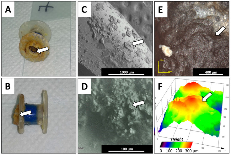
Figure 3.
Macroscopic and microscopic views of the used silicone voice prosthesis from one representative VP collected from a patient from Group 5. Panels (A,B) show a general view of the used representative prosthesis, investigated using microscopic methods, with biofilm formations on the esophageal and tracheal flange surfaces and the prosthesis’ valve. Panels (C,D) show examples of the microscopic topography (SEM) from the representative prosthesis’ polymeric surfaces, showing biofilm formations. Panels (E,F) show the microscopic topography characterization (CLM microscope) of polymeric surfaces with biofilm formations, with 3D height visualization (white arrows show biofilm formations).

