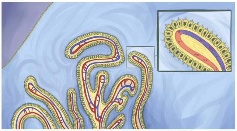
Figure 2.
A choroid plexus suspended within a ventricle. The choroid plexuses are highly vascularized structures that house choroid plexus epithelial cells (CPECs). These cells are specialized ependymal cells that perform a variety of key functions within the central nervous system. CPECs are situated above fenestrated choroidal vessels that permit the rapid entry of fluid, solutes, and macromolecules which include endogenous and exogenous proteins (insert). However, tight junctions towards the paracellular apex between CPECs restrict the free exchange of material between blood and cerebrospinal fluid (CSF) and constitute a blood-CSF barrier. Two main paths have been proposed for protein flux across the CPECs: transcytosis following either fluid phase or receptor-mediated endocytosis and diffusion through the paracellular barrier. Additionally, CPECs possess developed microvilli and cilia on their apical membranes that are important instruments for CPECs to conduct their physiological functions. Above the depicted choroid plexuses are multiciliated ependymal cells that form a semipermeable barrier within the ventricular system. Brain parenchyma resides directly under the ependymal cell barrier and is depicted by a shade of purple. Ependymal cells play a pivotal role in regulating the movement of cerebrospinal fluid within the ventricular system.

