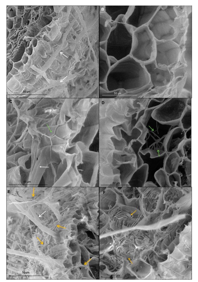
Figure 3.
Micrographs of spring wheat seedlings (cv. Bombona) taken on scanning electron microscopy (SEM): (A,B)—control roots; (C,D)—roots treated with T. atroviride AN35, (E,F)—roots treated with T. cremeum AN392. White arrows indicate root hairs. Green arrow indicate hyphae of T. atroviride AN35. Yellow arrow indicate to hyphae of T. cremeum AN392.

