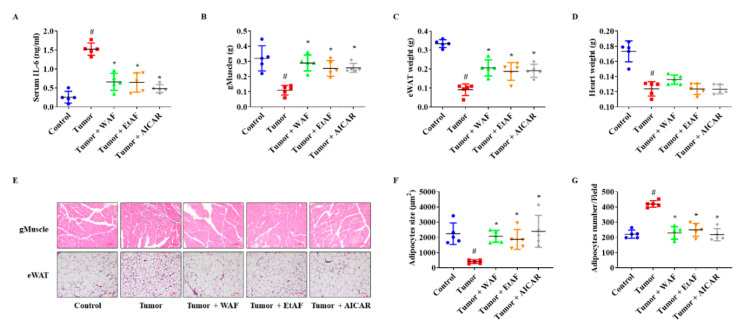
Figure 3.
Effects of AF in serum IL-6, muscle weight, and adipose tissue from CT-26 tumor-bearing mice. The serum IL-6 level was determined after sacrificing the mice (A). The weights of gMuscle (B), eWAT (C), and heart (D) were measured after sacrificing the mice. The H&E staining images of gMuscle and eWAT. The magnitude is ×200 and the scale bar is 100 µm (E). H&E-stained areas of eWAT were measured using ImageJ (F). Adipocytes numbers were counted by randomly selecting fields (G). All values are mean ± SD. # p < 0.05, significantly different from control group; * p < 0.05, significantly different from tumor group.

