Abstract
The Simbu serogroup is one of the serogroups that belong to the Orthobunyavirus genus of the family Peribunyaviridae. Simbu serogroup viruses are transmitted mainly by Culicoides biting midges. Meager information is available on Simbu serogroup virus infection in ruminants in Sudan. Therefore, in this study, serological surveillance of Simbu serogroup viruses in cattle in seven states in Sudan was conducted during the period from May, 2015, to March, 2016, to shed some light on the prevalence of this group of viruses in our country. Using a cross-sectional design, 184 cattle sera were collected and tested by a commercial SBV ELISA kit which enables the detection of antibodies against various Simbu serogroup viruses. The results showed an overall 86.4% prevalence of antibodies to Simbu serogroup viruses in cattle in Sudan. Univariate analysis showed a significant association (p=0.007) between ELISA seropositivity and states where samples were collected. This study suggests that Simbu serogroup virus infection is present in cattle in Sudan. Further epizootiological investigations on Simbu serogroup viruses infection and virus species involved are warranted.
1. Introduction
The Simbu serogroup is one of the serogroups that belong to the Orthobunyavirus genus of the family Peribunyaviridae. Virus members such as Akabane virus (AKAV), Aino virus (AINOV), Sathuperi virus (SATV), Schmallenberg virus (SBV), and Shamonda virus (SHAV) are prevalent in Oceania, Australia, Africa, and Asia [1–3]. Several Simbu serogroup viruses have been shown to cross the placenta and result in outbreaks of abortion, stillbirth, and malformations [4–9]. Simbu serogroup viruses are transmitted mainly by Culicoides biting midges [10, 11]. The congenital malformations seen at birth are recognized as congenital arthrogryposis-hydranencephaly syndrome (CAHS) affecting the musculoskeletal and nervous systems, respectively, and related to the pregnancy stage at which the dam is infected. In cattle, severe brain deformities may happen if the dam is infected between 76 and 106 days of pregnancy [4, 10].
In Sudan, Simbu serogroup viruses such as AKAV have been reported based on serological evidence in sheep, goats, and cattle in different ecological zones [12, 13]. Elhassan et al. [13] reported a significant association (p=0.03) between AKAV ELISA positivity and reproductive disorders (abortion and infertility).
Owing to the meager data available on Simbu serogroup viruses infection in ruminants in Sudan, this survey was carried out to detect anti-Simbu serogroup viruses IgG antibodies in cattle sera samples obtained in seven states in Sudan during the period from May, 2015, to March, 2016.
2. Materials and Methods
2.1. Study Area
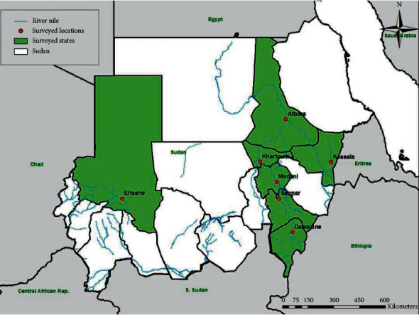
The survey which was conducted in seven states in Sudan aimed to cover the most of the country. Selection of these locations was based on them being the main potential areas for livestock rearing. A cross-sectional survey that included seven states (Blue Nile (Damazine), El Gezira (Madani), Kassala (Kassala), Khartoum (Khartoum), North Darfur (Elfashir), River Nile (Atbara), and Sennar (Sennar) States) of Sudan was conducted during the period from May, 2015, to March, 2016 (Figure 1). Selection of farms was made randomly, and the formal mechanism used was lottery. In each area, samples were collected from, at least, four groups of dairy cattle that were kept apart. Collection of animal samples was reviewed and in accordance with the animal welfare code of Sudan. Five ml of blood per animal were collected from 184 adults, apparently healthy dairy cattle. Sera were obtained by centrifugation at 1500 rpm/min for 10 minutes and kept at −20°C until tested.
Figure 1.

Map of Sudan showing states (green) and locations (red) where samples were collected.
2.2. Simbu Serogroup Enzyme-linked Immunosorbent Assay (ELISA)
A commercial SBV ELISA kit (IDEXX Laboratories, USA) which enables the detection of antibodies against various Simbu serogroup viruses was used to detect anti-Simbu serogroup viruses in diluted serum samples (1/10) according to the manufacturer's instructions. The specificity and sensitivity of the ELISA kit is 99.5% and 98.1%, respectively [14]. The sample optical densities (OD) were measured by using a microplate ELISA reader (Asys Expert Plus, Austria) at 450 nm. The sample to positive control ratio (S/P ratio) was, then, determined using the formula stated in the kit brochure. The cutoff value of antibody titer is ≥40%, i.e., all samples which have an S/P ratio ≥40 are considered positive as indicated in the kit literature.
2.3. Statistical Analysis
Risk factor with more than two categorical levels such as state was tested individually using univariate logistic regression. Differences in antibodies to Simbu serogroup viruses between cattle and state where samples were collected were evaluated using the Chi-square (χ2) test. Statistical differences between all possible pairs of groups were defined as p < 0.05. Statistical analysis was performed using SPSS version 20 (SPSS Inc., Chicago, U.S.A.).
3. Results
Antibodies to Simbu serogroup viruses were detected in cattle in all areas tested with varying prevalences. The seroprevalence rates in cattle ranged from 69.2% in North Darfur to 100% in Kassala and Sennar States. The prevalence rates were the highest in Kassala and Sennar (100%), River Nile (88%), Blue Nile (85.7%), and El Gezira (83.3%) and moderate in Khartoum (76.9%) and North Darfur (69.2%) states with an overall prevalence of 86.4%. Univariable logistic regression revealed a significant association (p=0.007) between ELISA seropositivity and state where samples were collected (Table 1).
Table 1.
Univariate analysis for the association of origin of collected samples (State) and seropositivity for Simbu serogroup viruses in cattle in seven states in Sudan during the period from May, 2015, to March, 2016.
| State | No of tested cattle | No positive | Prevalence rate in cattle (%) | p value |
|---|---|---|---|---|
| Blue Nile | 21 | 18 | 85.7 | 0.007∗ |
| El Gezira | 30 | 25 | 83.3 | |
| Kassala | 30 | 30 | 100 | |
| Khartoum | 26 | 20 | 76.9 | |
| North Darfur | 26 | 18 | 69.2 | |
| River Nile | 25 | 22 | 88 | |
| Sennar | 26 | 26 | 100 | |
| Total | 184 | 159 | 86.4 |
∗Significantly different
4. Discussion
There is a report of detection of antibodies to AKAV (a member of Simbu serogroup viruses) in livestock in Sudan since 1996 [12]. The present study further indicates that cattle are commonly exposed to Simbu serogroup viruses in Sudan with an overall seropositivity of 86.4%. In the current study, the overall seropositivity of Simbu serogroup viruses detected in cattle (86.4%) was lower than the estimated overall seropositivity (91.2%) of Simbu serogroup viruses reported in Nigeria [15] but was higher than that reported in cattle in Tanzania [16]. This result shows that Simbu serogroup viruses are endemic in counties in Africa that share ecological and meteorological drivers of arbovirus spread and circulation.
A high Simbu serogroup viruses seroprevalence in cattle was reported in different European countries. Seroprevalence of SBV within the herd was up to 100% and 70–100% in Germany and Netherlands, respectively [17, 18]. In the present study, the overall seropositivity of Simbu serogroup viruses is similar to the overall seropositivity of SBV in Europe: 79–94% in France [19], 90.8% in Belgium [20], and 72.5% in the Netherlands [18]. In Africa, serological screening suggests the presence of SBV in cattle, sheep, and goats in Mozambique with an overall 100% prevalence rate in cattle [21].
The differences in prevalence rates between the states herein reported may be attributed to local ecological factors, type of management and practices, flock or herd size, and insect vector activity that might influence the rates of transmission and infection with Simbu serogroup viruses. Thus, the amount of rainfall, humidity, and the plant coverage could influence the survival, abundance, and species of Culicoides and their activity in a specific area. Stocking rates and flock size, as well as rearing systems (grazing vs. feed lot feeding) could also affect the transmission rates of the virus (es) present in an area. Generally, arboviruses transmission and infectivity can be greatly enhanced when all of the components mentioned above in addition to immune status of host livestock and viral properties are favorable [22].
These results also support the high prevalence of AKAV (29.4%) that has previously been reported in Sudanese dairy cattle [13]. However, the much higher overall seroprevalence (86.4%) reported in the present study may indicate that the ELISA kit used is able to detect antibodies to Simbu serogroup viruses other than AKAV. This notion is supported by Oluwayelu et al. [15] who showed that all seropositive samples tested by a Simbu serogroup ELISA test were found positive for antibodies against, at least, one of the three other Simbu serogroup viruses (SBV, SHAV, and Simbu virus (SIMV)) using a serum neutralization test (SNT). Virus neutralization tests (VNT) are, thus, the best approach to distinguish antibodies against respective Simbu serogroup viruses. Otherwise, AKAV ELISA-kit results previously reported in Sudan [13] would also verify the seroprevalence of other Simbu serogroup viruses.
5. Conclusions
It could be concluded that Simbu serogroup viruses are widely circulating in Sudan. Finally, further epizootiological and virological investigations on Simbu serogroup viruses infection in cattle and other farm animals at the country level are important to identify the actual virus species from the vertebrate and invertebrate hosts and to determine its genetic relationships with the Simbu serogroup viruses circulating in Europe and Africa.
Acknowledgments
The authors thank the staff of the Immunology Unit, Central Laboratory, Ministry of Higher Education and Scientific Research, for helping to centrifuge and separate serum from blood samples and for facilitating this study. This work was supported by the Central Laboratory, Ministry of Higher Education and Scientific Research, Khartoum, Sudan.
Data Availability
Data used to support the findings of this study are available from the corresponding author upon request.
Conflicts of Interest
The authors declare that there are no conflicts of interest regarding the publication of this paper.
References
- 1.Zeller H., Bouloy M. Infections by viruses of the families bunyaviridae and filoviridae. Revue Scientifique et Technique de l’OIE. 2000;19(1):79–91. doi: 10.20506/rst.19.1.1208. [DOI] [PubMed] [Google Scholar]
- 2.Parsonson I. M., Della-Porta A. J., Snowdon W. A. Congenital abnormalities in newborn lambs after infection of pregnant sheep with akabane virus. Infection and Immunity. 1977;15(1):254–262. doi: 10.1128/iai.15.1.254-262.1977. [DOI] [PMC free article] [PubMed] [Google Scholar]
- 3.Della-Porta A. J., O’halloran M. L., Parsonson I. M., et al. Akabane disease: isolation of the virus from naturally infected ovine foetuses. Australian Veterinary Journal. 1977;53(1):51–52. doi: 10.1111/j.1751-0813.1977.tb15825.x. [DOI] [PubMed] [Google Scholar]
- 4.St George T. D., Standfast H. A. Simbu group viruses with teratogenic potential. In: Monath T. P., editor. The Arboviruses: Epidemiology and Ecology. Boca Raton, FL, USA: CRC Press; 1989. pp. 146–166. [Google Scholar]
- 5.Tsuda T., Yoshida K., Ohashi S., et al. Arthrogryposis, hydranencephaly and cerebellar hypoplasia syndrome in neonatal calves resulting from intrauterine infection with Aino virus. Veterinary Research. 2004;35(5):531–538. doi: 10.1051/vetres:2004029. [DOI] [PubMed] [Google Scholar]
- 6.Yanase T., Aizawa M., Kato T., Yamakawa M., Shirafuji H., Tsuda T. Genetic characterization of aino and peaton virus field isolates reveals a genetic reassortment between these viruses in nature. Virus Research. 2010;153(1):1–7. doi: 10.1016/j.virusres.2010.06.020. [DOI] [PubMed] [Google Scholar]
- 7.Hoffmann B., Scheuch M., Höper D., et al. Novel Orthobunyavirus in cattle, Europe, 2011. Emerging Infectious Diseases. 2012;18(3):469–472. doi: 10.3201/eid1803.111905. [DOI] [PMC free article] [PubMed] [Google Scholar]
- 8.Golender N., Brenner J., Valdman M., et al. Malformations caused by shuni virus in ruminants, Israel, 2014-2015. Emerging Infectious Diseases. 2015;21(12):2267–2268. doi: 10.3201/eid2112.150804. [DOI] [PMC free article] [PubMed] [Google Scholar]
- 9.Brenner J., Rotenberg D., Jaakobi S., et al. What can akabane disease teach us about other arboviral diseases? Veterinaria Italiana. 2016;52:353–362. doi: 10.12834/VetIt.547.2587.2. [DOI] [PubMed] [Google Scholar]
- 10.De Regge N. Akabane, aino and schmallenberg virus-where do we stand and what do we know about the role of domestic ruminant hosts and culicoides vectors in virus transmission and overwintering? Current Opinion in Virology. 2017;27:15–30. doi: 10.1016/j.coviro.2017.10.004. [DOI] [PubMed] [Google Scholar]
- 11.Hirashima Y., Kitahara S., Kato T., Shirafuji H., Tanaka S., Yanase T. Congenital malformations of calves infected with shamonda virus, Southern Japan. Emerging Infectious Diseases. 2017;23(6):993–996. doi: 10.3201/eid2306.161946. [DOI] [PMC free article] [PubMed] [Google Scholar]
- 12.Mohamed M. E., Mellor P. S., Taylor W. P. Akabane virus: serological survey of antibodies in livestock in the Sudan. Revue d’´eLevage et de M´edecine V´et´erinaire des Pays Tropicaux. 1996;49(4):285–288. [PubMed] [Google Scholar]
- 13.Elhassan A. M., Mansour M. E., Shamon A. A., El Hussein A. M. A serological survey of akabane virus infection in cattle in Sudan. ISRN Veterinary Science. 2014;2014:4. doi: 10.1155/2014/123904.123904 [DOI] [PMC free article] [PubMed] [Google Scholar]
- 14.IDEXX. IDEXX Schmallenberg antibody (Ab) test. 2013. http://www.idexx.com/pubwebresources/pdf/en_us/livestock-poultry/schmallenberg-ab-test-us-ss.pdf.
- 15.Oluwayelu D., Wernike K., Adebiyi A., Cadmus S., Beer M. Neutralizing antibodies against simbu serogroup viruses in cattle and sheep, Nigeria, 2012–2014. BMC Veterinary Research. 2018;14:p. 277. doi: 10.1186/s12917-018-1605-y. [DOI] [PMC free article] [PubMed] [Google Scholar]
- 16.Mathew C., Klevar S., Elbers A. R. W., et al. Detection of serum neutralizing antibodies to simbu sero-group viruses in cattle in Tanzania. BMC Veterinary Research. 2015;11:p. 208. doi: 10.1186/s12917-015-0526-2. [DOI] [PMC free article] [PubMed] [Google Scholar]
- 17.Wernike K., Silaghi C., Nieder M., Pfeffer M., Beer M. Dynamics of Schmallenberg virus infection within a cattle herd in Germany, 2011. Epidemiology & Infection. 2013;1–4 doi: 10.1017/S0950268813002525. [DOI] [PMC free article] [PubMed] [Google Scholar]
- 18.Elbers A. R., Loeffen W. L., Quak S., et al. Seroprevalence of schmallenberg virus antibodies among dairy cattle, The Netherlands, winter 2011–2012. Emerging Infectious Diseases. 2012;18:1065–1071. doi: 10.3201/eid1807.120323. [DOI] [PMC free article] [PubMed] [Google Scholar]
- 19.Zanella G., Raballand C., Durand B., et al. Likely introduction date of schmallenberg virus into France according to monthly serological surveys in cattle. Transboundary Emerging Diseases. 2015;62(5):76–79. doi: 10.1111/tbed.12198. [DOI] [PubMed] [Google Scholar]
- 20.Garigliany M. M., Bayrou C., Kleijnen D., Cassart D., Desmecht D. Schmallenberg virus in domestic cattle, Belgium, 2012. Emerging Infectious Diseases. 2012;18:1512–1514. doi: 10.3201/eid1809.120716. [DOI] [PMC free article] [PubMed] [Google Scholar]
- 21.Blomström A. L., Stenberg H., Scharin I., et al. Serological screening suggests presence of schmallenberg virus in cattle, sheep and goat in the zambezia province, Mozambique. Transboundary and Emerging Diseases. 2014;61(4):289–292. doi: 10.1111/tbed.12234. [DOI] [PMC free article] [PubMed] [Google Scholar]
- 22.Kyoungah J., Tadashi Y., Kun-Kyu L., Joong-Bok L. Seroprevalence of bovine arboviruses belonging to genus Orthobunyavirus in South Korea. The Journal of Veterinary Medical Science. 2018;80(10):1619–1623. doi: 10.1292/jvms.17-0542. [DOI] [PMC free article] [PubMed] [Google Scholar]
Associated Data
This section collects any data citations, data availability statements, or supplementary materials included in this article.
Data Availability Statement
Data used to support the findings of this study are available from the corresponding author upon request.


