An incorrect version of Figure 5 appeared in the June 2020 issue on page 1744.
The correct version of Figure 5 is published on this page.
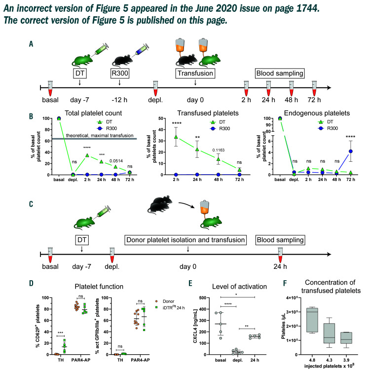
Figure 5.
Platelet transfusion efficacy and donor platelet function analysis of iDTRPlt mice. (A) Graphical overview for comparison of platelet transfusion. DT treatment started 7 days prior to transfusion and R300 treatment 12 hours prior to transfusion. Blood was taken at basal and depleted state, and 2, 24, 48, and 72 hours after transfusion. (B) Percentage of total, transfused, and endogenous platelet counts, relative to initial counts. Transfused platelets were labeled with an anti-GPIbβ-Dylight649 antibody. Theoretical, maximal transfusion is depicted as grey area. n = 5. (C) Graphical overview of donor platelet function evaluation. DT treatment started 7 days prior to transfusion and blood was taken at basal and depleted state, and 24 h after transfusion. (D) Comparison of percentage of CD62P+ and activated GPIIb/IIIa+ platelets in whole blood, freshly drawn from donors and after circulating for 24 hours in iDTRPlt mice. n = 4-9. (E) Concentration of plasma CXCL4 of iDTRPlt mice at basal and depleted levels, and 24 h after platelet transfusion. n = 5 (F) Concentration of circulating exogenous platelets after transfusion of indicated numbers of platelets. n = 5-10.
Supplementary Material
Associated Data
This section collects any data citations, data availability statements, or supplementary materials included in this article.


