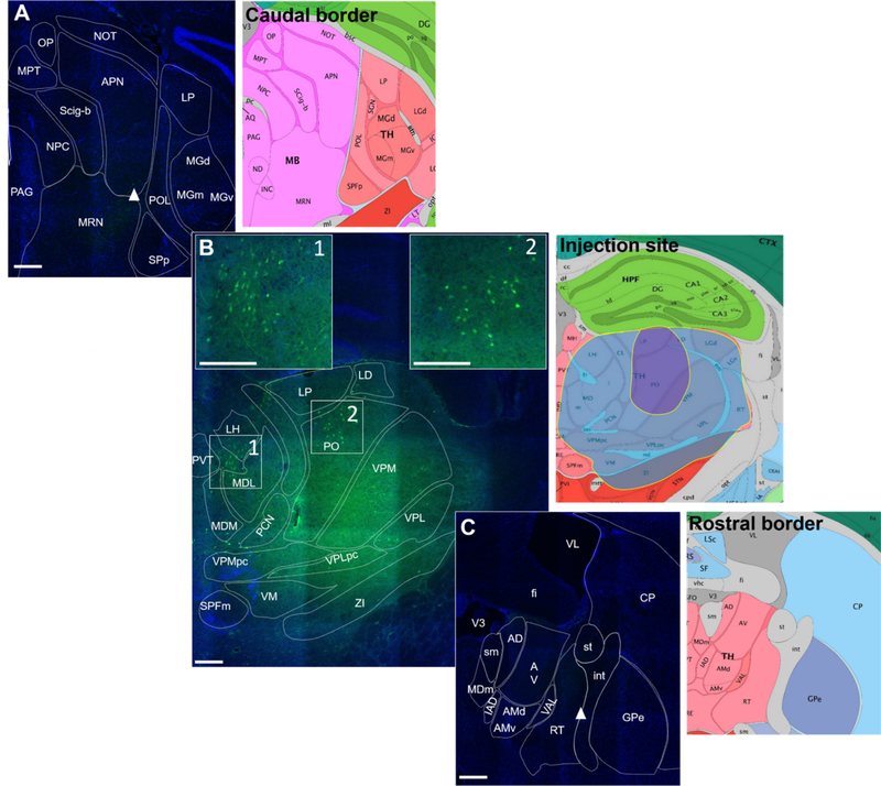
Figure 4. Fluoro-15-CGRP injection site targeting.
A. Caudal border of detectable signal from fluoro-15-CGRP (green) staining of a C57BL/6J mouse coronal brain section counterstained with DAPI (blue). Right panel is the Allen Mouse Brain Atlas coronal image representative of the brain slice (image 86/132). B. Fluoro-15-CGRP (1 μg) at the injection site. Boxes 1 and 2 represent magnified insets showing clusters of neurons that appear to have accumulated fluoro-15-CGRP signal. Right panel is the Allen Mouse Brain Atlas coronal image representative of the brain slice (image 74/132). The largest (blue shading) and smallest (purple shading) spread of the signal among the 5 mice are indicated. Shown here is the largest spread of the injections performed. C. Rostral border of detectable signal. Right panel is the representative Allen Mouse Brain Atlas coronal image (image 60/132). Image credit: Allen Institute. Scale bar = 100 μm for all images.

