Abstract
Fracture-dislocation of the lower lumbar spine, which is commonly caused by high-impact trauma and can lead to instability in the spine, is relatively rare. Surgical treatment is indicated to restore spinal balance, weight-bearing ability, and decompression of the neural elements. There are various available surgical options, including the posterior-only or anterior-only approaches, or a combination of them. However, there is still no definite classification and treatment strategy for fracture-dislocation of the lower lumbar spine. In this report, we describe a 65-year-old man presenting cauda equina syndrome caused by a fracture-dislocation of L5 combined with multi-level traumatic spondylolisthesis of the lower lumbar spine. The patient was treated via the posterior-only approach with neural decompression and anterior reconstruction with posterior instrumentation. We discuss the reasons why the posterior-only approach was decided upon and several meaningful points during the surgery in detail.
Keywords: Fifth lumbar vertebra, Corpectomy, Posterior approach, Fracture-dislocation, Lumbosacral
INTRODUCTION
Fracture-dislocation of the lower lumbar spine, which is commonly caused by high-impact trauma and can lead to instability in the spine, is relatively rare. Surgical treatment is indicated to restore spinal balance, weight-bearing ability, and decompression of the neural elements. Various surgical options are available, such as the posterior-only or anterior-only approaches, or a combination of them.3) However, insufficient studies on the classification and treatment of fracture-dislocation of the lower lumbar spine have been conducted due to the relatively low frequency and variety of these injuries compared to the thoracolumbar spine. Herein, we present a case of cauda equina syndrome caused by a fracture-dislocation of L5 combined with multi-level traumatic spondylolisthesis of the lower lumbar spine treated via the posterior-only approach.
CASE REPORT
A 65-year-old man arrived at our emergency center complaining of back pain, weakness in both legs of grade 1/5, and absence of anal tone after his back was hit by a falling heavy steel structure at his construction workplace. A computed tomography (CT) scan revealed a burst fracture of the L5 body with canal compromise, fracturing of the posterior elements including bilateral pedicles, facet joints, and spinous processes from L3 to L5, and spondylolisthesis of L4 on L5 (FIGURE 1). Magnetic resonance imaging confirmed the injuries to the bony structures identified from the CT scan, as well as massive ventral epidural hematoma with disrupted posterior structures, including ligamentum flavum, facet joints, muscle, and fascia. The thecal sac was compressed on the ventral epidural space by the retropulsed bone fragments of the fractured L5 body and an epidural hematoma between L3 and L5 (FIGURE 2).
FIGURE 1. Computed tomography scan revealing a burst fracture of the L5 body with canal compromise, fractures of the posterior elements including bilateral pedicles, facet joints, and spinous processes from L3 to L5, and spondylolisthesis of L4 on L5.
FIGURE 2. Magnetic resonance imaging confirming the same injuries of the bony structures identified on the computed tomography scan along with (A) massive ventral epidural hematoma (black asterisk) with disrupted posterior structures including ligamentum flavum, facet joints, muscle, and fascia and (B) compression of the thecal sac by the retropulsed bone fragments of fracture L5 body and epidural hematoma (blue asterisk) between L3 and L5.
We planned surgical treatment via the posterior-only approach. The patient was positioned in the prone position with extended hip posture on a Jackson spine operating table. As apparent on the intraoperative lateral fluoroscopy, spondylolisthesis of L4 on L5 was aggravated compared to the preoperative CT scan (FIGURE 3A), and so the patient was finally moved to the knee-chest position. However, although a slight reduction of the spondylolisthesis of L4 on L5 was achieved after the position change, complete reduction was not obtained (FIGURE 3B). En-block total laminectomies were performed from L3 to L5 with releasing the fibrous tissues around the fractured bony structures after removal of the fractured spinous processes. There was massive bleeding from the fractured pedicles and epidural spaces with cerebrospinal fluid leakage, and thus meticulous bleeding control and repair of the dural defect were performed.
FIGURE 3. Intraoperative lateral fluoroscopy showing (A) aggravation of spondylolisthesis of L4 on L5 compared to the initial Computed tomography scan in the prone position with extended hip posture, (B) a slight reduction in spondylolisthesis of L4 on L5 after the position change with knee-chest position, and (C) re-aggravation of the spondylolisthesis of L4 on L5 after removal of the posterior structures and laminectomies.
We identified re-aggravation of the spondylolisthesis of L4 on L5 after removal of the posterior structures and laminectomies (FIGURE 3C). We planned the insertion of an instrument with a reduction pedicle screw and rod construct between the normal vertebrae above and below the fracture sites followed by reduction of the spondylolisthesis. Pedicle screws were inserted from L1 to S1, which included L3–5 with fractured and removed pedicles, and the rod was installed between L1, L2, and S1 on the left side, and the same procedure was completed on the other side. In L3, L4, and L5, where the pedicles were removed due to traumatic spondylolysis, the pedicle screws were inserted directly into the body. The anteriorly displaced vertebral bodies of L3, L4, and L5 were slowly elevated by positioning the bilateral rods in the pedicle heads with locking head nuts while simultaneously checking the condition of the pedicle screws inserted to the vertebral bodies. Complete reduction of the spondylolisthesis was almost achieved. Corpectomy of the fractured L5 body and insertion of an expandable cage filled with autologous bone chips via the space between L5 and S1 nerve roots were achieved. The surgical procedures were finished by adding bilateral iliac screws (FIGURE 4).
FIGURE 4. Intraoperative lateral fluoroscopy showing (A) almost complete reduction of spondylolisthesis by slowly elevating the vertebral bodies of L3, L4, and L5 with positioning of the bilateral rods in the pedicle heads while checking the condition of pedicle screws inserted to the vertebral bodies simultaneously, (B) corpectomy of the fractured L5 body and insertion of the expandable cage filled with autologous bone chips via the space between L5 and S1 nerve roots (the surgery was completed by adding bilateral iliac screws) and (C) an intraoperative view.
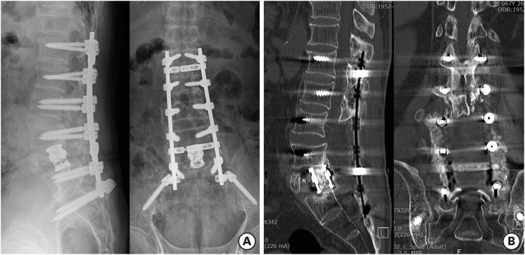
A few days after the operation, the patient showed partial improvement in leg weakness. Replacement was performed for the bilateral rods with fatigue fractures on L5–S1 identified at postoperative 11 months, which showed no displacement of cage and related symptom. Complete bone fusion was identified after 18 months of follow-up with the administration of teriparatide to enhance bone fusion (FIGURE 5). The patient was able to walk without assistance and showed no bladder or bowel symptoms.
FIGURE 5. (A) Radiographs and (B) computed tomography scan showing the corrected lumbosacral curve, the interbody fusion state and lateral fusion mass between L4 and S1 at 18 months post-operation.
DISCUSSION
We treated this patient with an L5 burst fracture combined with traumatic spondylolisthesis from L3 to L5 via the posterior-only approach to obtain direct neural decompression and reconstruction of the unstable lumbosacral spine. The patient was initially positioned in the prone position with extended hip posture on a Jackson spine operating table for restoration of lumbar lordosis with screw fixation. However, the posture worsened spondylolisthesis of L4 on L5, and a slight reduction was finally achieved after changing to knee-chest position. We think that an unstable burst fracture of L5 with multi-level traumatic spondylolisthesis showing hyper-lordotic change in L4–5 was related with hyperextension injury mechanism, which can be aggravated by the extension of lumbar spine under the prone position with hip extension. In this situation, knee-chest position with hip flexion was helpful to avoid aggravation of spondylolisthesis and allow easier access for corpectomy of L5.
Corpectomy of L5 is required for neural decompression with removing retropulsed bone fragments and mechanical anterior support using an expandable cage. Usually, corpectomy and reconstruction of L5 are considered to be challenging procedures and are performed through combined anterior and posterior approaches with high morbidity, a long hospital stay, and a long rehabilitation period.5) The anterior approach to L5 is particularly risky due to bifurcation of the common iliac vessels in front of it, and subjecting them to injury during an anterior approach can result in catastrophic complications.2) However, L5 corpectomy via the posterior-only approach avoids the possible morbidities associated with the anterior approach, with much less operative time and faster patient rehabilitation.6) In the case of burst fractures with retropulsed bone fragments, the posterior-only approach is beneficial for direct decompression of neural elements while removing encroaching bony fragments.7) Especially, the posterior-only approach is more feasible for a burst fracture of L5 combined with traumatic spondylolisthesis of the above segments compared to the combined anterior-posterior approach. Correction of malalignment on the lumbosacral spine with long-segment fixation is required before corpectomy and cage insertion, which is helpful for obtaining proper location of the cage to provide adequate stability under the corrected lumbosacral alignment.
The lumbosacral junction is a transition zone between the mobile lumbar spine and the relatively fixed sacrum, where high lordosis can subject the implants to higher loads and greater stress with a high rate of pseudoarthrosis.4,9) Rigid long-level posterior fixation including the pelvis with proper placement of an expandable cage filled with autologous bone chips to support anterior column provides good fixation that can tolerate maximum loads over the implants until the bone has healed. Placing a mesh or expandable cage after corpectomy via the posterior-only approach at this level is difficult due to the inherent lordosis of the lumbosacral junction and the narrow space between the L5 and S1 roots for placement of the cage compared to the same procedure on the other lumbar spine. Gentle manipulation of the cage insertion between the nerve roots is required to avoid traction injuries of the nerve roots. In this case, the space between the L5 and S1 nerve roots was insufficient for inserting a bigger cage in which to place the ventral portion of the vertebral body to restore ideal lumbosacral lordosis and to support the anterior column. We decided to use a small expandable cage and locate it at the posterior portion of the vertebral body with the remaining anterior portion of the fractured L5 body. This offered minimum traction of the nerve roots and relatively low lordotic curvature of the lumbosacral junction of the patient. Fortunately, interbody fusion centered in the expandable cage and formation of a lateral fusion mass with restoration of lumbosacral lordosis were achieved, and there was also healing of the bone defects on the fractured pedicles. We think that the effect of teriparatide on bone healing and union in the fracture sites enhanced successful bone fusion and healing. There have already been several studies presenting the effects of teriparatide for bone formation, fusion, and defect healing.8,10,11)
Due to the low occurrence of fracture-dislocation in lumbar spine injury, relevant studies describing its treatment have so far been limited to case reports and are insufficient. There are also no clear classification and treatment guidelines. Aihara et al.1) suggested the classification of fracture-dislocation of the fifth lumbar vertebra into 5 types, of which type 5 (dislocation of the L5 body with fracture of the body and/or pedicle, with or without injury of the lamina and/or facet) is similar to our case. They recommended multi-level posterior fusion using pedicle screws and interbody fusion as necessary for type 5. However, the above classification is limited to fracture-dislocation of the fifth lumbar vertebra, which may be difficult for applying to complex injury beyond the fifth lumbar vertebra as shown in this case. Therefore, extended classification based on a larger number of participants is required to describe complex injuries of the lumbosacral spine and to recommend treatment guidelines.
CONCLUSION
We report a rare case of fracture-dislocation of L5 combined with multi-level traumatic spondylolisthesis of the lower lumbar spine treated via the posterior-only approach. In this case, the approach was more feasible and is recommended for direct neural decompression, correction of malalignment, decreased operation time, and avoiding complications related to the anterior-only approach.
Footnotes
Funding: This work was supported by the National Research Foundation (NRF) of Korea grant funded by the Korea government (MSIP; Ministry of Science, ICT & Future Plan (2019M3E5D1A02068142).
Conflict of Interest: The authors have no financial conflicts of interest.
References
- 1.Aihara T, Takahashi K, Yamagata M, Moriya H. Fracture-dislocation of the fifth lumbar vertebra. A new classification. J Bone Joint Surg Br. 1998;80:840–845. doi: 10.1302/0301-620x.80b5.8657. [DOI] [PubMed] [Google Scholar]
- 2.Brau SA, Delamarter RB, Schiffman ML, Williams LA, Watkins RG. Vascular injury during anterior lumbar surgery. Spine J. 2004;4:409–412. doi: 10.1016/j.spinee.2003.12.003. [DOI] [PubMed] [Google Scholar]
- 3.Carminucci A, Assina R, Hernandez RN, Goldstein IM. Direct midline posterior corpectomy and fusion of a lumbar burst fracture with retrospondyloptosis. World Neurosurg. 2017;99:809.e11–809.e14. doi: 10.1016/j.wneu.2016.12.129. [DOI] [PubMed] [Google Scholar]
- 4.Cunningham BW, Lewis SJ, Long J, Dmitriev AE, Linville DA, Bridwell KH. Biomechanical evaluation of lumbosacral reconstruction techniques for spondylolisthesis: an in vitro porcine model. Spine. 2002;27:2321–2327. doi: 10.1097/00007632-200211010-00004. [DOI] [PubMed] [Google Scholar]
- 5.Dai LY, Jiang LS. Anterior-only instrumentation and grafting after L5 corpectomy for non-traumatic lesions. Acta Orthop Belg. 2010;76:94–99. [PubMed] [Google Scholar]
- 6.Elnady B, Shawky A, Abdelrahman H, Elmorshidy E, El-Meshtawy M, Said GZ. Posterior only approach for fifth lumbar corpectomy: indications and technical notes. Int Orthop. 2017;41:2535–2541. doi: 10.1007/s00264-017-3570-7. [DOI] [PubMed] [Google Scholar]
- 7.Jun DS, Yu CH, Ahn BG. Posterior direct decompression and fusion of the lower thoracic and lumbar fractures with neurological deficit. Asian Spine J. 2011;5:146–154. doi: 10.4184/asj.2011.5.3.146. [DOI] [PMC free article] [PubMed] [Google Scholar]
- 8.Lawrence JP, Ennis F, White AP, Magit D, Polzhofer G, Drespe I, et al. Effect of daily parathyroid hormone (1-34) on lumbar fusion in a rat model. Spine J. 2006;6:385–390. doi: 10.1016/j.spinee.2005.10.010. [DOI] [PubMed] [Google Scholar]
- 9.Lee YP, Ghofrani H, Regev GJ, Garfin SR. A retrospective review of long anterior fusions to the sacrum. Spine J. 2011;11:290–294. doi: 10.1016/j.spinee.2011.02.004. [DOI] [PubMed] [Google Scholar]
- 10.Liu Y, Levack AE, Marty E, Or O, Samuels BP, Redko M, et al. Anabolic agents: what is beyond osteoporosis? Osteoporos Int. 2018;29:1009–1022. doi: 10.1007/s00198-018-4507-8. [DOI] [PMC free article] [PubMed] [Google Scholar]
- 11.Tseng YY, Su CH, Lui TN, Yeh YS, Yeh SH. Prospective comparison of the therapeutic effect of teriparatide with that of combined vertebroplasty with antiresorptive agents for the treatment of new-onset adjacent vertebral compression fracture after percutaneous vertebroplasty. Osteoporos Int. 2012;23:1613–1622. doi: 10.1007/s00198-011-1730-y. [DOI] [PMC free article] [PubMed] [Google Scholar]


