Abstract
Cancer cells take advantage of NETosis to escape host immune surveillance and mediate metastasis. The pharmacological targeting of NETosis may prove beneficial in maximizing the response to cancer immunotherapy.
Subject terms: Cancer, Oncology
In a recent issue of Immunity, Teijeira et al. provide some crucial evidence that provides an important piece of the puzzle representing the immune-escaping strategies of cancer.1
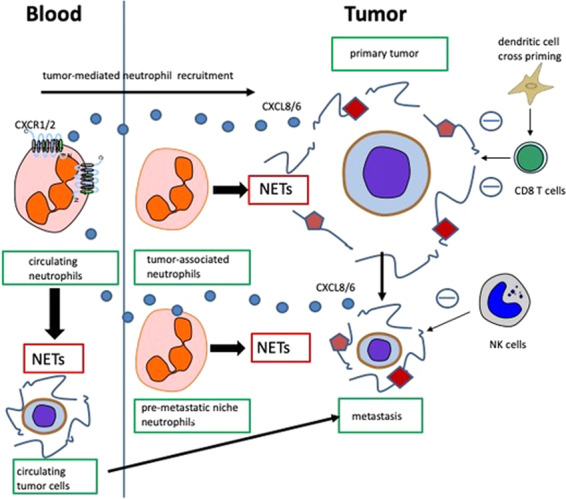
It is well established that a complex network of innate and adaptive immune effector mechanisms, including immune cells with phagocytic or cytotoxic activity and soluble factors, such as antibodies, chemokines, cytokines, antimicrobial peptides, and toxins, such as extracellular DNA and histones, protect us from infections and cancer.2 Nevertheless, pathogens and tumor cells have developed sophisticated mechanisms to escape from immunological surveillance, and the overall process is referred to as ‘immuno-editing’.3 In their manuscript, Teijeira and colleagues report that neutrophil extracellular traps (NETs) have the potential to limit the immune response against cancer by coating malignant cells. NETs are composed of extracellular web-like DNA decorated with antimicrobial proteins, which are released from activated neutrophils during NETosis. While NETosis plays essential roles in the elimination of microorganisms, excessive formation of NETs can harm the host.4 By binding pathogens, NETs prevent their spread, ensuring increased local concentrations of toxic factors. Previous data on the connection between NETs and cancer have shown that tumor-associated neutrophils also induce NET formation, but the implications have not been clarified. Neutrophils have emerged as an important component of the tumor microenvironment5 and may have a dual function in cancer spanning from anticancer properties based on the direct killing of cancer cells or stimulation of the immune system against cancer to cancer favoring abilities, such as the trap structures of NETs that can promote angiogenesis, extracellular matrix remodeling, proliferation, and migration of cancer cells, as well as constitute a physical barrier between cancer cells and immunocompetent cells (Fig. 1; refs. 5,6). In particular, Teijeira and colleagues found that tumor-secreted CXCR1 and CXCR2 ligands, such as IL-8, induce the extrusion of NETs, which in turn impairs the contact of immune cytotoxic cells with tumor cells, ultimately favoring their survival and metastatic potential.1
Fig. 1.

A schematic representation of neutrophil recruitment, NET formation and resistance to innate and adaptive lymphoid cell-mediated immune responses in primary and metastatic cancer
Nucleated cells present self-peptides derived from the processing of endogenous proteins through major histocompatibility complex (MHC) class I and contribute to peripheral tolerance.7 Cytotoxic CD8 T lymphocytes can recognize and lyse cells presenting non-self-peptides on MHC class I, such as tumoral cells presenting tumor-associated antigens or infected cells presenting microbial epitopes.7 Teijeira et al. reported that cancer takes advantage of the NET coating to avoid recognition by cytotoxic immune cells by inducing NETosis in tumor infiltrating neutrophils.1 Proteins associated with NETs may further enhance this strategy, as observed for pentraxin 3 (PTX3). PTX3, a multimeric glycoprotein with antimicrobial activity belonging to the long pentraxin family, is stored in neutrophil granules and released after proinflammatory signals8 and is one of the proteins decorating NETs. PTX3 is predominantly involved in fighting bacteria through direct opsonization and complement activation, but other functions have been proposed, including reduction of the detrimental effects of histone cytotoxicity during NETosis.9 Interestingly, PTX3 can restrict the cross-presentation of tumoral antigens by dendritic cells to CD8 T lymphocytes,10 thus suggesting a further mechanism by which NETs could interfere with immune cytotoxicity against cancer.
NETosis also seems to be involved in metastasis. Previously, neutrophils were reported to engage with circulating tumor cells in the bloodstream and to favor their implantation,5 and Teijeira et al.1 demonstrated that NETosis inhibition with DNase I or a PAD4 inhibitor significantly reduced lung micro-metastasis in an NK-dependent manner. Of interest, NETosis inhibition minimally delayed tumor progression while synergizing with anti-PD1 plus anti-CTLA4 checkpoint inhibitors in a CD8-dependent manner,1 thus suggesting a therapeutic role for NET inhibitors as putative adjuvants in cancer immunotherapy.
Immunosurveillance of infectious agents and malignant cells largely depends on NK cells and cytotoxic T cells, which specifically kill target cells after the polarized release of cytotoxic granules. Both cell types are subject to numerous immune evasion strategies that have evolved over time and result in the disarming or sequestration of immune cells from the pathological lesion.2 Immune checkpoint blockade overcomes the immunosuppressive status of the tumor microenvironment to enhance antigen-specific cytotoxic immunity. The present results suggest that NET inhibition could maximize the efficacy of immune checkpoint inhibitors in the treatment of cancer.
Competing interests
The authors declare no competing interests.
References
- 1.Teijeira A, et al. CXCR1 and CXCR2 chemokine receptor agonists produced by tumors induce neutrophil extracellular traps that interfere with immune cytotoxicity. Immunity. 2020;52:856–71 e8. doi: 10.1016/j.immuni.2020.03.001. [DOI] [PubMed] [Google Scholar]
- 2.Koch J, Tesar M. Recombinant antibodies to arm cytotoxic lymphocytes in cancer immunotherapy. Transfus. Med Hemother. 2017;44:337–350. doi: 10.1159/000479981. [DOI] [PMC free article] [PubMed] [Google Scholar]
- 3.Dunn GP, Old LJ, Schreiber RD. The three Es of cancer immunoediting. Annu Rev. Immunol. 2004;22:329–360. doi: 10.1146/annurev.immunol.22.012703.104803. [DOI] [PubMed] [Google Scholar]
- 4.Masuda S, et al. NETosis markers: quest for specific, objective, and quantitative markers. Clin. Chim. Acta. 2016;459:89–93. doi: 10.1016/j.cca.2016.05.029. [DOI] [PubMed] [Google Scholar]
- 5.Jaillon, S. et al. Neutrophil diversity and plasticity in tumour progression and therapy. Nat. Rev. Cancer. 2020. In press. 10.1038/s41568-020-0281-y. [DOI] [PubMed]
- 6.Garley M, Jablonska E, Dabrowska D. NETs in cancer. Tumour Biol. 2016;37:14355–14361. doi: 10.1007/s13277-016-5328-z. [DOI] [PubMed] [Google Scholar]
- 7.Cruz FM, Colbert JD, Merino E, Kriegsman BA, Rock KL. The biology and underlying mechanisms of cross-presentation of exogenous antigens on MHC-I molecules. Annu. Rev. Immunol. 2017;35:149–176. doi: 10.1146/annurev-immunol-041015-055254. [DOI] [PMC free article] [PubMed] [Google Scholar]
- 8.Doni A, et al. The long pentraxin PTX3 as a link between innate immunity, tissue remodeling, and cancer. Front Immunol. 2019;10:712. doi: 10.3389/fimmu.2019.00712. [DOI] [PMC free article] [PubMed] [Google Scholar]
- 9.Daigo K, Takamatsu Y, Hamakubo T. The protective effect against extracellular histones afforded by long-pentraxin PTX3 as a regulator of NETs. Front Immunol. 2016;7:344. doi: 10.3389/fimmu.2016.00344. [DOI] [PMC free article] [PubMed] [Google Scholar]
- 10.Baruah P, et al. The pattern recognition receptor PTX3 is recruited at the synapse between dying and dendritic cells, and edits the cross-presentation of self, viral, and tumor antigens. Blood. 2006;107:151–158. doi: 10.1182/blood-2005-03-1112. [DOI] [PubMed] [Google Scholar]


