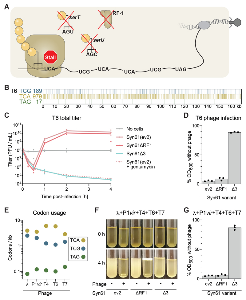
Fig. 2. Lytic phage propagation and cell lysis is obstructed in Syn61Δ3.
(A) Schematic of viral infection of Syn61Δ3. Deletion of serU (encoding tRNASer CGA), serT (encoding tRNASer UGA), and prfA (encoding RF1) makes the UCG, UCA, and UAG codons unreadable and the ribosome will stall at these codons within an mRNA that contains them, as shown here for a viral mRNA.
(B) Schematic of the number of TCG, TCA, and TAG codons and their positions in the genome of T6 phage.
(C) Cultures were infected with T6 phage at a multiplicity of infection (MOI) of 5 x 10⁻², and the total titer (intracellular phage plus free phage) was monitored over 4 hours. PFU, plaque-forming units. Treatment with gentamicin was used to ablate protein synthesis, providing a control for cells that cannot synthesize viral proteins or produce new viral particles.
(D) T6 efficiently lyses Syn61 variants but not Syn61Δ3. Cultures were infected as in panel (C) and OD600 was measured after 4 hours.
(E) Number of the indicated codons per kilobase in each indicated phage.
(F and G) Syn61Δ3 survives simultaneous infection of multiple phage: (F) photos of the culture at the indicated time points after infection (+) or in the absence of infection (-). Cultures were infected with phage λ, P1, T4, T6, and T7, each with an MOI = 1 x 10⁻². (G) OD600 of the cultures was measured after 4 hours. All experiments were performed in three independent replicates, the dots represent the independent replicates and the line (panel (C)) or bar [panels (D) and (G)] represents the mean. The photo (F) is a representative of data from three independent replicates.

