Abstract
Purpose
The purpose of the present study was to evaluate the individual susceptibility of four different types of OXIS contact areas (open [O], point [X], straight [I], and curved [S]) to approximal caries in children.
Methods
A retrospective cohort study was performed using clinical photographs and cone-beam computed tomography images of children, available from January 1, 2014, to August 31, 2015, showing the presence of at least one caries-free contact area between the primary molars. A single calibrated examiner scored 1,102 selected contacts using OXIS criteria from the occlusal view and subsequently evaluated the same contacts with a minimum follow-up period of one year for the presence of approximal caries.
Results
Of the 1,102 contacts, 259 (23.5 percent) were found to be carious or restored due to approximal caries. Multivariate logistic regression analysis showed that only the type of contact played a significant role in caries prevalence (P<0.05). The odds ratios of OXIS contacts for the development of approximal caries were: S contact—147.4 (95 percent confidence interval [95% CI] equals 19.7 to 1101.7); I contact—24.5 (95% CI equals 3.4 to 177.9); X contact—1.1 (95% CI equals 1.0 to 12.5); and O contact—1.00 (reference).
Conclusions
Among the OXIS contacts, the S type was most susceptible to approximal caries due to its complex morphology. The broad contact areas, namely, I and S types, are at greater risk for approximal caries in primary molars than O and X contacts.
Keywords: Dental Caries, Retrospective Cohort, Deciduous Teeth, Contact Area, Child
The types of interproximal contact areas of primary molars were first described in a cross-sectional study (2018), which reported four different types of contact areas: open (O); point (X); straight (I); and curved (S). 1 Hence, the OXIS classification was proposed. 1 Subsequently, several studies 2–4 have used the OXIS classification to evaluate the contact types of primary molars. Muthu et al. reported the clinical existence of OXIS among preschool-aged children, thus confirming the classification. 2 Kirthiga et al. examined 1,343 contacts from existing cone-beam computed tomography (CBCT) images of children and concluded that the contact area observed at the occlusal level determined the overall type of contact based on OXIS criteria. 3
The World Health Organization has identified caries as one of the most prominent chronic diseases in the world, affecting from 60 to 95 percent of children in both developed and developing countries, with especially high rates in Latin America and Asia. 5 Among the different types of caries in children, approximal caries is significant because of its propensity for a rapid rate of progression and the difficulty in determining the presence or absence of a lesion. This could be attributed to the peculiar morphologic characteristics of the primary tooth, specifically, less enamel and dentin thickness. 6 The prevalence of approximal caries, as observed in a previous study, was 4.95 percent among three-year-olds, 5.2 percent among four-year-olds, and 10.53 percent among five-year-olds. 7 In a previous study of caries experience among eight-year-olds, the distal surface on primary first molars was found to be most prone to caries, followed by the occlusal surfaces of primary first and second molars. 8 Approximal caries has been considered a challenge in terms of controlling carious lesions in primary teeth due to limited salivary access and the large area of contact between adjacent teeth. 6
Previous studies 9–13 of types of contact areas between primary molars evaluated the association of two types of contacts (open and closed) with approximal caries and concluded that the risk for approximal caries in the posterior primary dentition is increased if contact points are closed rather than open. A recent longitudinal study was conducted to evaluate the predictive power of the morphology of the distal surface of primary first molars and the mesial surface of primary second molars for caries development in young children, which identified four approximal variants: (1) concave-concave; (2) concave-convex; (3) convex-concave; and (4) convex-convex. Another noteworthy conclusion from the same study was that the morphology of approximal surfaces in primary molar teeth, particularly when both surfaces were concave, significantly influenced the risk of caries development. 14
Previous studies conducted concerning OXIS contacts have suggested that limited movement between adjacent teeth limits the accessibility to mechanical cleaning, which could lead to greater plaque accumulation, 2 thus altering the vulnerability of these contacts to dental caries. Although studies with OXIS contacts have been performed previously, 2–4 there is no evidence regarding their association with dental caries. A well-designed longitudinal study was needed to determine the relationship between variations in contact areas of primary molars and their caries predisposition. The present study hypothesized that the broad contact areas, namely, I and S, could be at greater risk for approximal caries than the O and X contacts.
Hence, the purpose of this study was to evaluate the individual susceptibility of four different types of OXIS contact areas (open [O], point [X], straight [I], and curved [S]) to approximal caries in children using existing electronic data-bases of clinical photographs.
Methods
Ethical considerations
This study was conducted following STROBE (Strengthening the Reporting of Observational Studies in Epidemiology) guidelines. Approval from the Research Ethics Committee of Children’s Dental Centre (CDC), Seoul, South Korea was obtained.
Study population and sampling frame
The present retrospective cohort study was conducted using electronic data of children in the form of clinical photographs and CBCT images, which were available at a private dental center in Seoul, South Korea, from January 1, 2014, to August 31, 2015. At this center, CBCT images were taken for the diagnosis of dental pathologies such as impacted supernumerary teeth, disorders in teeth eruption, and localization of unerupted teeth and for temporomandibular investigation. The inclusion criteria for this study were as follows: clinical photographs and CBCT images of good quality taken between January 1, 2014, and August 31, 2015, with the presence of at least one caries-free contact area between primary molars; at least one documented follow-up visit focused on the selected contact area (with the help of a clinical photograph/radiograph); a minimum of one year between the baseline and follow-up visits; if there were multiple clinical follow-up visits of a particular child, a maximum of three successive visits was included. The exclusion criteria were as follows: less than one year between baseline and follow-up visits; clinical photographs of children with special health care needs; and contact areas with caries, restorations, or crowns (stainless steel or zirconia), primary molars with developmental malformation of shapes/sizes of teeth, and erupted premolars in baseline photographs.
Calibration of the examiner
Before the start of the study, a single pediatric dentist was trained and calibrated under the supervision of an expert to clinically evaluate the contact areas over two months. The detailed process of calibration and intra-examiner variability has been described elsewhere. 1
In total, 1,295 children visited the CDC between January 1, 2014, and August 31, 2015. Among them, CBCT images and clinical photographs were present for 1,285 children.
Initial screening of CBCT images for inclusion
To ensure image standardization, all CBCT images were chosen from a single machine (Planmeca ProMax 3D Mid) with a standard field of view (80 mm by 80 mm), a voxel size of 0.40 mm, 90 kV and 12 mA, an exposure time of 12 seconds, and a slice thickness of 0.4 mm. CBCT images were analyzed with the built-in Romexis 3.5.2 digital imaging software (Planmeca, Helsinki, Finland), on a 15.6-inch Samsung LCD screen with an Intel CoreTM i3 2.4 GHz processor and 500 GB of memory at a resolution of 1,280 by 1,024 pixels. All CBCT images were observed and evaluated via a benchmark by a previously calibrated examiner. Observations were performed with no time restrictions. The examiner was free to scroll through the total length of the crown to detect approximal caries. The initial screening was performed in two stages. In the first stage, the quality of the CBCT images was assessed. Of the 1,285 CBCT images available, 58 CBCT images were excluded due to poor quality. In the second stage, 1,227 CBCT images with 4,908 contact areas were screened according to the inclusion criteria. Among them, 3712 contact areas were eliminated due to the presence of a carious contact. Therefore 332 CBCT images with 1,196 caries-free contact areas were present for further assessment with clinical photographs.
Initial screening of clinical photographs for inclusion
Clinical photographs were acquired using an Olympus digital camera (E-M10 II, Olympus Corp., Tokyo, Japan). All photographs were taken by two trained dental hygienists. Since there were two photographs (one each for the maxilla and mandible) for each child, 664 clinical photographs (from 332 CBCT images) were available for the initial screening process. At the first stage, the quality of the clinical photographs was assessed. Of the 664 clinical photographs, 52 were excluded because of poor quality. In the second stage, 612 clinical photographs with 1,166 caries-free contacts were screened, among which 64 contacts were eliminated due to the absence of a followup visit within a minimum of one year. Finally, 562 clinical photographs (281 maxillary and 281 mandibular) with 1,102 contacts (at least one intact noncarious contact area) were deemed eligible for assessment.
Baseline assessment of the contact areas with clinical photographs
The 1,102 selected contacts were scored from the clinical photographs, according to the OXIS criteria, as O-shaped, X-shaped, I-shaped, and S-shaped from the occlusal view. 5 In addition to the type of contact, data was also extracted for other dependent variables (e.g., age, gender, and type of arch).
Diagnosis of the outcome at the follow-up visits
The follow-up assessment of the children with the selected 1,102 contacts began in September 2019 and was completed in October 2019. The electronic records, available until August 31, 2019, were utilized for the assessment. The diagnosis of approximal caries for each contact was performed by a single examiner using clinical photographs of the children. If there was a cavitated lesion on the distal surface of the primary first molar or the mesial surface of the primary second molar, the outcome was considered to be present. In the absence of a clinical photograph, an intraoral periapical radiograph or orthopantomogram was evaluated, if present, for the presence of approximal caries between the primary molars. If any of the primary molars presented with a restoration/stainless steel crown/zirconia crown in the clinical photograph or radiograph, the caries status of these teeth, based on existing records, was taken into consideration. If there was approximal caries on the distal surface of the primary first molar or the mesial surface of the primary second molar, the outcome was marked as present. If dental caries was recorded at multiple outcomes for the same contact area, it was considered only once within the particular time frame.
Statistical analysis
All statistical calculations were conducted using SPSS 21.0 software (IBM Corp., Armonk, N.Y.,USA). A P-value of ≤0.05 was considered statistically significant. Univariate logistic regression analysis was used to assess the strength of association between the independent variables, namely the type of contact, type of arch, right or left side, age, gender, and the dependent variable (dental caries). Multiple logistic regression analysis calculated the impact of all independent variables on the dependent variable. The outcome was expressed as an odds ratio with 95 percent confidence intervals.
Results
Five hundred and sixty-two clinical photographs of 281 children (aged 2.1 to 10.0 years) with 1,102 contacts (at least one intact noncarious contact area per child) were deemed eligible for assessment. The mean age of the included participants was 5.02 years (±1.54 standard deviation [SD]).
Prevalence and frequency of the contacts
Of the 1,102 contacts, the observed prevalence of O, X, I, and S types was 80 (7.2 percent), 141 (12.8 percent), 750 (68.1 percent), and 131 (11.9 percent), respectively. The most common contact observed was I, followed by X, S, and O.
Caries prevalence period determination
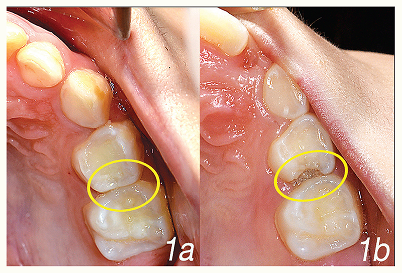
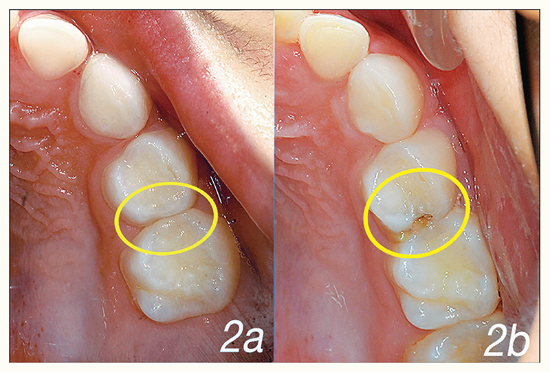
The mean time (days) between the initial and first follow-up examinations of the children with caries-affected contacts was 722 days (±285.5 SD, approximately 24 months). The shortest duration observed for caries to occur was 365 days (12 months), and the maximum duration observed was 1,580 days (52 months). The average duration for caries to occur on the specific types of OXIS contacts was as follows: 1,301 days for an O contact; 1,235 days for an X contact; 728 days for an I contact; and 511 days for an S contact. Figure 1 depicts “I” or straight type of contact between the primary maxillary left molars at baseline (1a) and after 19 months (1b). Figure 2 depicts “S” or curved type of contact between the primary maxillary left molars at baseline (2a) and after 24 months (2b).
Figure 1. “I” or straight type of contact between the primary maxillary left molars at (a) baseline and (b) after 19 months.
Figure 2. “S” or curved type of contact between the primary maxillary left molars at (a) baseline and (b) after 24 months.
Assessment of the outcome
Of the 345 follow-up assessments (281 first-visit follow-ups, 41 second-visit follow-ups, and 23 third-visit follow-ups), 292 were performed based on clinical photographs and 53 were performed based on radiographs. The main outcome of interest in the present study was the susceptibility of OXIS contacts to approximal caries between the primary molars. Of the 1,102 contacts, 259 were found to be carious or restored due to approximal caries. The remaining 843 contacts were sound. Of the carious contacts, 122 were carious or restored on both approximal surfaces (mesial surface of the primary second molar and distal surface of the primary first molar). In the other 137 carious contacts, there were 99 mesial (primary second molar) surfaces and 38 distal surfaces showing the presence of approximal caries or restorations in the form of a stainless steel crown, zirconia crown, or a composite restoration. Among these 259 carious or restored contacts, one contact was an O type (among 80 contacts), two were an X type (among 141 contacts), 176 were an I type (among 750 contacts), and 80 were an S type (among 131 contacts). The adjusted and unadjusted odds ratios of the OXIS contacts, regarding the prevalence of caries, are shown in the Table. In the univariate logistic regression analysis, the I (P=0.002) and S (P<0.001) types of contacts were found to be significantly associated with dental caries in children. There was also a significant association present concerning the type of arch (P=0.016). However, no significant findings were observed when the right and left sides were compared (P=0.831) and when genders were compared (P=0.178). The multivariate logistic regression analysis showed that only the type of contact played a significant role in caries prevalence. None of the other predictive variables included in the multivariate analysis (e.g., type of arch, side, gender, and age) had a significant influence on the development of approximal caries (Table).
Table. Analyses of Approximal Caries With Distribution Among Categories of Independent Variables.
| Variable | Total (N) | Number of contacts with caries present N (%) | Number of contacts with caries absent N (%) | Unadjusted odds ratio (95% CI) | P-value* | Adjusted odds ratio (95% CI) | P-value* |
|---|---|---|---|---|---|---|---|
| Type of contact | |||||||
| O | 80 | 1 (1.2) | 79 (98.8) | Ref | |||
| X | 141 | 2 (1.4) | 139 (98.6) | 1.14 (0.10-12.74) | 0.917 | 1.11 (0.99-12.45) | 0.933 |
| I | 750 | 176 (23.5) | 574 (76.5) | 24.22 (3.35-175.36) | 0.002 | 24.54 (3.39-177.90) | 0.002 |
| S | 131 | 80 (61.1) | 51 (38.9) | 123.92 (16.712-918.74) | <0.001 | 147.39 (19.72-1101.65) | <0.001 |
| Arch type | |||||||
| Maxilla | 553 | 147 (56.8) | 406 (48.2) | 1.413 (1.07-1.87) | 0.016 | 0.758 (0.54-1.06) | 0.102 |
| Mandible | 549 | 112 (43.2) | 437 (51.8) | Ref | |||
| Side | |||||||
| Right | 551 | 128 (49.4) | 423 (50.2) | 1.031 (0.78-1.36) | 0.831 | 0.99 (0.73-1.35) | 0.967 |
| Left | 551 | 131 (50.6) | 420 (49.8) | Ref | |||
| Gender | |||||||
| Male | 598 | 150 (25.1) | 448 (74.9) | 1.213 (0.92-1.61) | 0.178 | 1.25 (0.92-1.70) | 0.155 |
| Female | 504 | 109 (21.6) | 395 (78.4) | Ref | |||
| Age (years) | |||||||
| 2-4 | 281 | 66 (23.5) | 215 (76.5) | 1.299 (0.67-2.52) | 0.441 | 1.32 (0.64-2.73) | 0.454 |
| 4.1-6 | 549 | 125 (22.8) | 424 (77.2) | 1.247 (0.66-2.36) | 0.496 | 0.407 | |
| 6.1-8 | 204 | 55 (27.0) | 149 (73.0) | 1.562 (0.79-3.08) | 0.198 | 1.343 (0.67-2.70) | 0.116 |
| 8.1-10 | 68 | 13 (19.1) | 55 (80.9) | Ref | 1.82 (0.86-3.83) | ||
Abbreviation in this table: 95% CI= 95 percent confidence intervals.
P-value <0.05=significant.
Discussion
The present study is the first of its kind to evaluate the association of OXIS contacts with approximal caries in primary molars using a retrospective cohort design. The results of the present study agree with the hypothesis that, based on the morphology of the OXIS contact types, the I and S types may be at greater risk for approximal caries than the O and X types. This could be attributed to the increased plaque accumulation that may occur below the contact area in these types when compared with the O and X types. Further, difficulty in the maintenance of oral hygiene by routine mechanical cleaning methods can lead to the retention of plaque that has accumulated in I and S contact types. This could be because the routine mechanical cleansing method (toothbrushing) occurs using bristles that may not cover the contact area thoroughly. Hence, using dental floss might be beneficial in the presence of I and S contact types. The S contact type has been found to contribute to 61.1 percent of approximal caries, primarily attributable to the design complexity of the contact: The concavity followed by the convexity in this design forms a favorable niche for plaque accumulation. A previous study in this area concluded that, if both surfaces are concave, the plaque accumulation is supposed to be higher than if only one surface is concave. 14
Previous studies 9–13 investigated the association between two types of contacts (i.e., open and closed) with approximal caries in children. The major drawback of these studies was their cross-sectional study design, which meant that the contact present before the occurrence of caries development was unknown. Hence, no major conclusion could be drawn from these studies. However, one recently conducted prospective cohort study 14 concluded that, if both surfaces are concave, the risk of the development of approximal caries is significantly influenced. The concave surface could be considered equivalent to the S-shaped contact, which has a maximum risk for the development of approximal caries.
The present study used existing electronic databases in the form of clinical photographs to evaluate the prevalence of OXIS contact areas. The previous studies conducted in this area used various other methods (e.g., existing CBCT images, clinical examinations, or sectional die models) to score OXIS contacts. 1–4 A recent study was conducted to correlate the types of OXIS contact areas by CBCT with those by clinical photographs using existing electronic databases. The study revealed a correlation of 0.958, indicating that a two-dimensional evaluation was sufficient to score the types of OXIS contact areas. Hence, clinical photographs were used as the reference point to score the contact areas. 3
Of the 1,102 contacts, 259 were caries-affected (23.5 percent). A carious contact was considered when either the mesial surface of a primary second molar or the distal surface of a primary first molar was found to be carious. Regarding caries prevalence, the results of this study agreed with those of another study, 14 where a radiographic examination showed a 28.3 percent incidence of carious lesions (59 of 208 approximal surfaces were caries-affected).
Logistic regression was used to calculate the odds ratios (ORs) and corresponding confidence intervals, with the children as the clustered variable. The ORs of the S (147.39) and I contacts (24.54) were much higher when compared to the X contact (1.11). Thus, the analyses clearly showed that, among OXIS contacts, S and I contacts are more susceptible to the development of approximal caries. A previous study in this area showed that gender, the morphology of focus surfaces, and the morphology of adjacent surfaces were the only variables of significance for approximal caries development in primary molars. 11 In the present study, however, gender was not found to be a significant factor for approximal caries development.
A significant result from the present study is the duration needed for approximal caries to occur among the four types of OXIS contacts. The maximum duration for the development of approximal caries was taken by the O contact, followed by the X, I, and S type contacts. However, the risk for approximal caries development was found to be maximum for the S contact followed by I, X, and O contacts. The S type had the maximum risk for approximal caries formation with the shortest time.
Strengths and limitations
The primary strength of this study is that it is the first to document the susceptibility of OXIS contacts to approximal caries with a significant sample size. In addition, the center where the study was conducted had an exceptional database of records for children who visited the hospital for dental treatment concerning baseline and follow-up visits. However, the present study’s findings must be interpreted in the context of its limitations. The use of clinical photographs as the first reference point could have played a role in the underdiagnosis of noncavitated lesions. The present study’s findings cannot be extrapolated to all children in South Korean or ethnic populations, since the sample was drawn from only one private hospital. Finally, the role of confounding factors in the prevalence of dental caries could not be measured, due to the present study’s design.
Implications for research
Forthcoming studies should focus on the association of OXIS contacts with early childhood caries employing a prospective cohort design so that the role of confounders can be taken into consideration. The identification of the type of contact as a risk factor for approximal caries has increased the need for this to be considered as a potential risk factor for caries risk assessment. Furthermore, it has been corroborated that tooth morphology is under strong genetic control, which will also determine the types of contact between the approximal surfaces of primary molars. Therefore, future research to determine the degree of this genetic control can help in the assessment of the risk susceptibility of these surfaces, thus aiding in the prevention of early childhood caries.
Conclusions
Based on this study’s results, the following conclusions can be made:
Among the OXIS contacts, the S type was the most susceptible to the development of approximal caries. This was followed by I, X, and O contacts.
Clinicians should be aware of these variations in the approximal contacts while rendering care to children.
Further research is recommended to understand the susceptibility of OXIS contacts in different ethnic populations as a risk factor for approximal caries.
Acknowledgments
The authors wish to thank Miss Somi Park, BS, Technician in Radiology, Children’s Dental Centre, Seoul, South Korea for her assistance in the process of retrieval of CBCT images from the database and Mrs. Gayathri, MSc, MPhil, Senior Lecturer in Statistics, Department of Allied Health Sciences, Sri Ramachandra Institute of Higher Education & Research, Chennai, India, for her assistance in statistical analysis. This study was funded by the Wellcome Trust DBT India Alliance (IA/CPHE/17/1/503352).
References
- 1.Kirthiga M, Muthu MS, Kayalvizhi G, Krithika C. Proposed classification for interproximal contacts of primary molars using CBCT: A pilot study. Wellcome Open Res. 2018;3:98. doi: 10.12688/wellcomeopenres.14713.1. [DOI] [PMC free article] [PubMed] [Google Scholar]
- 2.Muthu MS, Kirthiga M, Kayalvizhi G, Mathur VP. OXIS classification of interproximal contacts of primary molars and its prevalence in 3- to 4-year-old children. Pediatr Dent. 2020;42(3):197–202. [PubMed] [Google Scholar]
- 3.Kirthiga M, Muthu MS, Cheuon Lee JJ, et al. Prevalence and correlation of OXIS contacts using cone-beam computed tomography images (CBCT) and photographs. Int J Paediatr Dent. 2021;31(4):520–7. doi: 10.1111/ipd.12687. [DOI] [PubMed] [Google Scholar]
- 4.Walia T, Kirthiga M, Brigi C, et al. Interproximal contact areas of primary molars based on OXIS classification: A two-centre cross-sectional study. Wellcome Open Res. 2021;5:285. doi: 10.12688/wellcomeopenres.16424.1. [DOI] [PMC free article] [PubMed] [Google Scholar]
- 5.Petersen PE. The World Oral Health Report 2003: Continuous improvement of oral health in the 21st century-the approach of the WHO Global Oral Health Programme. Community Dent Oral Epidemiol. 2003;31(Suppl 1):3–23. doi: 10.1046/j..2003.com122.x. [DOI] [PubMed] [Google Scholar]
- 6.Virajsilp V, Thearmontree A, Aryatawong S, Paiboonwarachat D. Comparison of proximal caries detection in primary teeth between laser fluorescence and bitewing radiography. Pediatr Dent. 2005;27(6):493–9. [PubMed] [Google Scholar]
- 7.Bruzda-Zwiech A, Filipińska R, Borowska-Strugińska B, Żądzińska E, Wochna-Sobańska M. Caries experience and distribution by tooth surfaces in primary molars in the pre-school child population of Lodz, Poland. Oral Health Prev Dent. 2015;13(6):557–66. doi: 10.3290/j.ohpd.a34371. [DOI] [PubMed] [Google Scholar]
- 8.Kuzmina IN, Kuzmina E, Ekstrand KR. Dental caries among children from Solntsevsky: A district in Moscow, 1993. Community Dent Oral Epidemiol. 1995;23(5):266–70. doi: 10.1111/j.1600-0528.1995.tb00246.x. [DOI] [PubMed] [Google Scholar]
- 9.Allison PJ, Schwartz S. Interproximal contact points and proximal caries in posterior primary teeth. Pediatr Dent. 2003;25(4):334–40. [PubMed] [Google Scholar]
- 10.Warren JJ, Slayton RL, Yonezu T, Kanellis MJ, Levy SM. Interdental spacing and caries in the primary dentition. Pediatr Dent. 2003;25(2):109–13. [PubMed] [Google Scholar]
- 11.Subramaniam P, Babu GKL, Nagarathna J. Interdental spacing and dental caries in the primary dentition of 4-to 6-year-old children. J Dent (Tehran) 2012;9(3):207–14. [PMC free article] [PubMed] [Google Scholar]
- 12.Parfitt GJ. Conditions influencing the incidence of occlusal and interstitial caries in children. J Dent Child. 1956;23:31–9. [Google Scholar]
- 13.Ben-Basset Y, Harari D, Brin I. Occlusal traits in a group of school children in an isolated society in Jerusalem. Br J Orthod. 1997;24(3):229–35. doi: 10.1093/ortho/24.3.229. [DOI] [PubMed] [Google Scholar]
- 14.Cortes A, Martignon S, Qvist V, Ekstrand KR. Approximal morphology as predictor of approximal caries in primary molar teeth. Clin Oral Invest. 2018;22(2):951–9. doi: 10.1007/s00784-017-2174-3. [DOI] [PubMed] [Google Scholar]


