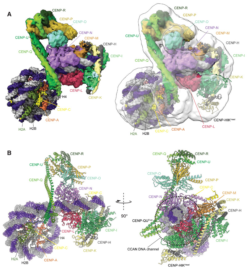
Figure 2. Structure of the CCANΔT-CENP-ANuc complex.
(A) Right panel shows the consensus CCANΔT-5 CENP-ANuc cryo-EM map (transparent white) overlaid onto the composite CCANΔT-CENP-ANuc cryo-EM density map based on individual cryo-EM maps for the CENP-ANuc-CENP-CN and CCANΔT-DNA reconstructions. Left panel: composite map alone. (B) Two orthogonal views of the CCANΔT-CENP-ANuc complex depicted in cartoon representation for protein and space filling for DNA. CENP-ANuc has a diameter of 11 nm.

