Abstract
Poly(A) tails, present on almost every eukaryotic mRNA, were discovered over 50 years ago. Early experiments led to the hypothesis that poly(A) tails and the cytoplasmic poly(A)-binding protein promote translation and prevent mRNA degradation, but the details remained unclear. More recent data suggest that the role of poly(A) tails is much more complex: poly(A)-binding protein can stimulate poly(A) tail removal (deadenylation), and the poly(A) tails of stable, highly-translated mRNAs at steady state are much shorter than expected. Moreover, the rate of translation elongation impacts deadenylation. Together, the interplay between poly(A) tails, poly(A)-binding protein, translation, and mRNA decay plays a major role in regulating gene expression. In this review, we summarize recent work that is revolutionizing the field and changing our perspective on the roles of poly(A) tails in the cytoplasm. Specifically, we discuss the roles of poly(A) tails in translation and mRNA stability, and how poly(A) tails are removed by exonucleases (deadenylases) including Ccr4-Not and Pan2-Pan3. We also cover major topics of current research, including how deadenylation rate is determined, the integration of deadenylation with other cellular processes, and the function of the cytoplasmic poly(A)-binding protein. Finally, we provide an outlook for the future of research in this area.
I. Introduction
Early studies on eukaryotic messenger RNA (mRNA) were quick to note that these emissaries of the genetic code are polyadenylated on their 3′-end1–6. Poly(A) tails are present on almost every eukaryotic mRNA, with the only known exception being some mammalian histone transcripts. Poly(A) tails are added co-transcriptionally in the nucleus and are required for the export of mature mRNAs into the cytoplasm (Figure 1). Eukaryotic transcripts receive a poly(A) tail with an average length of ~200 nt in mammals7 and ~70 nt in yeast8.
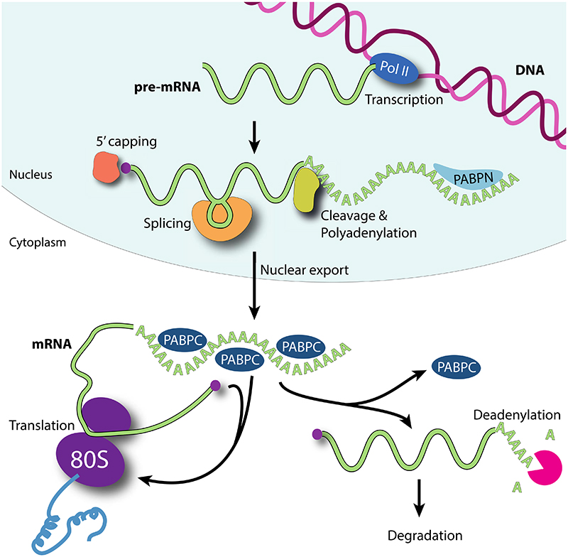
Figure 1. mRNA Poly(A) tails function as master regulators of gene expression in the cytoplasm.
In the nucleus, pre-mRNAs are transcribed by RNA polymerase II (Pol II) and processed, including 5′-capping, splicing and 3′-cleavage and polyadenylation. Nuclear poly(A) binding proteins (PABPN) function in the nucleus to control poly(A) tail addition. Mature, polyadenylated mRNAs are exported into the cytoplasm. Cytoplasmic poly(A)- binding protein (PABPC) binds poly(A) and promotes translation by the 80S ribosome. Poly(A) tails and PABPC also influence mRNA stability: removal or shortening of the poly(A) tail (deadenylation) releases PABPC and leads to mRNA degradation.
The poly(A) tail contributes to both the translational status and stability of mRNAs, and it functions as a master regulator of gene expression in the cytoplasm (Figure 1). Specifically, it can act synergistically with the 7-methylguanosine cap (m7Gppp) on the 5′-end of the mRNA to stimulate translation9. Accordingly, a transcript with a short poly(A) tail has reduced translation and is also a substrate for removal of the 5′ cap (decapping). Thus poly(A) shortening (deadenylation) ultimately triggers translation repression and also subsequent mRNA decay. The rate of deadenylation can be regulated in response to cellular cues, in a transcript-specific manner. Moreover, in specific cases, poly(A) tails can be extended in the cytoplasm to reactivate translationally-repressed transcripts or maintain their stability. The dynamic nature of poly(A) tails therefore is vital in regulating gene expression. This is important in almost every aspect of eukaryotic biology, including in early development, the inflammatory response and synaptic plasticity. Consistent with a conserved and critical role in gene expression, many viral transcripts either have a poly(A) tail, or use alternate mechanisms that functionally substitute for a poly(A) tail10,11. Likewise, metazoan replication-dependent histone mRNAs substitute poly(A) with a unique 3′ ribonucleoprotein (RNP) complex12.
Overall, post-transcriptional regulation of mRNAs – including changes in the length of the poly(A) tail – is a quintessential aspect of gene expression that determines the proteome’s overall architecture. Transcripts are differentially regulated, resulting in large variations in translation efficiencies and mRNA stabilities. For instance, translation efficiency can vary >1000-fold between transcripts, while mRNA half-lives vary by a similar order of magnitude13–17.
Recent insights from new sequencing techniques, biochemical reconstitution and structural biology are now providing new molecular insights into poly(A) tail biology. We now know that poly(A) tail lengths are not as uniform as once hypothesized, they can contain non-A nucleotides, and the aforementioned connection between tail length, translation efficiency and mRNA stability is not straightforward.
Here, we review the roles of poly(A) tails in the cytoplasm – specifically in translation and mRNA stability – and the mechanisms of poly(A) tail removal by the Ccr4-Not and Pan2-Pan3 deadenylation complexes. We discuss how the rate of deadenylation is controlled, how deadenylation interfaces with other cellular processes, and the role of the major cytoplasmic poly(A)-binding protein (PABPC – Pab1 in yeast, PABPC1 in humans). We start out by describing key historical findings. We then discuss recent discoveries that extend our understanding of the roles of poly(A) tails in the cytoplasm and that are now leading to a resurgent interest in the field. Finally, we provide a perspective where we discuss future areas of focus on cytoplasmic mRNA translation and decay.
II. Poly(A) tails in mRNA translation
Pioneering work in the 1970s demonstrated that poly(A) tails are important for efficient translation. For example, sea urchin eggs were shown to have a large burst of cytoplasmic poly(A) addition shortly after fertilization18,19: Maternal mRNAs were found to be translationally quiescent, often with very short poly(A) tails20–23 and these are extended at oocyte activation, coincident with expression of their cognate protein products24,25. And perhaps most dramatically, cytoplasmic polyadenylation was shown to be both necessary and sufficient for the activation of the proto-oncogene c-mos, driving meiotic maturation of frog oocytes, and suggesting that it was the newly-formed poly(A) tail that turned on translation26,27. At about the same time, it was shown that in synaptic junctions, some transcripts are translationally silent and have short poly(A) tails, much like maternal mRNAs in oocytes28. Synaptic stimulation results in both polyadenylation and concomitant translational activation of these dendritic mRNAs. Collectively these data suggested that mRNA translation is broadly influenced by regulated control of poly(A) tail length29,30.
The storage and activation of maternal mRNAs in oocytes and neuronal mRNAs is analogous in almost every molecular detail, including the protein factors that mediate these processes. In both cases, the length of the poly(A) tail correlates with translation efficiency – the longer the poly(A) tail, the more efficiently it is translated31,32. Thus, poly(A) tail metabolism is dynamic and a critical node in gene expression that is leveraged in multiple biological contexts. The discovery of cytoplasmic polyadenylation opened up a new field and the details have been reviewed extensively elsewhere33,34.
It is now clear that the poly(A) tail at the 3′ end of the mRNA can influence translation initiation at the 5′ end. However, despite a litany of information spanning 30 years demonstrating that poly(A) enhances translation, a clear mechanistic explanation is lacking. This is partly because methods to study poly(A) tail lengths were slow to develop and in vitro translation systems do not always recapitulate poly(A) effects. Additionally, homeostatic mechanisms balance out gene expression levels when they are disrupted in cells, further complicating the interpretation of in vivo mechanistic studies: Disruption of one pathway may be compensated for by modulation of another. In addition, the role of poly(A) tails is likely different in embryonic and post-embryonic cells17, as will be discussed below. But perhaps most importantly, a major factor known to mediate the effects of poly(A) – cytoplasmic poly(A) binding protein (PABPC) – has been difficult to study.
A. Role of PABPC
In the cytoplasm, poly(A) tails are bound by PABPC35, which was discovered at about the same time as mRNAs were found to bear a poly(A) tail36,37. There is one cytoplasmic PABPC in yeast (Pab1), but multiple isoforms in mammals: PABPC1 is the best-studied mammalian isoform and likely the most abundant in most cell types38. Other mammalian isoforms include PABPC4 which may act in a transcript-specific manner, embryonic ePABP and the testes-specific tPABP38. PABPC is highly conserved in eukaryotes and has four N-terminal RNA Recognition Motif (RRM) domains that bind poly(A) RNA with a nanomolar affinity39 (Figure 2A). RRMs 1 and 2 have a higher affinity and specificity for poly(A) than RRMs 3 and 440,41. A crystal structure of RRMs 1 and 2 showed that RRM1 is located 3′ of RRM2 on the RNA42 (Figure 2A). The RRMs are followed by a proline-rich linker and a C-terminal Mademoiselle (MLLE) domain (Figure 2A). The MLLE domain recognizes a peptide motif called poly(A)-interacting motif 2 (PAM2) which is found in a number of partner proteins that regulate poly(A) tail dynamics39. Nuclear-localized poly(A)-binding proteins (PABPN1 humans, Nab2 in yeast) have a different domain architecture to cytoplasmic PABPCs and influence the process of polyadenylation. PABPN function will not be covered here43.
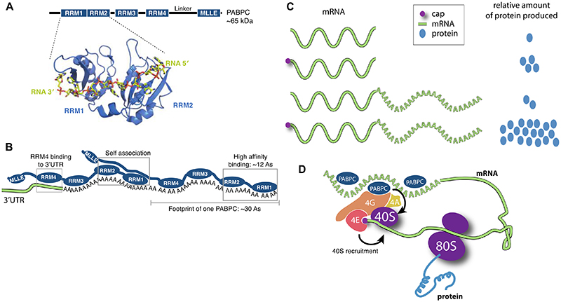
Figure 2. mRNA poly(A) tails stimulate translation.
A Structure of eukaryotic poly(A)- binding protein (PABPC – Pab1 in yeast, PABPC1 in mammals). Domain diagram of the conserved PABPC is shown with the position of the four RNA-recognition motif (RRM) domains and the Mademoiselle (MLLE) domain indicated38,39. Below, a crystal structure of RRM1-RRM2 (cartoon) of yeast Pab1 bound to poly(A) RNA (sticks) is shown (PDB 1CVJ)42. B Arrangement of PABPC on RNA. Two PABPC molecules can bind a 60 nt poly(A) tail. RRMs 1 and 2 have the highest affinity and specificity for poly(A) and require ~12 As for high affinity binding40,41. Full-length PABPC footprints ~30 nt and adjacent PABPC molecules interact with each other35,45,191. RRM4 may bind to the 3′-UTR130. C The mRNA 5′-cap (magenta circle) and 3′-poly(A) tail act synergistically to stimulate gene expression in eukaryotes. The relative amount of protein produced from reporter mRNAs with and without 5′ cap and poly(A) tail in plant, animal and yeast cells are depicted9. D Closed-loop model. The eukaryotic translation initiation factor 4E (eIF4E) binds the 5′-cap. eIF4G binds both eIF4E and PABPC, as well as the RNA helicase eIF4A, and this is thought to stimulate recruitment of the small (40S) ribosomal subunit. 40S assembles with a large (60S) ribosomal subunit on a start codon to form a translation-competent 80S ribosome.
PABPC requires ~12 A residues for high-affinity binding (via RRMs 1 and 2), but physically covers ~30 As35 (Figure 2B). Longer tails can bind more PABPC and a poly(A) tail of 90 nt can bind three molecules44. Interaction between adjacent PABPC molecules promotes co-operative binding to poly(A) RNA, facilitating multimerization45. However, recent data17 suggest that PABPC concentrations in cells may be limiting, and that steady-state poly(A) tail length in cells does not necessarily correlate with the amount of PABPC associated46. This will be discussed in more detail below45,47,48.
Most of our understanding of the function of PABPC comes from early studies in yeast. In S. cerevisiae, bypass suppressors of PAB1 mutants have been identified and are divided into two classes. First are mutations in genes encoding large (60S) ribosomal subunits and 60S biogenesis factors49. The other class of PAB1 suppressors are found in genes that encode factors involved in mRNA degradation, including decapping regulators (PAT1, LSM1, and DCP1) and exonucleases (XRN1 and RRP6)50,51. While these genetic findings are consistent with a role for PABPC in translation, they also point to a vital role for PABPC (and poly(A) tails) in mRNA stability. PABPC is both necessary and sufficient for the roles of poly(A) in mediating transcript translation and stability52–54 and this will be discussed in more detail below.
B. The closed-loop model
The m7Gppp cap at the 5′ end of the mRNA also plays an important role in regulating translation. The cap binds directly to translation factors to promote initiation55. Synergy between the 5′ cap and the 3′ poly(A) tail further stimulates this process, as demonstrated by monitoring translation of reporter mRNAs in plant, animal and yeast cells9. In these experiments, the combination of both cap and tail on the same mRNA led to a strongly enhanced translation efficiency compared to mRNAs with a single modification (Figure 2C).
How does the 3′-end of the mRNA stimulate translation initiation at the 5′-end? Mechanistically, it was shown that the eukaryotic translation initiation factor (eIF) 4E recognizes the 5′ cap55. eIF4E also interacts with another translation initiation factor, eIF4G, which, in turn, binds PABPC56. Thus, mRNAs can form a ‘closed-loop’ with direct physical communication between the 5′ cap and the 3′ poly(A) tail57 (Figure 2D). The interaction of eIF4G with PABPC stabilizes the eIF4E-cap interaction58. And similarly, PABPC interaction with poly(A) RNA stabilizes its interaction with eIF4G58. PABPC also stimulates eIF4A, another translation initiation factor, by enhancing its ATPase and helicase activity59. Together, the cap–eIF4E–eIF4G–PABPC–poly(A) complex is thought to stimulate translation, at least in part, by recruiting the small (40S) ribosomal subunit60–62 (Figure 2D).
It is tantalizing to compare the mRNA closed loop in translation to that of a rolling circle in DNA replication. In DNA replication, circular molecules of DNA can undergo a unidirectional process of replication that greatly enhances production, with DNA polymerase going round and round the circle ad infinitum. In terms of mRNA translation, the situation is different; while DNA replication can be continuous, translation is discontinuous with distinct steps of initiation, elongation, and termination for each polypeptide produced. Nevertheless, an mRNA closed-loop would result in translation termination occurring in close physical proximity to the 5′ end of the mRNA, potentially allowing for rapid recycling of ribosomes back to the initiation codon of the same transcript. Circularization of the transcript could also act as a quality control mechanism to ensure that translation does not initiate on a partially degraded mRNA.
Direct biochemical data demonstrate that PABPC and eIF4G can mediate circularization of an mRNA63,64 but this ‘closed-loop’ may not be as simple as was originally documented. For example, single molecule imaging of RNA molecules supports a model where mRNAs are not fully extended, but the 5′- and 3′-ends of transcripts are often not in close enough proximity to be physically connected by eIF4E, eIF4G and PABPC65. Some of these inconsistencies between in vitro and in-cell work might be explained by differences in experimental setup. Still, while the ‘closed-loop’ model may apply to some mRNAs, it may be dynamic and may not apply to all transcripts, in all biological contexts66. There is ample evidence for communication between the 5′- and 3′-ends of mRNAs, but whether this is mediated by a closed-loop and the nature of the relationship between the cap and tail is still somewhat ambiguous. Some of the key discrepancies in this model will be discussed below but we refer the reader to a recent review that extensively discusses this topic67.
III. Poly(A) tails in mRNA stability
Early experiments showed that poly(A) tails are shortened exonucleolytically in a time-dependent manner68–70. mRNAs injected into Xenopus oocytes with a poly(A) tail of at least 32 nt were translated as efficiently as mRNAs with long (150 nt) poly(A) tails71. In contrast, poly(A) tails of 16 nt or less were not translated71 and tails shorter than 30 As are not often observed in cells72. Together, these data suggested that poly(A) tails stabilize mRNAs (allowing their translation), and a minimum length of ~30 nt is generally required to confer stability. Satisfyingly, this minimum length corresponds with the footprint of PABPC. Some mRNAs, however, are stable and efficiently translated despite bearing a very short (<20 nt) or no poly(A) tail, including histone mRNAs, some viral transcripts, and mRNAs with a conserved sequence that limits poly(A) tails to lengths shorter than 20 nucleotides10–12,73. Efficiently translated mRNAs without poly(A) tails are most likely exceptions.
How do poly(A) tails confer stability? One hypothesis was that by binding poly(A), PABPC protects the 3′-end of the mRNA, preventing access by exonucleases. In agreement with this, a series of experiments including transcriptional pulse-chase and in vitro reconstitution showed that there is an ordered progression of mRNA degradation, where poly(A) tail shortening, or deadenylation, is required to release PABPC before mRNA decay can proceed74–76. Addition of excess poly(A) RNA into an in vitro degradation system sequesters PABPC, thereby exposing the poly(A) tail on reporter RNAs and resulting in their destabilization77. Conversely, addition of excess PABPC to in vitro assays inhibits deadenylation78–80.
In a general model of canonical mRNA decay, the poly(A) tail is first shortened to 10–12 nt, then the 5′-cap is removed (decapping) and the transcript is degraded in a 5′–3′ direction by Xrn1 or in a 3–5′ direction by the cytoplasmic exosome81–84 (Figure 3). Deadenylation is hypothesized to be the rate limiting step for canonical mRNA decay81. Deadenylation rate varies across different transcripts in a sequence-dependent manner and this can result in vastly different half-lives74,81,85–88.
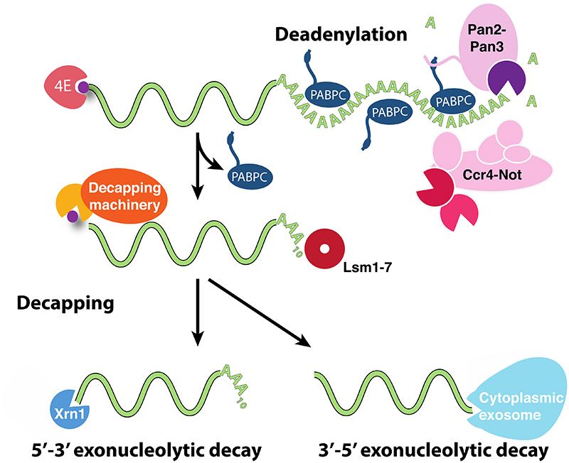
Figure 3. Eukaryotic mRNA deadenylation and decay.
Before an mRNA can be degraded, its poly(A) tail is removed by the Pan2-Pan3 and/or Ccr4-Not deadenylation complexes. This releases PABPC and may weaken the association of the eukaryotic translation initiation factor 4E (eIF4E) with the 5′-cap (purple circle). The decapping machinery can then access and remove the 5′-cap. Lsm1-7 can associate with oligo-A or 3′ uridylated tails to help recruit the decapping machinery. Decapping is followed by degradation of the mRNA in the 5′-3′ direction by Xrn1 or in the 3′-5′ direction by the cytoplasmic exosome.
A physical interaction between the 5′- and 3′-ends of mRNAs via eIF4E-eIF4G-PABPC in a ‘closed-loop’ provides a possible explanation for how the poly(A) tail influences decapping. These interactions stabilize eIF4E on the cap58, and could prevent decapping enzyme association. In agreement with this, eIF4E and the decapping complex compete for cap access, at least in vitro89,90. Moreover, it has also been suggested that PABPC in humans can interact directly with the 5′ cap91. Stabilization of mRNA by PABPC, therefore, could be due to simple steric inhibition of decapping enzyme association. Consistent with this, deadenylated mRNAs with short oligo(A) tails do not efficiently bind PABPC but are capable of binding the heptameric Lsm1–7 complex of proteins, which in turn physically binds the decapping complex thereby connecting the 5′- and 3′-ends92–94.
Taken together, the 5′-end of the mRNA appears to contribute to poly(A) control of mRNA stability and mRNA translation. However, some data do not agree with this. For example, deletion of PAB1 in yeast results in an increase in the steady state length of poly(A) tails49,51 and a reduced rate of poly(A) tail shortening51. The opposite effect would be expected if Pab1 simply acts to block 3′ exonucleases from accessing the poly(A) tail. Similarly, PABPC1 is required for efficient deadenylation in a mouse extract system95. These in vivo experiments on PABPC are complicated by its pleotropic effects and needed to be integrated with data addressing how poly(A) tails are removed. This will be further discussed below.
IV. Modulation of poly(A) tail length
Poly(A) tails are shortened or removed by dedicated exonucleases that act specifically on the adenosines (deadenylases). Two protein complexes account for the major cytoplasmic deadenylation activities in eukaryotes: Pan2-Pan3 and Ccr4-Not (Table 1).
Table 1. Major eukaryotic deadenylases.
| H. sapiens | S. cerevisiae | S. pombe | ||
|---|---|---|---|---|
| Pan2-Pan3 | PAN2 | Pan2 | DEDD exonuclease | |
| PAN3 | Pan3 | RNA binding, scaffold | ||
| Ccr4-Not | CNOT1 | Not1 | Not1 | Scaffold |
| CNOT2 | Not2 | Not2 | NOT box scaffold | |
| CNOT3 | Not3, Not5 | Not3 | NOT box scaffold | |
| (CNOT4) | Not4 | Mot2 | RING E3 ligase | |
| CNOT6/6L | Ccr4 | Ccr4 | EEP exonuclease | |
| CNOT7/8 | Pop2 | Caf1 | DEDD exonuclease | |
| CNOT9 | Caf40 | Rcd1 | Protein-protein interaction | |
| CNOT10 | ||||
| CNOT11 | RNA binding | |||
| Caf130 | ||||
A. Deadenylation enzymes
Pan2-Pan3 was first identified through fractionation of yeast extracts and it contains a DEDD/RNaseD-type exonuclease in its Pan2 subunit96–98. Within the complex, two copies of the Pan3 protein form an asymmetric homodimer which acts as a scaffold for one Pan2 molecule99–101. The genes encoding Pan2 and Pan3 are not essential in yeast: deletion of either gene results in longer poly(A) tails at steady state but does not eliminate deadenylation because Pan2-Pan3 is partially redundant with Ccr4-Not97,98.
Pan2-Pan3 is recruited to poly(A) RNA through at least three interactions: a Pan3 N-terminal zinc finger specifically binds adenosines99, a PAM2 motif in Pan3 binds PABPC102, and additional regions of the complex interact with the PABPC:poly(A) RNP44. Thus, by recruiting Pan2-Pan3 to poly(A) RNA, Pab1 stimulates deadenylation. This has been observed both in vivo and using fully purified components in vitro99,103. The exonuclease active site of Pan2 additionally selects for poly(A) RNA because it recognizes an intrinsic, single-stranded helical conformation of RNA that is uniquely formed by poly(A) (Box 1)104 Pan2-Pan3 can be recruited to specific transcripts via an interaction with the GW182 protein within the miRNA-induced silencing complex (miRISC)105,106, but it remains unclear how or if Pan2-Pan3 is specifically recruited to other transcripts.
Box 1. The structure of poly(A) RNA.
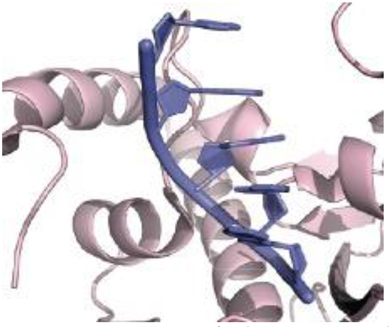
What makes poly(A) RNA unique? Biophysicists have long known that poly(A) has unique properties among polyribonucleotides. Poly(A) RNA forms a single-stranded A-form-like helix (Box 1 Figure) whose structure was originally proposed from a crystal structure of two adenosines in a stacked arrangement192. More recent crystal structures with longer segments of RNA directly show that poly(A) RNA adopts this helical configuration when bound to Pan2104,193. Although Pan2 has nucleotide specificity, no base-specific contacts were observed in the co-crystal structure. Instead, Pan2 (and likely Caf1) contacts the ribophosphate backbone and specifically recognizes poly(A) RNA through this unique A-form helical structure.
The structure of poly(A) is not only recognized by nucleases but also by ribosomes that have translated through the 3′-UTR into the poly(A) tail, for example on mRNAs lacking a translation termination codon194,195. This contributes to activation of a quality control pathway that eliminates those mRNAs and their associated polypeptide chains.
Box 1 Figure.
Structure of poly(A) RNA (purple) bound to the Pan2 exonuclease domain (pink) (PDB 6R9J)42. The poly(A) RNA forms an A-form like, single-stranded helix where the bases are stacked on top of each other.
The second major deadenylase, Ccr4-Not, is a 0.5 MDa complex with seven core subunits including two exonucleases: Caf1 (also known as Pop2, CNOT7 or CNOT8), a DEDD-type exonuclease, and Ccr4 (CNOT6, CNOT6L), an EEP-type exonuclease79,107–110). Lack of Caf1 or Ccr4 in yeast results in slowed and incomplete deadenylation107,111. The Not1 subunit acts as a scaffold to assemble Ccr4-Not and is the only subunit essential for viability in yeast, possibly because Not1 acts to integrate several non-redundant functions of Ccr4-Not112,113. The Not2, Not3 and Not5 subunits are linked to decapping114,115 and Caf40/Rcd1/Not9 is required for interaction with several RNA-binding proteins and RNA116–118. Finally, Not4 is an E3 ubiquitin ligase that monoubiquitinates ribosomal proteins (as will be discussed below) and may also promote protein degradation119–122. Ccr4-Not is specifically recruited to transcripts by RNA-binding proteins (discussed in detail below).
A third deadenylase, PARN (poly(A)-specific exoribonuclease), has also been identified123–125. PARN is not found universally in all eukaryotes (orthologues have been identified in vertebrates only). It may have more specialized roles for instance in the maturation and stability of snoRNAs, piRNAs or miRNAs126–129, and will not be discussed further.
1. Biphasic model of deadenylation
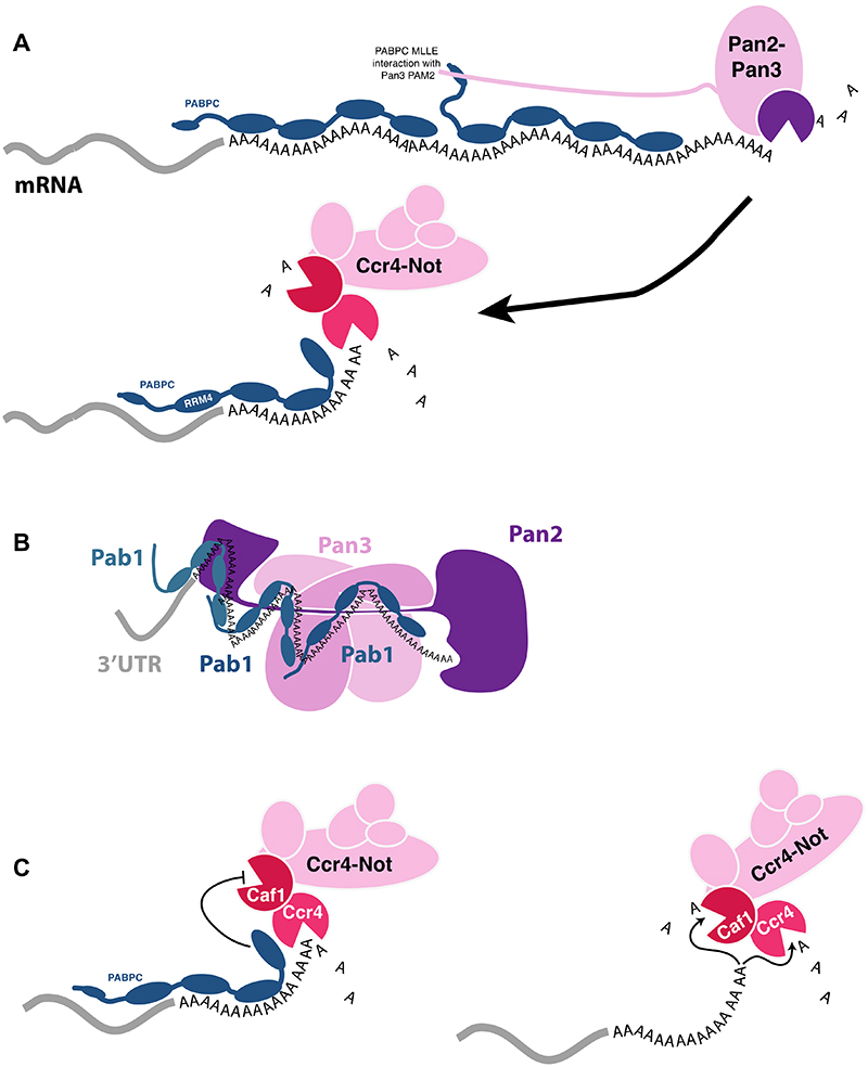
A major question is why there are multiple deadenylation enzymes in the cell. Do they target different sets of mRNAs or do they respond to different stimuli? One hypothesis is that the major deadenylation complexes act in a biphasic (or sequential) model of deadenylation where Pan2-Pan3 removes the distal part of the poly(A) tail and Ccr4-Not removes adenosines more proximal to the 3′-untranslated region (UTR) (Figure 4A).
Figure 4. Deadenylation by Pan2-Pan3 and Ccr4-Not.
A Sequential (or biphasic) model of deadenylation. In this model, Pan2-Pan3 preferentially removes the distal part of the poly(A) tail. A PABPC-interacting motif 2 (PAM2) within an intrinsically-disordered segment of Pan3 interacts with the C-terminal mademoiselle (MLLE) domain in PABPC. Ccr4-Not removes the poly(A) tail that is more proximal to the 3′-UTR. The most 5′ PABPC protein may be positioned on the poly(A) tail such that its RRM4 is located on the 3′-UTR of the mRNA. B Model of the Pan2-Pan3-Pab1-poly(A) complex. A cryoEM structure (PDB 6R5K)44 shows that Pan2-Pan3 contacts the interface between adjacent Pab1 molecules, providing an explanation for why it preferentially functions on longer poly(A) tails. In the structure, three Pab1 molecules are bound to a 90 nt poly(A) tail. C Ccr4 is a general deadenylase that degrades poly(A), even when it is bound by PABPC. Caf1 is a specialized deadenylase that degrades naked poly(A) and is blocked by PABPC130.
Deletion of Pan2 in yeast results in longer poly(A) tails at steady state97, consistent with a defect in removing the distal poly(A) sequences. In contrast, deletion of Ccr4 results in an accumulation of poly(A) tails with a length of 20–40 nt, and changes the endpoint of deadenylation from ~10 nt to ~20 nt107,130, suggesting that it has a major role in removing poly(A) sequences proximal to the 3′-UTR. Deletion of both Pan2 and Ccr4 exacerbates the defects in deadenylation107. Similar results were obtained using in vitro studies44. Experiments in mammalian cells126,131 also showed that PAN2 is important for a slower, initial phase of deadenylation involving removal of the distal part of the longer mammalian poly(A) tail (200–110 nt), whereas CCR4 affects a second, fast phase to remove the final 110 nt of the poly(A) tail. The activity of the Ccr4-Not complex may be more processive than Pan2-Pan3 in many cases, and this may account for its more rapid deadenylation. Thus, Pan2 and Ccr4 have partially overlapping functions but they appear to act predominantly at different points in poly(A) tail removal. Pan2-Pan3 does not appear to substantially affect mRNA half-life and may be more important under specific cellular circumstances.
A recent cryoEM structure of the yeast Pan2-Pan3 complex provides an explanation for why it preferentially acts on the distal (not the proximal) poly(A) tail 44. In the structure, a 90 nt poly(A) RNA is bound by three Pab1 molecules. The poly(A)-Pab1 RNP winds across the surface of Pan2-Pan3 in a zigzag configuration (Figure 4B). Adjacent Pab1 molecules interact with each other and with the deadenylase complex. Since Pan2-Pan3 recognizes the Pab1 oligomerization interface, it preferentially acts on poly(A) RNA that is long enough to accommodate multiple Pab1 molecules. This model therefore suggests that as the poly(A) tail is shortened, its affinity for Pan2-Pan3 progressively decreases. It will be interesting to determine how the mammalian Pan2-Pan3 achieves a similar function on longer poly(A) tails.
Both Pan2-Pan3 and Ccr4-Not act specifically on poly(A) sequences. Therefore, they remove the poly(A) tail and do not act on the 3′-UTR. mRNA decay occurs only when the poly(A) tail becomes very short (<10 nt). Therefore, in the sequential model, Ccr4-Not plays a more important role than Pan2-Pan3 in triggering mRNA decay: Pan2-Pan3 initiates deadenylation and Ccr4-Not completes it. In agreement with this, knockdown of Ccr4-Not components in mammalian cells results in an overall increase in mRNA half-lives but knockdown of Pan2-Pan3 does not have a major effect126.
2. Separable roles of Ccr4 and Caf1
But what are the roles of the two different nucleases within Ccr4-Not? In budding yeast, Ccr4 deletion has a greater effect on deadenylation than deletion of Cafl 107,108,130. However, as mentioned above, homeostatic mechanisms can compensate for disruption of gene expression in vivo, complicating interpretation of these experiments.
Fully reconstituted systems to study deadenylation have helped to clarify the roles of Caf1 and Ccr4. Using purified, recombinant S. pombe proteins, it is possible to make point mutations in active site residues to dissect the roles of individual nucleases130,132. In these in vitro experiments, both Ccr4 and Caf1 shorten poly(A) tails. Point mutations in either nuclease did not strongly influence deadenylation activity on naked poly(A) RNA. However, Caf1 did not act on Pab1-bound RNA. In contrast, Ccr4 binds Pab1 and can release it from poly(A) tails. Similar conclusions were obtained for human Ccr4-Not126. Together, these data suggest that the two nucleases in Ccr4-Not play different functional roles: Caf1 degrades naked poly(A) RNA and is blocked by PABPC, whereas Ccr4 is able to release PABPC to act on PABPC-bound poly(A) RNA (Figure 4C). Moreover, Caf1 acts in a translation-dependent manner whereas Ccr4 is not dependent on translation per se130.
This work showed that PABPC does not simply act to block the 3′-end of the RNA. In fact, PABPC stimulates both Pan2 and Ccr4, explaining why deletion of Pab1 results in a reduced rate of poly(A) tail shortening in yeast. These data also suggest that there may be regulatory mechanisms to control which nuclease is used.
B. What controls deadenylation rate?
The deadenylation rates and half-lives of different transcripts can vary by >1000-fold 16. This raises the question of how poly(A) tails are differentiated by deadenylases to allow transcript-specific mRNA decay. The answer to this lies, unsurprisingly, not in the poly(A) tail itself but in other parts of the transcript. First, specific sequences (often in the 3′-UTR) are recognized by RNA-binding proteins that recruit deadenylases to specific transcripts. Second, RNA sequence affects translation elongation rate, which is also a major determinant of mRNA half-life. A recent model suggested that mRNA sequence accounts for almost 60% of the variation in mRNA half-lives133: 3′-UTR motifs explain 5.5% of the variation while codon usage explains 55%. These two factors will be discussed in detail below.
Most of the known regulatory processes for deadenylation act through the Ccr4-Not complex. It remains unclear whether Pan2-Pan3 activity is regulated (e.g. by translation), whether Pan2-Pan3 is targeted to specific transcripts by multiple different RNA adapter proteins, or whether it acts as a general factor to ‘trim’ long poly(A) tails. Given that P an2-Pan3 is conserved across evolution, we expect it to play a major, as-yet-undiscovered role – perhaps on a subset of transcripts, or only in response to specific situations.
1. Targeted deadenylation
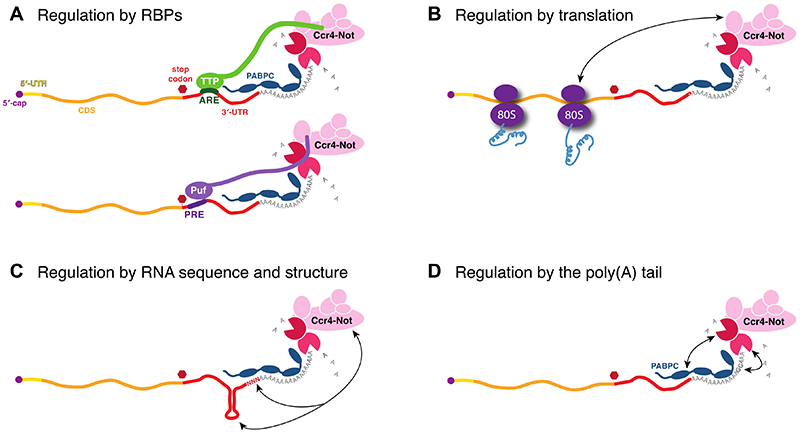
It is well-established that Ccr4-Not is recruited to specific mRNAs through association with RNA-binding proteins that act as adapters between specific sequences in the 3′-UTR and the deadenylase enzymes (Figure 5A). Widely-studied examples include Tristetraprolin/TTP/Tis11d family members that recognize AU-rich elements (AREs)134,135 and Pumilio/Puf proteins that use a modular system to recognize Pumilio-response elements (PREs)136–139. The microRNA-induced silencing complex (miRISC) also mediates targeted deadenylation as its GW182 subunit interacts directly with the Pan2-Pan3 and Ccr4-Not complexes140. Many other RNA adapters have been identified in recent years including Nanos, Roquin, and YTHDF2116,117,132,141.
Figure 5. Factors that influence deadenylation rate.
The deadenylation rate of Ccr4-Not is modulated by several factors. A RNA-binding proteins, such as Tristetraprolin (TTP) and Puf/Pumilio proteins act as RNA adapters to tether Ccr4-Not to specific transcripts, accelerating their deadenylation139,142. TTP binds to AU-rich elements (AREs) in the 3′- untranslated region (UTR)134,135 and Puf proteins bind to Pumilio-recognition elements (PRE)136. B Slow translation elongation rate results in more rapid deadenylation, likely through direct interactions between Ccr4-Not and the ribosome122. C RNA structure in the 3′- UTR and changes in the final nucleotides of the 3′-UTR influence deadenylation, possibly by altering Ccr4-Not or PABPC affinity for the 3′-UTR15,118,132,170. D Insertion of nucleotides other than adenosine into the poly(A) tail can stall deadenylation by Ccr4-Not and Pan2- Pan3104,176. In all panels, the 5′-cap is depicted as a purple circle; 5′-UTR is in yellow; coding sequence (CDS) is in orange; stop codon is a red hexagon; 3′-UTR is in red; and PABPC is in blue. Pan2-Pan3 deadenylation rate may also be influenced by the same factors.
The RNA adapter proteins that mediate targeted deadenylation often contain extended intrinsically-disordered regions (IDRs), in addition to their RNA-binding domain. Short motifs within these IDRs interact with Ccr4-Not, and often multiple motifs from the same RNA adapter interact with one deadenylase complex suggesting complicated multipartite interfaces142–146. The interacting sequences within IDRs are difficult to identify because they are not highly-conserved at the sequence level and binding mechanisms of orthologous proteins sometimes vary between species147.
Artificially tethering RNA adapters to reporter transcripts in cells results in increased deadenylation and RNA decay117. Similarly, using in vitro fully reconstituted systems, RNA adapters including TTP- and Puf-family proteins substantially accelerate deadenylation on target RNAs139,142. RNA adapter proteins therefore act as molecular tethers between mRNAs and deadenylation complexes. By physically tethering enzyme and substrate together, deadenylation rate is increased in a transcript-specific manner and deadenylation activity is more processive. This is highly tunable since regulatory mechanisms can modulate affinities between the RNA adapter and mRNA, and between the RNA adapter and the deadenylation complex. For example, phosphorylation of TTP disrupts binding to Ccr4-Not to stabilize mRNAs involved in the inflammatory response in cells148. There are a number of other mechanisms for regulating how RNA adapters affect deadenylation, for example through collaboration or competition for RNA binding sites by RNA adapters149–151.
2. Translation and deadenylation rate
A growing body of data suggests that translation impacts poly(A) tail dynamics (Figure 5B). For instance, inhibition of translation initiation in either cis or trans enhances the deadenylation rate of long-lived transcripts90,152–154. Moreover, translation elongation rate can impact deadenylation rate from yeast to humans, and this is mediated by codon optimality130,155–165. Codon optimality is the concept that each of the 61 codons is not read by the ribosome at the same rate. Subtle distinctions in tRNA concentration, composition and flanking codon identity can alter decoding kinetics and these effects are additive across a transcript, setting the overall elongation rate for the mRNA166. Surprisingly, this rate is read by the mRNA deadenylation and decapping complexes; mRNAs hosting slow moving ribosomes are targeted for rapid deadenylation and decapping in a Caf1-dependent manner, whereas mRNAs with relatively fast ribosome movement evade poly(A) tail shortening and decapping more effectively. It is important to note that the classic definition of codon optimality, i.e. variability in tRNA concentration is not the only feature that can slow elongation in such a way that elicits faster poly(A) shortening and decapping. Indeed, other features such as tRNA charging, tRNA and mRNA modifications, amino acid identity and concentration, mRNA sequence and structure, codon context, polypeptide composition within the ribosome exit channel, and folding of the nascent polypeptide may also slow translational elongation in such a way to be read by the deadenylase and decapping complexes166.
The emerging picture is that the deadenylase and decapping complexes senses a particular ribosome conformation which is the result of slowed elongation. There are now several documented interactions of Ccr4-Not with the translational apparatus. The Ccr4-Not complex binds and ubiquitinates (through Not4) the Nascent-Associated polypeptide Complex (NAC) on the ribosome167. Moreover, Not4 binds and ubiquitinates the ribosomal protein eS7 in response to stress168,169. Lastly, Not5 (CNOT3 in mammals) was recently shown to bind to translating ribosomes with empty A- and E-sites, a condition that occurs on transcripts with nonoptimal codon content122. Importantly, deletion of the Not5 domain that binds the ribosomal E-site or mutation of the ubiquitination sites in ribosomal protein eS7 stabilizes transcripts that contain non-optimal codons, and prevents association of the decapping activator Dhh1122. Thus, Not5 binding to the ribosome and Not4-mediated ubiquitination of eS7 likely directly detect and/or signal slow elongation to the deadenylation and decapping machinery.
Together, these data suggest that a key feature in regulating transcript-specific deadenylation is the monitoring of distinct ribosome activities or states by the deadenylase itself. A slowly elongating ribosome may, in fact, be another type of RNA adapter that connects mRNAs with Ccr4-Not. Thus, it is possible that almost all cellular deadenylation occurs through a conceptually similar recruitment step by an RNA adapter – either an RNA-binding protein, a ribosome, or another unknown factor – and deadenylation is too slow to play a major role in mRNA decay in the absence of an RNA adapter.
3. Other contributing factors
A large-scale study of mRNA half-life in yeast, as well as studies in human cells, implicated RNA sequence and structure as major determinants of mRNA stability15,170. For example, inclusion of poly(U) within the 3′-UTR (which can base-pair with poly(A) and prevent Pab1 binding) or other stem-loop structures within the 3′-UTR results in increased mRNA half-life (Figure 5C). Stem-loops may block deadenylase recruitment to single-stranded RNA132 or may alter the binding of PABPC RRM4 to the 3′-UTR130. Also, small variations in the final 3 nt of a transcript due to alternative polyadenylation can result in isoforms that have ≥2-fold change in half-lives15 (Figure 5C). In agreement, RNA sequence can have a direct effect on deadenylation rate in vitro132. Thus, the direct recruitment of deadenylase complexes to specific sequences may also have an influence on deadenylation rate118.
Other protein factors regulate the interplay between deadenylases and PABPC to control deadenylation. Many of these regulators have PAM2 motifs that allow direct interaction with the MLLE domain of PABPC39. For example TOB proteins interact with Ccr4-Not and also contain a PAM2 motif that binds PABPC to promote deadenylation171. The interaction between TOB2 and PABPC is regulated by phosphorylation172. LARP1 and LARP4 bind directly to poly(A) RNA and to PABPC1 via a PAM2 motif173. In doing so, they protect mRNAs from deadenylation, possibly by stabilizing PABPC binding174. The translation termination factor eRF3 interacts with PABPC through a PAM2 motif, thereby linking the poly(A) tail with translation175. Other poly(A) regulatory proteins also contain PAM2 motifs including GW182 and PABPC-interacting protein 1 (PAIP1) and PAIP239. Together, PAM2-containing proteins contribute to poly(A) tail dynamics.
Finally, insertion of ribonucleotides other than adenosine may slow deadenylation in a transcript-specific manner, at least in part because guanosine disrupts the helical conformation of poly(A) RNA (Figure 5D; Box 1). Sequencing methods have shown that non-adenosine residues are sometimes found in poly(A) tails in cells, and the presence of guanosine correlates with mRNA stability176–178. Guanosine is the least efficiently removed by the deadenylation machinery in vitro104. Cytosines and uracils are removed by Pan2-Pan3 slightly less efficiently than guanosine but Ccr4-Not is more selective104. Interestingly, viruses can promote addition of non-As into poly(A) tails of viral transcripts, protecting them from the host deadenylation machinery179. 3′ oligo-uridyl tails are often found on transcripts downstream of short (<25 nt) poly(A) tails, consistent with uridylation acting as a decay signal176,180.
V. Reconsidering mRNA metabolism
There has been a recent resurgence in interest in poly(A) tail biology. Developments in sequencing methods, protein expression technology and cryoEM have allowed new investigations into the length and composition of poly(A) tails, and the mechanisms of deadenylase complexes.
A. Poly(A) tail length
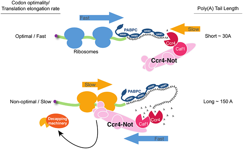
Until recently, an understanding of how poly(A) tail length regulates mRNA metabolism remained limited, in part because analysis of poly(A) tail length was difficult to perform on a transcriptome-wide level. New developments in sequencing methodologies (e.g. TAILseq, mTAILseq, PALseq, FLAMseq, PATseq, Nanopore sequencing) now enable both the length and sequence of poly(A) tails to be analyzed31,32,176,178,181,182. The range of poly(A) tail lengths was found to be large, and poly(A) tails that were much longer (>250 nt in human) than expected were found178,182. However, very long tails are relatively rare. Surprisingly, multiple studies found that the length of poly(A) tails of highly-translated, stable RNAs is relatively short (~30 As) at steady state32 (Figure 6). In contrast, poorly translated mRNAs have comparatively long poly(A) tails. Overall, the modal poly(A) tail length is 30 nt in yeast and 50–100 nt in other eukaryotes including human, mouse, Drosophila and C. elegans. This is consistent with earlier results showing that stable transcripts in Dictyostelium can have relatively short poly(A) tails of 40–60 nt183. Poly(A) tail lengths in vivo and in vitro are phased in ~30 nt increments, consistent with multiple PABPCs bound to the poly(A) tail32,44,126,130.
Figure 6. Summary of recent insights into gene regulation by poly(A) tails.
Translation efficiency is depicted by ‘fast’ or ‘slow’ moving ribosomes on transcripts with optimal or non-optimal codons, respectively. Poly(A) tails can be short (~30 As) on stable, highly- translated mRNAs. The concentration of cytoplasmic poly(A)-binding protein (PABPC) may also affect the role of poly(A) tails in gene expression. Ccr4-Not may ′read’ translation elongation rate by detecting ribosomes containing empty A- and E-sites and this recruits the decapping machinery.
These findings have a number of important implications. First, since highly-translated transcripts have short poly(A) tails of ~30 adenosines, a length that would accommodate only a single PABPC, it suggests that one PABPC is sufficient to promote efficient translation. Second, it was previously assumed that a longer poly(A) tail correlated with increased mRNA stability. However, these new data on poly(A) tail length showed that transcripts with poly(A) tails that have been shortened to ~30 nt can also be stable. In fact, steady-state poly(A) tail length and mRNA half-life were found to be poorly or inversely correlated, and PABPC occupancy on mRNAs does not correlate with steady-state poly(A) tail length46,184. However, a study examining the kinetics of deadenylation instead of steady-state tail length does show a correlation between mRNA half-life and deadenylation rate16. Third, these findings provided a strong link between translation efficiency and deadenylation, supporting the idea that translation rate is directly related to mRNA stability.
Finally, the global phasing of poly(A) tail length in 30 nt increments suggested that PABPC does not bind randomly across the poly(A) tail. Instead, the first PABPC binds at the junction of the 3′-UTR and the poly(A) tail. Subsequent PABPC molecules bind downstream of this initial PABPC and any adenosines not bound by PABPC are likely removed rapidly. In vitro reconstitution of deadenylation agrees with this – one Pab1 molecule binds at the junction of the 3′-UTR and poly(A) tail, and Ccr4-Not releases one Pab1 molecule at a time during deadenylation126,130. RRM4 of the Pab1 molecule most proximal to the transcript body may bind the 3′-UTR130, particularly when the poly(A) tail length is insufficient to accommodate RRM4 binding (Figure 4A). In agreement with this, RRM4 is highly-conserved but is not selective for poly(A)40. Although Ccr4-Not activity removes the final part of the poly(A) tail, and is often thought of as being ‘faster’ than Pan2-Pan3, it is clear that it does not act equally on all short poly(A) tails because stable, highly-translated transcripts are more resistant to deadenylation. The mechanisms of such resistance remain unclear. Fast deadenylation by Ccr4-Not may only occur when Ccr4-Not is tethered to a transcript by an RNA adapter protein.
In summary, it is now clear that poly(A) tail length is not only regulated in a transcript-specific manner, but poly(A) tail metabolism also controls gene expression in a more complex, transcript-specific way that likely depends on translation. Whether other factors, e.g. mRNA localization, also act as determinants of poly(A) tail function remains to be tested. With new tools available, experiments to test this are now possible.
B. PABPC
Despite an omnipresence in translation and mRNA decay, the roles of PABPC remain largely enigmatic. How does PABPC stimulate translation? Do all mRNAs form closed loops? If not, how do the 5′- and 3′-ends of the mRNA communicate? How many PABPC molecules are loaded on an average poly(A) tail and how are they arranged on the poly(A) tail? Is PABPC binding different on different RNAs? What is the effect of PABPC stoichiometry on deadenylation?
A complete molecular understanding of PABPC loading onto poly(A) would facilitate an understanding of its function. The recent structure of Pan2-Pan3 bound to a Pab1-poly(A) RNP combined with the earlier structure of Pab1 RRMs 1 and 2 provides important new insight into the overall arrangement of Pab1 on poly(A) RNA42,44. However, due to the limited resolution of the cryoEM structure, the precise molecular details of the interactions (e.g. which amino acids in Pab1 contact RNA and adjacent Pab1 molecules) remain unclear. This will be important to clarify to allow perturbation and evaluation of the importance of Pab1 oligomerization in gene expression.
In terms of function, the closed-loop model for how PABPC controls mRNA metabolism is enticing and has made its way into numerous textbooks as canon. But the described interaction between PABPC and eIF4G does not seem to fully capture the role of PABPC in either controlling mRNA decapping or translation. In yeast, the loss of Pab1 results in premature decapping while the mRNA still has a long poly(A) tail. However, a similar phenotype is not observed when eIF4G function is impaired in a wild-type Pab1 background or when Pab1 is tethered to mRNAs; deadenylation is still a prerequisite to mRNA decapping90, 185. Thus, Pab1 is required to render decapping dependent on deadenylation. Blocking translation initiation in either cis or trans also does not change the coupling between deadenylation and decapping89,152,153. Depletion of PABPC in mammalian cells destabilizes mRNAs and this phenotype can be rescued by the addition of PABPC mutants that cannot bind eIF4G17. Thus, simply breaking the PABPC–eIF4G interaction does not abrogate the ability of PABPC to stabilize mRNA.
Perhaps a simple model of PABPC in contact with eIF4G does not completely capture what is truly occurring on mRNA. Consistent with this, at least two distinct closed-loop states were found to exist on an mRNAs64: The ‘classic’ closed-loop is mediated by PABPC and eIF4G (with other initiation factors) while a second closed-loop is mediated by PABPC and an interaction with the 80S ribosome and translational termination factors (eRF3 and eRF1). PABPC serves an important role in translation termination, leading to discrimination of normal termination events from aberrant events186. Thus, it is emerging that communication between the 5′- and 3′-ends may occur through mechanisms that do not solely rely on the PABPC-eIF4G-mediated closed loop and instead may involve other factors including the ribosome67. The translating mRNP architecture is complex and the true role for PABPC in protecting mRNA from degradation and promoting translation remains unknown. To gain further insight into the importance and prevalence of the closed-loop model, we will require new experiments that monitor how translation and mRNA stability are modulated by mutations that disrupt the closed loop without disrupting PABPC or eIF4E association, ideally in a cellular environment.
Recent work suggests that PABPC concentration in cells might also be critical for mediating poly(A) effects on translation and stability17. In early oocytes, PABPC concentrations in the cell are limited; this results in longer-tailed mRNAs outcompeting shorter-tailed mRNAs for PABPC binding so the transcripts with longer tails are more robustly expressed. In contrast, in many somatic cells PABPC concentrations are not limiting and it is therefore presumed that most poly(A) RNA is bound by PABPC. Unlike in oocytes, in many somatic cells deadenylated mRNAs are unstable, and PABPC does not strongly influence translation efficiency. Together, these data help to rationalize why poly(A) tail length has different effects in different cell types, i.e. poly(A) tail length correlates with translation efficiency only in specialized cells.
Consistent with these findings, PABPC levels are known to be tightly regulated within the cell. The PABPC gene encodes an extensive A-rich sequence in its 5′-UTR. This poly(A) stretch binds to the PABPC protein, and represses its own translation through steric inhibition of ribosome joining187. Thus, an autoregulatory feedback determines the ratio of PABPC to poly(A) tails within the cell. This may be critical in determining how poly(A) tail length mediates post-transcriptional regulation. Future work should further address how PABPC levels are controlled.
The influence of PABPC on deadenylation also remains unclear. The Ccr4 enzyme can release PABPC from RNA but Caf1 cannot126,130. Is this regulated? What is the effect of PABPC stoichiometry on Ccr4-Not activity and how does this influence mRNA stability? PABPC has very high affinity for poly(A) RNA – one could imagine that there are active mechanisms to remove PABPC from RNA and remodel its distribution over the transcriptome. If PABPC straddles the 3′-UTR and poly(A) tail130, some 3′-UTR sequences might be more favorable for PABPC binding, especially under situations where the PABPC concentration is limiting. There is also some evidence that active translation affects the stability of PABPC binding130. Elucidating the details of these processes will be important.
C. Coupling 5′ and 3′ RNA degradation
All RNA processing events (i.e. pre-mRNA splicing, ribosome maturation, miRNA biogenesis, etc.) are hallmarked by a series of ribonucleoprotein transitions, with one complex serving as the foundation upon which the next complex is built. In mRNA splicing, for example, the order of ribonucleoprotein transitions ensures precise intron excision and exon ligation188. But considering the process of mRNA deadenylation and decapping, there is little information on how over 30 polypeptides interact with each other and with the mRNA. It can be assumed that higher order RNP assembly is, at least in part, responsible for how the rates of both deadenylation and decapping are achieved. Moreover, it is likely that ordered complex assembly also helps ensure that decapping doesn’t occur prior to deadenylation.
Deadenylation and decapping appear to directly communicate with each other via the ribosome. First, the deadenylase components Not1, Not2, Not4, and Not5 have all been shown to be required for efficient mRNA decapping114,122. Second, the decapping protein Dhh1 (DDX6 in humans) is also known to communicate with Ccr4-Not through Not1189. Third, Dhh1 and the deadenylase proteins Not4 and Not5 (CNOT3 in humans) physically bind the ribosome122,190. Lastly, Not5 binding to the ribosome is required for Dhh1 binding to the ribosome122. This latter observation provides the first evidence that the deadenylase directly influences the decapping complex. Of interest Not5 can only associate with ribosomes having vacant A- and E-sites and this may explain how Ccr4-Not ‘reads’ translation elongation rate (Figure 6).
It is tantalizing to speculate that both deadenylase and decapping proteins not only bind to unique ribosome states, but also stabilize them, thereby facilitating downstream factor assembly and committing the bound mRNA for decay - this is perhaps exemplified by the observation that Dhh1 ribosome binding depends on Not5 ribosome binding. We anticipate that understanding how the decapping and deadenylase complexes interface and interact with the translating ribosome will be critical in solving the mystery of how the mRNA 3′-end controls the 5′-end.
VI. Conclusions and future perspective
It has been over 50 years since mRNAs were shown to be polyadenylated at their 3′-end. In this time, poly(A) has emerged as a central regulator of mRNA translation and stability. And because of numerous advances, dogmatic notions are being challenged by new tools, structural analysis, and biochemical reconstitution experiments. For instance, the relationship between poly(A) tail length and translation efficiency is being reconsidered. The pervasive “closed-loop” model is being challenged. And major advances in understanding deadenylase function have emerged that are providing insight into biology.
We view that the next few years promise to bring a renaissance into the study of post- transcriptional mRNA regulation, especially as mediated by the poly(A) tail. Moving forward, we see several major goals as being important:
-
-
To document and understand the regulation of deadenylation rates.
-
-
To understand how ribonucleoprotein context (i.e. RNA sequence, structure, codon content, RNA modifications, etc.) modulates deadenylase activity.
-
-
To understand the specificity and roles of the various deadenylase enzymes.
-
-
To gain a clearer picture of the translating mRNP and the events involved in stimulating translation and mRNA stability.
-
-
To understand the differential roles of PABPC isoforms and determine if they provide regulatory flexibility.
-
-
To fully understand how PABPC functions in mediating 5′ events.
-
-
To revisit how poly(A) length controls mRNA metabolism.
-
-
Moreover, we must gain a more detailed view of how poly(A) metabolism functions in unique biological contexts and cell types.
-
-
Lastly, we must understand how the deadenylase and its various components interface with the ribosome.
The emergence of mRNA-based therapeutics and vaccines will undoubtably advance this area, as basic biology is leveraged to advance human health. These applications are dependent on understanding the cytoplasmic life of an mRNA and manipulating it accordingly for therapeutic efficacy. We anticipate, therefore, that this tail is far from over.
Acknowledgements
We thank Drs. Allan Jacobson, Marvin Wickens, Feng He, Terence Tang, Eva Absmeier, Jay Leipheimer, Sophie Martin, Ryan Kawalerski and members of the Passmore and Coller labs for helpful insight and discussion. This review would not have been possible without contributions from many individuals. This field has been rich in discovery, collaboration, and friendship. We thank all of you and apologize for any oversights we may have made. In particular, we dedicate this review to the memory of Drs. Elisa Izaurralde and Richard Jackson; their seminal discoveries and keen intellect have inspired us all.
Funding is provided to L.A.P. by the European Union’s Horizon 2020 research and innovation programme (ERC grant No. 725685) and the Medical Research Council, as part of United Kingdom Research and Innovation (MRC grant No. MC_U105192715); and to J.C. by the National Institutes of Health (USA; GM118018 and GM125086) and Bloomberg Philanthropies.
Footnotes
Competing Interests
The authors declare no competing interests.
Literature Cited
- 1.Edmonds M, Abrams R. Polynucleotide biosynthesis: formation of a sequence of adenylate units from adenosine triphosphate by an enzyme from thymus nuclei. J Biol Chem. 1960;235:1142–1149. [PubMed] [Google Scholar]
- 2.Hadjivassiliou A, Brawerman G. Polyadenylic acid in the cytoplasm of rat liver. J Mol Biol. 1966;20:1–7. doi: 10.1016/0022-2836(66)90112-4. [DOI] [PubMed] [Google Scholar]
- 3.Edmonds M, Caramela MG. The isolation and characterization of adenosine monophosphate-rich polynucleotides synthesized by Ehrlich ascites cells. J Biol Chem. 1969;244:1314–1324. [PubMed] [Google Scholar]
- 4.Burr H, Lingrel JB. Poly A sequences at the 3’termini of rabbit globin mRNAs. Nature New Biol. 1971;233:41–43. doi: 10.1038/newbio233041a0. [DOI] [PubMed] [Google Scholar]
- 5.Lim L, Canellakis ES. Adenine-rich polymer associated with rabbit reticulocyte messenger RNA. Nature. 1970;227:710–712. doi: 10.1038/227710a0. [DOI] [PubMed] [Google Scholar]
- 6.Kates J, Beeson J. Ribonucleic acid synthesis in vaccinia virus. II. Synthesis of polyriboadenylic acid. J Mol Biol. 1970;50:19–33. doi: 10.1016/0022-2836(70)90101-4. [DOI] [PubMed] [Google Scholar]
- 7.Edmonds M, Vaughan MH, Nakazato H. Polyadenylic acid sequences in the heterogeneous nuclear RNA and rapidly-labeled polyribosomal RNA of HeLa cells: possible evidence for a precursor relationship. Proc Natl Acad Sci U S A. 1971;68:1336–1340. doi: 10.1073/pnas.68.6.1336. [DOI] [PMC free article] [PubMed] [Google Scholar]
- 8.McLaughlin CS, Warner JR, Edmonds M, Nakazato H, Vaughan MH. Polyadenylic acid sequences in yeast messenger ribonucleic acid. J Biol Chem. 1973;248:1466–1471. [PubMed] [Google Scholar]
- 9.Gallie DR. The cap and poly(A) tail function synergistically to regulate mRNA translational efficiency. Genes Dev. 1991;5:2108–2116. doi: 10.1101/gad.5.11.2108. [DOI] [PubMed] [Google Scholar]
- 10.Vende P, Piron M, Castagné N, Poncet D. Efficient translation of rotavirus mRNA requires simultaneous interaction of NSP3 with the eukaryotic translation initiation factor eIF4G and the mRNA 3’ end. J Virol. 2000;74:7064–7071. doi: 10.1128/jvi.74.15.7064-7071.2000. [DOI] [PMC free article] [PubMed] [Google Scholar]
- 11.Hsu MT, Parvin JD, Gupta S, Krystal M, Palese P. Genomic RNAs of influenza viruses are held in a circular conformation in virions and in infected cells by a terminal panhandle. Proc Natl Acad Sci U S A. 1987;84:8140–8144. doi: 10.1073/pnas.84.22.8140. [DOI] [PMC free article] [PubMed] [Google Scholar]
- 12.Cakmakci NG, Lerner RS, Wagner EJ, Zheng L, Marzluff WF. SLIP1, a factor required for activation of histone mRNA translation by the stem-loop binding protein. Mol Cell Biol. 2008;28:1182–1194. doi: 10.1128/MCB.01500-07. [DOI] [PMC free article] [PubMed] [Google Scholar]
- 13.Holstege FC, et al. Dissecting the regulatory circuitry of a eukaryotic genome. Cell. 1998;95:717–728. doi: 10.1016/s0092-8674(00)81641-4. [DOI] [PubMed] [Google Scholar]
- 14.Wang Z-Y, et al. Transcriptome and translatome co-evolution in mammals. Nature. 2020;588:642–647. doi: 10.1038/s41586-020-2899-z. [DOI] [PMC free article] [PubMed] [Google Scholar]
- 15.Geisberg JV, Moqtaderi Z, Fan X, Ozsolak F, Struhl K. Global analysis of mRNA isoform half-lives reveals stabilizing and destabilizing elements in yeast. Cell. 2014;156:812–824. doi: 10.1016/j.cell.2013.12.026. [DOI] [PMC free article] [PubMed] [Google Scholar]
- 16.Eisen TJ, et al. The Dynamics of Cytoplasmic mRNA Metabolism. Mol Cell. 2020;77:786–799.:e10. doi: 10.1016/j.molcel.2019.12.005. [DOI] [PMC free article] [PubMed] [Google Scholar]
- 17.Xiang K, Bartel DP. The molecular basis of coupling between poly(A)-tail length and translational efficiency. bioRxiv. 2021:2021.01.18.427055. doi: 10.1101/2021.01.18.427055. [DOI] [PMC free article] [PubMed] [Google Scholar]
- 18.Wilt FH. Polyadenylation of maternal RNA of sea urchin eggs after fertilization. Proc Natl Acad Sci U S A. 1973;70:2345–2349. doi: 10.1073/pnas.70.8.2345. [DOI] [PMC free article] [PubMed] [Google Scholar]
- 19.Slater DW, Slater I, Gillespie D. Post-fertilization synthesis of polyadenylic acid in sea urchin embryos. Nature. 1972;240:333–337. doi: 10.1038/240333a0. [DOI] [PubMed] [Google Scholar]
- 20.Rosenthal ET, Tansey TR, Ruderman JV. Sequence-specific adenylations and deadenylations accompany changes in the translation of maternal messenger RNA after fertilization of Spisula oocytes. J Mol Biol. 1983;166:309–327. doi: 10.1016/s0022-2836(83)80087-4. [DOI] [PubMed] [Google Scholar]
- 21.Dworkin MB, Dworkin-Rastl E. Changes in RNA titers and polyadenylation during oogenesis and oocyte maturation in Xenopus laevis. Dev Biol. 1985;112:451–457. doi: 10.1016/0012-1606(85)90417-8. [DOI] [PubMed] [Google Scholar]
- 22.Dworkin MB, Shrutkowski A, Dworkin-Rastl E. Mobilization of specific maternal RNA species into polysomes after fertilization in Xenopus laevis. Proc Natl Acad Sci U S A. 1985;82:7636–7640. doi: 10.1073/pnas.82.22.7636. [DOI] [PMC free article] [PubMed] [Google Scholar]
- 23.Fox CA, Wickens M. Poly(A) removal during oocyte maturation: a default reaction selectively prevented by specific sequences in the 3’UTR of certain maternal mRNAs. Genes Dev. 1990;4:2287–2298. doi: 10.1101/gad.4.12b.2287. [DOI] [PubMed] [Google Scholar]
- 24.McGrew LL, Richter JD. Translational control by cytoplasmic polyadenylation during Xenopus oocyte maturation: characterization of cis and trans elements and regulation by cyclin/MPF. EMBO J. 1990;9:3743–3751. doi: 10.1002/j.1460-2075.1990.tb07587.x. [DOI] [PMC free article] [PubMed] [Google Scholar]
- 25.McGrew LL, Dworkin-Rastl E, Dworkin MB, Richter JD. Poly(A) elongation during Xenopus oocyte maturation is required for translational recruitment and is mediated by a short sequence element. Genes Dev. 1989;3:803–815. doi: 10.1101/gad.3.6.803. [DOI] [PubMed] [Google Scholar]
- 26.Sheets MD, Wu M, Wickens M. Polyadenylation of c-mos mRNA as a control point in Xenopus meiotic maturation. Nature. 1995;374:511–516. doi: 10.1038/374511a0. [DOI] [PubMed] [Google Scholar]
- 27.Barkoff A, Ballantyne S, Wickens M. Meiotic maturation in Xenopus requires polyadenylation of multiple mRNAs. EMBO J. 1998;17:3168–3175. doi: 10.1093/emboj/17.11.3168. [DOI] [PMC free article] [PubMed] [Google Scholar]
- 28.Wu L, et al. CPEB-mediated cytoplasmic polyadenylation and the regulation of experience-dependent translation of alpha-CaMKII mRNA at synapses. Neuron. 1998;21:1129–1139. doi: 10.1016/s0896-6273(00)80630-3. [DOI] [PubMed] [Google Scholar]
- 29.Doel MT, Carey NH. The translational capacity of deadenylated ovalbumin messenger RNA. Cell. 1976;8:51–58. doi: 10.1016/0092-8674(76)90184-7. [DOI] [PubMed] [Google Scholar]
- 30.Jacobson A, Favreau M. Possible involvement of poly(A) in protein synthesis. Nucleic Acids Res. 1983;11:6353–6368. doi: 10.1093/nar/11.18.6353. [DOI] [PMC free article] [PubMed] [Google Scholar]
- 31.Subtelny AO, Eichhorn SW, Chen GR, Sive H, Bartel DP. Poly(A)-tail profiling reveals an embryonic switch in translational control. Nature. 2014;508:66–71. doi: 10.1038/nature13007. [DOI] [PMC free article] [PubMed] [Google Scholar]
- 32.Lima SA, et al. Short poly(A) tails are a conserved feature of highly expressed genes. Nat Struct Mol Biol. 2017;24:1057–1063. doi: 10.1038/nsmb.3499. [DOI] [PMC free article] [PubMed] [Google Scholar]
- 33.Groppo R, Richter JD. Translational control from head to tail. Curr Opin Cell Biol. 2009;21:444–451. doi: 10.1016/j.ceb.2009.01.011. [DOI] [PMC free article] [PubMed] [Google Scholar]
- 34.Huang Y-S, Richter JD. Regulation of local mRNA translation. Curr Opin Cell Biol. 2004;16:308–313. doi: 10.1016/j.ceb.2004.03.002. [DOI] [PubMed] [Google Scholar]
- 35.Baer BW, Kornberg RD. The protein responsible for the repeating structure of cytoplasmic poly(A)-ribonucleoprotein. J Cell Biol. 1983;96:717–721. doi: 10.1083/jcb.96.3.717. [DOI] [PMC free article] [PubMed] [Google Scholar]
- 36.Blobel G. A protein of molecular weight 78,000 bound to the polyadenylate region of eukaryotic messenger RNAs. Proc Natl Acad Sci U S A. 1973;70:924–928. doi: 10.1073/pnas.70.3.924. [DOI] [PMC free article] [PubMed] [Google Scholar]
- 37.Baer BW, Kornberg RD. Repeating structure of cytoplasmic poly(A)- ribonucleoprotein. Proc Natl Acad Sci U S A. 1980;77:1890–1892. doi: 10.1073/pnas.77.4.1890. [DOI] [PMC free article] [PubMed] [Google Scholar]
- 38.Smith RWP, Blee TKP, Gray NK. Poly(A)-binding proteins are required for diverse biological processes in metazoans. Biochem Soc Trans. 2014;42:1229–1237. doi: 10.1042/BST20140111. [DOI] [PMC free article] [PubMed] [Google Scholar]
- 39.Xie J, Kozlov G, Gehring K. The ‘tale’ of poly(A) binding protein: the MLLE domain and PAM2-containing proteins. Biochim Biophys Acta. 2014;1839:1062–1068. doi: 10.1016/j.bbagrm.2014.08.001. [DOI] [PubMed] [Google Scholar]
- 40.Burd CG, Matunis EL, Dreyfuss G. The multiple RNA-binding domains of the mRNA poly(A)-binding protein have different RNA-binding activities. Mol Cell Biol. 1991;11:3419–3424. doi: 10.1128/mcb.11.7.3419. [DOI] [PMC free article] [PubMed] [Google Scholar]
- 41.Kühn U, Pieler T. Xenopus poly(A) binding protein: functional domains in RNA binding and protein-protein interaction. J Mol Biol. 1996;256:20–30. doi: 10.1006/jmbi.1996.0065. [DOI] [PubMed] [Google Scholar]
- 42.Deo RC, Bonanno JB, Sonenberg N, Burley SK. Recognition of polyadenylate RNA by the poly(A)-binding protein. Cell. 1999;98:835–845. doi: 10.1016/s0092-8674(00)81517-2. [DOI] [PubMed] [Google Scholar]
- 43.Wigington CP, Williams KR, Meers MP, Bassell GJ, Corbett AH. Poly(A) RNA-binding proteins and polyadenosine RNA: new members and novel functions. Wiley Interdiscip Rev RNA. 2014;5:601–622. doi: 10.1002/wrna.1233. [DOI] [PMC free article] [PubMed] [Google Scholar]
- 44.Schäfer IB, et al. Molecular Basis for poly(A) RNP Architecture and Recognition by the Pan2-Pan3 Deadenylase. Cell. 2019;177:1619–1631.:e21. doi: 10.1016/j.cell.2019.04.013. [DOI] [PMC free article] [PubMed] [Google Scholar]
- 45.Melo EO, Dhalia R, Martins de Sa C, Standart N, de Melo Neto OP. Identification of a C-terminal poly(A)-binding protein (PABP)-PABP interaction domain: role in cooperative binding to poly (A) and efficient cap distal translational repression. J Biol Chem. 2003;278:46357–46368. doi: 10.1074/jbc.M307624200. [DOI] [PubMed] [Google Scholar]
- 46.Rissland OS, et al. The influence of micro RNAs and poly(A) tail length on endogenous mRNA-protein complexes. Genome Biol. 2017;18:211. doi: 10.1186/s13059-017-1330-z. [DOI] [PMC free article] [PubMed] [Google Scholar]
- 47.Lin J, Fabian M, Sonenberg N, Meller A. Nanopore detachment kinetics of poly(A) binding proteins from RNA molecules reveals the critical role of C-terminus interactions. Biophys J. 2012;102:1427–1434. doi: 10.1016/j.bpj.2012.02.025. [DOI] [PMC free article] [PubMed] [Google Scholar]
- 48.Yao G, et al. PAB1 self-association precludes its binding to poly(A), thereby accelerating CCR4 deadenylation in vivo. Mol Cell Biol. 2007;27:6243–6253. doi: 10.1128/MCB.00734-07. [DOI] [PMC free article] [PubMed] [Google Scholar]
- 49.Sachs AB, Davis RW. The poly(A) binding protein is required for poly(A) shortening and 60S ribosomal subunit-dependent translation initiation. Cell. 1989;58:857–867. doi: 10.1016/0092-8674(89)90938-0. [DOI] [PubMed] [Google Scholar]
- 50.Brune C, Munchel SE, Fischer N, Podtelejnikov AV, Weis K. Yeast poly(A)- binding protein Pab1 shuttles between the nucleus and the cytoplasm and functions in mRNA export. RNA N Y N. 2005;11:517–531. doi: 10.1261/rna.7291205. [DOI] [PMC free article] [PubMed] [Google Scholar]
- 51.Caponigro G, Parker R. Multiple functions for the poly(A)-binding protein in mRNA decapping and deadenylation in yeast. Genes Dev. 1995;9:2421–2432. doi: 10.1101/gad.9.19.2421. [DOI] [PubMed] [Google Scholar]
- 52.Coller JM, Gray NK, Wickens MP. mRNA stabilization by poly(A) binding protein is independent of poly(A) and requires translation. Genes Dev. 1998;12:3226–3235. doi: 10.1101/gad.12.20.3226. [DOI] [PMC free article] [PubMed] [Google Scholar]
- 53.Gray NK, Coller JM, Dickson KS, Wickens M. Multiple portions of poly(A)- binding protein stimulate translation in vivo. EMBO J. 2000;19:4723–4733. doi: 10.1093/emboj/19.17.4723. [DOI] [PMC free article] [PubMed] [Google Scholar]
- 54.Blagden SP, et al. Drosophila Larp associates with poly(A)-binding protein and is required for male fertility and syncytial embryo development. Dev Biol. 2009;334:186–197. doi: 10.1016/j.ydbio.2009.07.016. [DOI] [PubMed] [Google Scholar]
- 55.Shirokikh NE, Preiss T. Translation initiation by cap-dependent ribosome recruitment: Recent insights and open questions. Wiley Interdiscip Rev RNA. 2018;9:e1473. doi: 10.1002/wrna.1473. [DOI] [PubMed] [Google Scholar]
- 56.Tarun SZ, Sachs AB. Association of the yeast poly(A) tail binding protein with translation initiation factor eIF-4G. EMBO J. 1996;15:7168–7177. [PMC free article] [PubMed] [Google Scholar]
- 57.Jacobson A, Peltz SW. Interrelationships of the pathways of mRNA decay and translation in eukaryotic cells. Annu Rev Biochem. 1996;65:693–739. doi: 10.1146/annurev.bi.65.070196.003401. [DOI] [PubMed] [Google Scholar]
- 58.Borman AM, Michel YM, Kean KM. Biochemical characterisation of cap-poly(A) synergy in rabbit reticulocyte lysates: the eIF4G-PABP interaction increases the functional affinity of eIF4E for the capped mRNA 5’-end. Nucleic Acids Res. 2000;28:4068–4075. doi: 10.1093/nar/28.21.4068. [DOI] [PMC free article] [PubMed] [Google Scholar]
- 59.Bi X, Goss DJ. Wheat germ poly(A)-binding protein increases the ATPase and the RNA helicase activity of translation initiation factors eIF4A, eIF4B, and eIF-iso4F. J Biol Chem. 2000;275:17740–17746. doi: 10.1074/jbc.M909464199. [DOI] [PubMed] [Google Scholar]
- 60.Kapp LD, Lorsch JR. The molecular mechanics of eukaryotic translation. Annu Rev Biochem. 2004;73:657–704. doi: 10.1146/annurev.biochem.73.030403.080419. [DOI] [PubMed] [Google Scholar]
- 61.Munroe D, Jacobson A. mRNA poly(A) tail, a 3’ enhancer of translational initiation. Mol Cell Biol. 1990;10:3441–3455. doi: 10.1128/mcb.10.7.3441. [DOI] [PMC free article] [PubMed] [Google Scholar]
- 62.Preiss T, Hentze MW. Dual function of the messenger RNA cap structure in poly(A)-tail-promoted translation in yeast. Nature. 1998;392:516–520. doi: 10.1038/33192. [DOI] [PubMed] [Google Scholar]
- 63.Wells SE, Hillner PE, Vale RD, Sachs AB. Circularization of mRNA by eukaryotic translation initiation factors. Mol Cell. 1998;2:135–140. doi: 10.1016/s1097-2765(00)80122-7. [DOI] [PubMed] [Google Scholar]
- 64.Amrani N, Ghosh S, Mangus DA, Jacobson A. Translation factors promote the formation of two states of the closed-loop mRNP. Nature. 2008;453:1276–1280. doi: 10.1038/nature06974. [DOI] [PMC free article] [PubMed] [Google Scholar]
- 65.Adivarahan S, et al. Spatial Organization of Single mRNPs at Different Stages of the Gene Expression Pathway. Mol Cell. 2018;72:727–738.:e5. doi: 10.1016/j.molcel.2018.10.010. [DOI] [PMC free article] [PubMed] [Google Scholar]
- 66.Costello J, et al. Global mRNA selection mechanisms for translation initiation. Genome Biol. 2015;16:10. doi: 10.1186/s13059-014-0559-z. [DOI] [PMC free article] [PubMed] [Google Scholar]
- 67.Vicens Q, Kieft JS, Rissland OS. Revisiting the Closed-Loop Model and the Nature of mRNA 5’-3’ Communication. Mol Cell. 2018;72:805–812. doi: 10.1016/j.molcel.2018.10.047. [DOI] [PMC free article] [PubMed] [Google Scholar]
- 68.Sheiness D, Puckett L, Darnell JE. Possible relationship of poly(A) shortening to mRNA turnover. Proc Natl Acad Sci U S A. 1975;72:1077–1081. doi: 10.1073/pnas.72.3.1077. [DOI] [PMC free article] [PubMed] [Google Scholar]
- 69.Merkel CG, Kwan S, Lingrel JB. Size of the polyadenylic acid region of newly synthesized globin messenger ribonucleic acid. J Biol Chem. 1975;250:3725–3728. [PubMed] [Google Scholar]
- 70.Gorski J, Morrison MR, Merkel CG, Lingrel JB. Poly(A) size class distribution in globin mRNAs as a function of time. Nature. 1975;253:749–751. doi: 10.1038/253749a0. [DOI] [PubMed] [Google Scholar]
- 71.Nudel U, Soreq H, Littauer UZ. Globin mRNA species containing poly(A) segments of different lengths. Their functional stability in Xenopus oocytes. Eur J Biochem. 1976;64:115–121. doi: 10.1111/j.1432-1033.1976.tb10279.x. [DOI] [PubMed] [Google Scholar]
- 72.Mercer JF, Wake SA. An analysis of the rate of metallothionein mRNA polypshortening using RNA blot hybridization. Nucleic Acids Res. 1985;13:7929–7943. doi: 10.1093/nar/13.22.7929. [DOI] [PMC free article] [PubMed] [Google Scholar]
- 73.Peng J, Schoenberg DR. mRNA with a <20-nt poly(A) tail imparted by the poly(A)- limiting element is translated as efficiently in vivo as long poly(A) mRNA. RNA N Y N. 2005;11:1131–1140. doi: 10.1261/rna.2470905. [DOI] [PMC free article] [PubMed] [Google Scholar]
- 74.Wilson T, Treisman R. Removal of poly(A) and consequent degradation of c-fos mRNA facilitated by 3’ AU-rich sequences. Nature. 1988;336:396–399. doi: 10.1038/336396a0. [DOI] [PubMed] [Google Scholar]
- 75.Swartwout SG, Kinniburgh AJ. c-myc RNA degradation in growing and differentiating cells: possible alternate pathways. Mol Cell Biol. 1989;9:288–295. doi: 10.1128/mcb.9.1.288. [DOI] [PMC free article] [PubMed] [Google Scholar]
- 76.Brewer G, Ross J. Poly(A) shortening and degradation of the 3’ A+U-rich sequences of human c-myc mRNA in a cell-free system. Mol Cell Biol. 1988;8:1697–1708. doi: 10.1128/mcb.8.4.1697. [DOI] [PMC free article] [PubMed] [Google Scholar]
- 77.Bernstein P, Peltz SW, Ross J. The poly(A)-poly(A)-binding protein complex is a major determinant of mRNA stability in vitro. Mol Cell Biol. 1989;9:659–670. doi: 10.1128/mcb.9.2.659. [DOI] [PMC free article] [PubMed] [Google Scholar]
- 78.Simón E, Séraphin B. A specific role for the C-terminal region of the Poly(A)-binding protein in mRNA decay. Nucleic Acids Res. 2007;35:6017–6028. doi: 10.1093/nar/gkm452. [DOI] [PMC free article] [PubMed] [Google Scholar]
- 79.Tucker M, Staples RR, Valencia-Sanchez MA, Muhlrad D, Parker R. Ccr4p is the catalytic subunit of a Ccr4p/Pop2p/Notp mRNA deadenylase complex in Saccharomyces cerevisiae. EMBO J. 2002;21:1427–1436. doi: 10.1093/emboj/21.6.1427. [DOI] [PMC free article] [PubMed] [Google Scholar]
- 80.Viswanathan P, Ohn T, Chiang Y-C, Chen J, Denis CL. Mouse CAF1 can function as a processive deadenylase/3′-5′-exonuclease in vitro but in yeast the deadenylase function of CAF1 is not required for mRNA poly(A) removal. J Biol Chem. 2004;279:23988–23995. doi: 10.1074/jbc.M402803200. [DOI] [PubMed] [Google Scholar]
- 81.Decker CJ, Parker R. A turnover pathway for both stable and unstable mRNAs in yeast: evidence for a requirement for deadenylation. Genes Dev. 1993;7:1632–1643. doi: 10.1101/gad.7.8.1632. [DOI] [PubMed] [Google Scholar]
- 82.Muhlrad D, Decker CJ, Parker R. Deadenylation of the unstable mRNA encoded by the yeast MFA2 gene leads to decapping followed by 5′-->3′ digestion of the transcript. Genes Dev. 1994;8:855–866. doi: 10.1101/gad.8.7.855. [DOI] [PubMed] [Google Scholar]
- 83.Hsu CL, Stevens A. Yeast cells lacking 5′-->3′ exoribonuclease 1 contain mRNA species that are poly(A) deficient and partially lack the 5’ cap structure. Mol Cell Biol. 1993;13:4826–4835. doi: 10.1128/mcb.13.8.4826. [DOI] [PMC free article] [PubMed] [Google Scholar]
- 84.Garneau NL, Wilusz J, Wilusz CJ. The highways and byways of mRNA decay. Nat Rev Mol Cell Biol. 2007;8:113–126. doi: 10.1038/nrm2104. [DOI] [PubMed] [Google Scholar]
- 85.Shyu AB, Belasco JG, Greenberg ME. Two distinct destabilizing elements in the c-fos message trigger deadenylation as a first step in rapid mRNA decay. Genes Dev. 1991;5:221–231. doi: 10.1101/gad.5.2.221. [DOI] [PubMed] [Google Scholar]
- 86.Muhlrad D, Parker R. Mutations affecting stability and deadenylation of the yeast MFA2 transcript. Genes Dev. 1992;6:2100–2111. doi: 10.1101/gad.6.11.2100. [DOI] [PubMed] [Google Scholar]
- 87.Chen CY, Chen TM, Shyu AB. Interplay of two functionally and structurally distinct domains of the c-fos AU-rich element specifies its mRNA-destabilizing function. Mol Cell Biol. 1994;14:416–426. doi: 10.1128/mcb.14.1.416. [DOI] [PMC free article] [PubMed] [Google Scholar]
- 88.Schiavi SC, et al. Multiple elements in the c-fos protein-coding region facilitate mRNA deadenylation and decay by a mechanism coupled to translation. J Biol Chem. 1994;269:3441–3448. [PubMed] [Google Scholar]
- 89.Schwartz DC, Parker R. mRNA decapping in yeast requires dissociation of the cap binding protein, eukaryotic translation initiation factor 4E. Mol Cell Biol. 2000;20:7933–7942. doi: 10.1128/mcb.20.21.7933-7942.2000. [DOI] [PMC free article] [PubMed] [Google Scholar]
- 90.Schwartz DC, Parker R. Mutations in translation initiation factors lead to increased rates of deadenylation and decapping of mRNAs in Saccharomyces cerevisiae. Mol Cell Biol. 1999;19:5247–5256. doi: 10.1128/mcb.19.8.5247. [DOI] [PMC free article] [PubMed] [Google Scholar]
- 91.Khanna R, Kiledjian M. Poly(A)-binding-protein-mediated regulation of hDcp2 decapping in vitro. EMBO J. 2004;23:1968–1976. doi: 10.1038/sj.emboj.7600213. [DOI] [PMC free article] [PubMed] [Google Scholar]
- 92.Lobel JH, Gross JD. Pdc2/Pat1 increases the range of decay factors and RNA bound by the Lsm1-7 complex. RNA N Y N. 2020;26:1380–1388. doi: 10.1261/rna.075812.120. [DOI] [PMC free article] [PubMed] [Google Scholar]
- 93.Montemayor EJ, et al. Molecular basis for the distinct cellular functions of the Lsm1-7 and Lsm2-8 complexes. RNA N Y N. 2020;26:1400–1413. doi: 10.1261/rna.075879.120. [DOI] [PMC free article] [PubMed] [Google Scholar]
- 94.He W, Parker R. Functions of Lsm proteins in mRNA degradation and splicing. Curr Opin Cell Biol. 2000;12:346–350. doi: 10.1016/s0955-0674(00)00098-3. [DOI] [PubMed] [Google Scholar]
- 95.Fabian MR, et al. Mammalian mi RNA RISC recruits CAF1 and PABP to affect PABP- dependent deadenylation. Mol Cell. 2009;35:868–880. doi: 10.1016/j.molcel.2009.08.004. [DOI] [PMC free article] [PubMed] [Google Scholar]
- 96.Sachs AB, Deardorff JA. Translation initiation requires the PAB-dependent poly(A) ribonuclease in yeast. Cell. 1992;70:961–973. doi: 10.1016/0092-8674(92)90246-9. [DOI] [PubMed] [Google Scholar]
- 97.Boeck R, et al. The yeast Pan2 protein is required for poly(A)-binding protein- stimulated poly(A)-nuclease activity. J Biol Chem. 1996;271:432–438. doi: 10.1074/jbc.271.1.432. [DOI] [PubMed] [Google Scholar]
- 98.Brown CE, Tarun SZ, Boeck R, Sachs AB. PAN3 encodes a subunit of the Pab1p-dependent poly(A) nuclease in Saccharomyces cerevisiae. Mol Cell Biol. 1996;16:5744–5753. doi: 10.1128/mcb.16.10.5744. [DOI] [PMC free article] [PubMed] [Google Scholar]
- 99.Wolf J, et al. Structural basis for Pan3 binding to Pan2 and its function in mRNA recruitment and deadenylation. EMBO J. 2014;33:1514–1526. doi: 10.15252/embj.201488373. [DOI] [PMC free article] [PubMed] [Google Scholar]
- 100.Schäfer IB, Rode M, Bonneau F, Schüssler S, Conti E. The structure of the Pan2- Pan3 core complex reveals cross-talk between deadenylase and pseudokinase. Nat Struct Mol Biol. 2014;21:591–598. doi: 10.1038/nsmb.2834. [DOI] [PubMed] [Google Scholar]
- 101.Jonas S, et al. An asymmetric PAN3 dimer recruits a single PAN2 exonuclease to mediate mRNA deadenylation and decay. Nat Struct Mol Biol. 2014;21:599–608. doi: 10.1038/nsmb.2837. [DOI] [PubMed] [Google Scholar]
- 102.Siddiqui N, et al. Poly(A) nuclease interacts with the C-terminal domain of polyadenylate-binding protein domain from poly(A)-binding protein. J Biol Chem. 2007;282:25067–25075. doi: 10.1074/jbc.M701256200. [DOI] [PubMed] [Google Scholar]
- 103.Lowell JE, Rudner DZ, Sachs AB. 3’-UTR-dependent deadenylation by the yeast poly(A) nuclease. Genes Dev. 1992;6:2088–2099. doi: 10.1101/gad.6.11.2088. [DOI] [PubMed] [Google Scholar]
- 104.Tang TTL, Stowell JAW, Hill CH, Passmore LA. The intrinsic structure of poly(A) RNA determines the specificity of Pan2 and Caf1 deadenylases. Nat Struct Mol Biol. 2019;26:433–442. doi: 10.1038/s41594-019-0227-9. [DOI] [PMC free article] [PubMed] [Google Scholar]
- 105.Braun JE, Huntzinger E, Fauser M, Izaurralde E. GW182 proteins directly recruit cytoplasmic deadenylase complexes to mi RNA targets. Mol Cell. 2011;44:120–133. doi: 10.1016/j.molcel.2011.09.007. [DOI] [PubMed] [Google Scholar]
- 106.Christie M, Boland A, Huntzinger E, Weichenrieder O, Izaurralde E. Structure of the PAN3 pseudokinase reveals the basis for interactions with the PAN2 deadenylase and the GW182 proteins. Mol Cell. 2013;51:360–373. doi: 10.1016/j.molcel.2013.07.011. [DOI] [PubMed] [Google Scholar]
- 107.Tucker M, et al. The transcription factor associated Ccr4 and Caf1 proteins are components of the major cytoplasmic mRNA deadenylase in Saccharomyces cerevisiae. Cell. 2001;104:377–386. doi: 10.1016/s0092-8674(01)00225-2. [DOI] [PubMed] [Google Scholar]
- 108.Chen J, Chiang Y-C, Denis CL. CCR4, a 3’-5’ poly(A) RNA and ss DNA exonuclease, is the catalytic component ofthe cytoplasmic deadenylase. EMBO J. 2002;21:1414–1426. doi: 10.1093/emboj/21.6.1414. [DOI] [PMC free article] [PubMed] [Google Scholar]
- 109.Finoux A-L, Séraphin B. In vivo targeting of the yeast Pop2 deadenylase subunit to reporter transcripts induces their rapid degradation and generates new decay intermediates. J Biol Chem. 2006;281:25940–25947. doi: 10.1074/jbc.M600132200. [DOI] [PubMed] [Google Scholar]
- 110.Thore S, Mauxion F, Séraphin B, Suck D. X-ray structure and activity of the yeast Pop2 protein: a nuclease subunit of the mRNA deadenylase complex. EMBO Rep. 2003;4:1150–1155. doi: 10.1038/sj.embor.7400020. [DOI] [PMC free article] [PubMed] [Google Scholar]
- 111.Daugeron MC, Mauxion F, Séraphin B. The yeast POP2 gene encodes a nuclease involved in mRNA deadenylation. Nucleic Acids Res. 2001;29:2448–2455. doi: 10.1093/nar/29.12.2448. [DOI] [PMC free article] [PubMed] [Google Scholar]
- 112.Maillet L, Tu C, Hong YK, Shuster EO, Collart MA. The essential function of Not1 lies within the Ccr4-Not complex. J Mol Biol. 2000;303:131–143. doi: 10.1006/jmbi.2000.4131. [DOI] [PubMed] [Google Scholar]
- 113.Bai Y, et al. The CCR4 and CAF1 proteins of the CCR4-NOT complex are physically and functionally separatedfrom NOT2, NOT4, and NOT5. Mol Cell Biol. 1999;19:6642–6651. doi: 10.1128/mcb.19.10.6642. [DOI] [PMC free article] [PubMed] [Google Scholar]
- 114.Alhusaini N, Coller J. The deadenylase components Not2p, Not3p, and Not5p promote mRNA decapping. RNA N Y N. 2016;22:709–721. doi: 10.1261/rna.054742.115. [DOI] [PMC free article] [PubMed] [Google Scholar]
- 115.Muhlrad D, Parker R. The yeast EDC1 mRNA undergoes deadenylation-independent decapping stimulated by Not2p, Not4p, and Not5p. EMBO J. 2005;24:1033–1045. doi: 10.1038/sj.emboj.7600560. [DOI] [PMC free article] [PubMed] [Google Scholar]
- 116.Sgromo A, et al. A CAF40-binding motif facilitates recruitment of the CCR4-NOT complex to mRNAs targeted by Drosophila Roquin. Nat Commun. 2017;8:14307. doi: 10.1038/ncomms14307. [DOI] [PMC free article] [PubMed] [Google Scholar]
- 117.Sgromo A, et al. Drosophila Bag-of-marbles directly interacts with the CAF40 subunit of the CCR4-NOT complex to elicit repression of mRNA targets. RNA N Y N. 2018;24:381–395. doi: 10.1261/rna.064584.117. [DOI] [PMC free article] [PubMed] [Google Scholar]
- 118.Raisch T, et al. Reconstitution of recombinant human CCR4-NOT reveals molecular insights into regulated deadenylation. Nat Commun. 2019;10:3173. doi: 10.1038/s41467-019-11094-z. [DOI] [PMC free article] [PubMed] [Google Scholar]
- 119.Albert TK, et al. Isolation and characterization of human orthologs of yeast CCR4-NOT complex subunits. Nucleic Acids Res. 2000;28:809–817. doi: 10.1093/nar/28.3.809. [DOI] [PMC free article] [PubMed] [Google Scholar]
- 120.Jiang H, Wolgast M, Beebe LM, Reese JC. Ccr4-Not maintains genomic integrity by controlling the ubiquitylation and degradation of arrested RNAPII. Genes Dev. 2019;33:705–717. doi: 10.1101/gad.322453.118. [DOI] [PMC free article] [PubMed] [Google Scholar]
- 121.Panasenko OO. The role of the E3 ligase Not4 in cotranslational quality control. Front Genet. 2014;5:141. doi: 10.3389/fgene.2014.00141. [DOI] [PMC free article] [PubMed] [Google Scholar]
- 122.Buschauer R, et al. The Ccr4-Not complex monitors the translating ribosome for codon optimality. Science. 2020;368 doi: 10.1126/science.aay6912. [DOI] [PMC free article] [PubMed] [Google Scholar]
- 123.Körner CG, Wahle E. Poly(A) tail shortening by a mammalian poly(A)-specific 3’-exoribonuclease. J Biol Chem. 1997;272:10448–10456. doi: 10.1074/jbc.272.16.10448. [DOI] [PubMed] [Google Scholar]
- 124.Körner CG, et al. The deadenylating nuclease (DAN) is involved in poly(A) tail removal during the meiotic maturation of Xenopus oocytes. EMBO J. 1998;17:5427–5437. doi: 10.1093/emboj/17.18.5427. [DOI] [PMC free article] [PubMed] [Google Scholar]
- 125.Aström J, Aström A, Virtanen A. In vitro deadenylation of mammalian mRNA by a HeLa cell 3’ exonuclease. EMBO J. 1991;10:3067–3071. doi: 10.1002/j.1460-2075.1991.tb07858.x. [DOI] [PMC free article] [PubMed] [Google Scholar]
- 126.Yi H, et al. PABP Cooperates with the CCR4-NOT Complex to Promote mRNA Deadenylation and Block PrecociousDecay. Mol Cell. 2018;70:1081–1088.:e5. doi: 10.1016/j.molcel.2018.05.009. [DOI] [PubMed] [Google Scholar]
- 127.Berndt H, et al. Maturation of mammalian H/ACA box sno RNAs: PAPD5-dependent adenylation and PARN-dependent trimming. RNA N Y N. 2012;18:958–972. doi: 10.1261/rna.032292.112. [DOI] [PMC free article] [PubMed] [Google Scholar]
- 128.Tang W, Tu S, Lee H-C, Weng Z, Mello CC. The RNase PARN-1 Trims pi RNA 3’ Ends to Promote Transcriptome Surveillance in C. elegans. Cell. 2016;164:974–984. doi: 10.1016/j.cell.2016.02.008. [DOI] [PMC free article] [PubMed] [Google Scholar]
- 129.Shukla S, Parker R. PARN Modulates Y RNA Stability and Its 3’-End Formation. Mol Cell Biol. 2017;37 doi: 10.1128/MCB.00264-17. [DOI] [PMC free article] [PubMed] [Google Scholar]
- 130.Webster MW, et al. mRNA Deadenylation Is Coupled to Translation Rates by the Differential Activities of Ccr4-Not Nucleases. Mol Cell. 2018;70:1089–1100.:e8. doi: 10.1016/j.molcel.2018.05.033. [DOI] [PMC free article] [PubMed] [Google Scholar]
- 131.Yamashita A, et al. Concerted action of poly(A) nucleases and decapping enzyme in mammalian mRNA turnover. Nat Struct Mol Biol. 2005;12:1054–1063. doi: 10.1038/nsmb1016. [DOI] [PubMed] [Google Scholar]
- 132.Stowell JAW, et al. Reconstitution of Targeted Deadenylation by the Ccr4-Not Complex and the YTH Domain Protein Mmi1. Cell Rep. 2016;17:1978–1989. doi: 10.1016/j.celrep.2016.10.066. [DOI] [PMC free article] [PubMed] [Google Scholar]
- 133.Cheng J, Maier KC, Avsec Ž, Rus P, Gagneur J. Cis-regulatory elements explain most of the mRNA stability variation across genes in yeast. RNA N Y N. 2017;23:1648–1659. doi: 10.1261/rna.062224.117. [DOI] [PMC free article] [PubMed] [Google Scholar]
- 134.Lykke-Andersen J, Wagner E. Recruitment and activation of mRNA decay enzymes by two ARE-mediated decay activation domains in the proteins TTP and BRF-1. Genes Dev. 2005;19:351–361. doi: 10.1101/gad.1282305. [DOI] [PMC free article] [PubMed] [Google Scholar]
- 135.Brooks SA, Blackshear PJ. Tristetraprolin (TTP): interactions with mRNA and proteins, and current thoughts on mechanisms of action. Biochim Biophys Acta. 2013;1829:666–679. doi: 10.1016/j.bbagrm.2013.02.003. [DOI] [PMC free article] [PubMed] [Google Scholar]
- 136.Goldstrohm AC, Hall TMT, McKenney KM. Post-transcriptional Regulatory Functions of Mammalian Pumilio Proteins. Trends Genet TIG. 2018;34:972–990. doi: 10.1016/j.tig.2018.09.006. [DOI] [PMC free article] [PubMed] [Google Scholar]
- 137.Wang X, Zamore PD, Hall TM. Crystal structure of a Pumilio homology domain. Mol Cell. 2001;7:855–865. doi: 10.1016/s1097-2765(01)00229-5. [DOI] [PubMed] [Google Scholar]
- 138.Wickens M, Bernstein DS, Kimble J, Parker R. A PUF family portrait: 3’UTR regulation as a way of life. Trends Genet TIG. 2002;18:150–157. doi: 10.1016/s0168-9525(01)02616-6. [DOI] [PubMed] [Google Scholar]
- 139.Goldstrohm AC, Hook BA, Seay DJ, Wickens M. PUF proteins bind Pop2p to regulate messenger RNAs. Nat Struct Mol Biol. 2006;13:533–539. doi: 10.1038/nsmb1100. [DOI] [PubMed] [Google Scholar]
- 140.Jonas S, Izaurralde E. Towards a molecular understanding of micro RNA-mediated gene silencing. Nat Rev Genet. 2015;16:421–433. doi: 10.1038/nrg3965. [DOI] [PubMed] [Google Scholar]
- 141.Du H, et al. YTHDF2 destabilizes m(6)A-containing RNA through direct recruitment of the CCR4-NOT deadenylase complex. Nat Commun. 2016;7:12626. doi: 10.1038/ncomms12626. [DOI] [PMC free article] [PubMed] [Google Scholar]
- 142.Webster MW, Stowell JA, Passmore LA. RNA-binding proteins distinguish between similar sequence motifs to promote targeted deadenylation by Ccr4-Not. eLife. 2019;8 doi: 10.7554/eLife.40670. [DOI] [PMC free article] [PubMed] [Google Scholar]
- 143.Weidmann CA, Goldstrohm AC. Drosophila Pumilio Protein Contains Multiple Autonomous Repression Domains That Regulate mRNAs Independently of Nanos and Brain Tumor. Mol Cell Biol. 2012;32:527–540. doi: 10.1128/MCB.06052-11. [DOI] [PMC free article] [PubMed] [Google Scholar]
- 144.Arvola RM, et al. Unique repression domains of Pumilio utilize deadenylation and decapping factors to accelerate destruction of target mRNAs. Nucleic Acids Res. 2020;48:1843–1871. doi: 10.1093/nar/gkz1187. [DOI] [PMC free article] [PubMed] [Google Scholar]
- 145.Fabian MR, et al. Structural basis for the recruitment of the human CCR4-NOT deadenylase complex by tristetraprolin. Nat Struct Mol Biol. 2013;20:735–739. doi: 10.1038/nsmb.2572. [DOI] [PMC free article] [PubMed] [Google Scholar]
- 146.Jonas S, Izaurralde E. The role of disordered protein regions in the assembly of decapping complexes and RNP granules. Genes Dev. 2013;27:2628–2641. doi: 10.1101/gad.227843.113. [DOI] [PMC free article] [PubMed] [Google Scholar]
- 147.Raisch T, et al. Distinct modes of recruitment of the CCR4-NOT complex by Drosophila and vertebrate Nanos. EMBO J. 2016;35:974–990. doi: 10.15252/embj.201593634. [DOI] [PMC free article] [PubMed] [Google Scholar]
- 148.Marchese FP, et al. MAPKAP kinase 2 blocks tristetraprolin-directed mRNA decay by inhibiting CAF1 deadenylase recruitment. J Biol Chem. 2010;285:27590–27600. doi: 10.1074/jbc.M110.136473. [DOI] [PMC free article] [PubMed] [Google Scholar]
- 149.Akira S, Maeda K. Control of RNA Stability in Immunity. Annu Rev Immunol. 2021;39:481–509. doi: 10.1146/annurev-immunol-101819-075147. [DOI] [PubMed] [Google Scholar]
- 150.Kundu P, Fabian MR, Sonenberg N, Bhattacharyya SN, Filipowicz W. HuR protein attenuates mi RNA-mediated repression by promoting mi RISC dissociation from thetarget RNA. Nucleic Acids Res. 2012;40:5088–5100. doi: 10.1093/nar/gks148. [DOI] [PMC free article] [PubMed] [Google Scholar]
- 151.Srikantan S, Tominaga K, Gorospe M. Functional interplay between RNA-binding protein HuR and micro RNAs. Curr Protein Pept Sci. 2012;13:372–379. doi: 10.2174/138920312801619394. [DOI] [PMC free article] [PubMed] [Google Scholar]
- 152.Beelman CA, Parker R. Differential effects of translational inhibition in cis and in trans on the decay of the unstable yeast MFA2 mRNA. J Biol Chem. 1994;269:9687–9692. [PubMed] [Google Scholar]
- 153.Muhlrad D, Decker CJ, Parker R. Turnover mechanisms of the stable yeast PGK1 mRNA. Mol Cell Biol. 1995;15:2145–2156. doi: 10.1128/mcb.15.4.2145. [DOI] [PMC free article] [PubMed] [Google Scholar]
- 154.Coller J, Parker R. General translational repression by activators of mRNA decapping. Cell. 2005;122:875–886. doi: 10.1016/j.cell.2005.07.012. [DOI] [PMC free article] [PubMed] [Google Scholar]
- 155.Bazzini AA, et al. Codon identity regulates mRNA stability and translation efficiency during the maternal-to-zygotic transition. EMBO J. 2016;35:2087–2103. doi: 10.15252/embj.201694699. [DOI] [PMC free article] [PubMed] [Google Scholar]
- 156.Presnyak V, et al. Codon optimality is a major determinant of mRNA stability. Cell. 2015;160:1111–1124. doi: 10.1016/j.cell.2015.02.029. [DOI] [PMC free article] [PubMed] [Google Scholar]
- 157.Radhakrishnan A, Green R. Connections Underlying Translation and mRNA Stability. J Mol Biol. 2016;428:3558–3564. doi: 10.1016/j.jmb.2016.05.025. [DOI] [PubMed] [Google Scholar]
- 158.Forrest ME, et al. Codon and amino acid content are associated with mRNA stability in mammalian cells. PloS One. 2020;15:e0228730. doi: 10.1371/journal.pone.0228730. [DOI] [PMC free article] [PubMed] [Google Scholar]
- 159.Harigaya Y, Parker R. Analysis of the association between codon optimality and mRNA stability in Schizosaccharomyces pombe. BMC Genomics. 2016;17:895. doi: 10.1186/s12864-016-3237-6. [DOI] [PMC free article] [PubMed] [Google Scholar]
- 160.de Freitas Nascimento J, Kelly S, Sunter J, Carrington M. Codon choice directs constitutive mRNA levels in trypanosomes. eLife. 2018;7 doi: 10.7554/eLife.32467. [DOI] [PMC free article] [PubMed] [Google Scholar]
- 161.Burow DA, et al. Attenuated Codon Optimality Contributes to Neural-Specific mRNA Decay in Drosophila. Cell Rep. 2018;24:1704–1712. doi: 10.1016/j.celrep.2018.07.039. [DOI] [PMC free article] [PubMed] [Google Scholar]
- 162.Mishima Y, Tomari Y. Codon Usage and 3’ UTR Length Determine Maternal mRNA Stability in Zebrafish. Mol Cell. 2016;61:874–885. doi: 10.1016/j.molcel.2016.02.027. [DOI] [PubMed] [Google Scholar]
- 163.Wu Q, et al. Translation affects mRNA stability in a codon-dependent manner in human cells. eLife. 2019;8 doi: 10.7554/eLife.45396. [DOI] [PMC free article] [PubMed] [Google Scholar]
- 164.Narula A, Ellis J, Taliaferro JM, Rissland OS. Coding regions affect mRNA stability in human cells. RNA N Y N. 2019;25:1751–1764. doi: 10.1261/rna.073239.119. [DOI] [PMC free article] [PubMed] [Google Scholar]
- 165.Hia F, et al. Codon bias confers stability to human mRNAs. EMBO Rep. 2019;20:e48220. doi: 10.15252/embr.201948220. [DOI] [PMC free article] [PubMed] [Google Scholar]
- 166.Hanson G, Coller J. Codon optimality, bias and usage in translation and mRNA decay. Nat Rev Mol Cell Biol. 2018;19:20–30. doi: 10.1038/nrm.2017.91. [DOI] [PMC free article] [PubMed] [Google Scholar]
- 167.Panasenko O, et al. The yeast Ccr4-Not complex controls ubiquitination of the nascent-associated polypeptide (NAC-EGD) complex. J Biol Chem. 2006;281:31389–31398. doi: 10.1074/jbc.M604986200. [DOI] [PubMed] [Google Scholar]
- 168.Ikeuchi K, et al. Collided ribosomes form a unique structural interface to induce Hel2- driven quality control pathways. EMBO J. 2019;38 doi: 10.15252/embj.2018100276. [DOI] [PMC free article] [PubMed] [Google Scholar]
- 169.Panasenko OO, Collart MA. Presence of Not5 and ubiquitinated Rps7A in polysome fractions depends upon the Not4 E3 ligase. Mol Microbiol. 2012;83:640–653. doi: 10.1111/j.1365-2958.2011.07957.x. [DOI] [PubMed] [Google Scholar]
- 170.Mauger DM, et al. mRNA structure regulates protein expression through changes in functional half-life. Proc Natl Acad Sci U S A. 2019;116:24075–24083. doi: 10.1073/pnas.1908052116. [DOI] [PMC free article] [PubMed] [Google Scholar]
- 171.Mauxion F, Faux C, Séraphin B. The BTG2 protein is a general activator of mRNA deadenylation. EMBO J. 2008;27:1039–1048. doi: 10.1038/emboj.2008.43. [DOI] [PMC free article] [PubMed] [Google Scholar]
- 172.Chen C-YA, Strouz K, Huang K-L, Shyu A-B. Tob2 phosphorylation regulates global mRNA turnover to reshape transcriptome and impact cell proliferation. RNA N Y N. 2020;26:1143–1159. doi: 10.1261/rna.073528.119. [DOI] [PMC free article] [PubMed] [Google Scholar]
- 173.Mattijssen S, Kozlov G, Fonseca BD, Gehring K, Maraia RJ. LARP1 and LARP4: up close with PABP for mRNA 3’ poly(A) protection and stabilization. RNA Biol. 2021;18:259–274. doi: 10.1080/15476286.2020.1868753. [DOI] [PMC free article] [PubMed] [Google Scholar]
- 174.Mattijssen S, Iben JR, Li T, Coon SL, Maraia RJ. Single molecule poly(A) tail-seq shows LARP4 opposes deadenylation throughout mRNA lifespan with most impact on short tails. eLife. 2020;9 doi: 10.7554/eLife.59186. [DOI] [PMC free article] [PubMed] [Google Scholar]
- 175.Funakoshi Y, et al. Mechanism of mRNA deadenylation: evidence for a molecular interplay between translation termination factor eRF3 and mRNA deadenylases. Genes Dev. 2007;21:3135–3148. doi: 10.1101/gad.1597707. [DOI] [PMC free article] [PubMed] [Google Scholar]
- 176.Chang H, Lim J, Ha M, Kim VN. TAIL-seq: genome-wide determination of poly(A) tail length and 3’ end modifications. Mol Cell. 2014;53:1044–1052. doi: 10.1016/j.molcel.2014.02.007. [DOI] [PubMed] [Google Scholar]
- 177.Lim J, et al. Mixed tailing by TENT4A and TENT4B shields mRNA from rapid deadenylation. Science. 2018;361:701–704. doi: 10.1126/science.aam5794. [DOI] [PubMed] [Google Scholar]
- 178.Legnini I, Alles J, Karaiskos N, Ayoub S, Rajewsky N. FLAM-seq: full-length mRNA sequencing reveals principles of poly(A) tail length control. Nat Methods. 2019;16:879–886. doi: 10.1038/s41592-019-0503-y. [DOI] [PubMed] [Google Scholar]
- 179.Kim D, et al. Viral hijacking of the TENT4-ZCCHC14 complex protects viral RNAs via mixed tailing. Nat Struct Mol Biol. 2020;27:581–588. doi: 10.1038/s41594-020-0427-3. [DOI] [PubMed] [Google Scholar]
- 180.Rissland OS, Norbury CJ. Decapping is preceded by 3’ uridylation in a novel pathway of bulk mRNA turnover. Nat Struct Mol Biol. 2009;16:616–623. doi: 10.1038/nsmb.1601. [DOI] [PMC free article] [PubMed] [Google Scholar]
- 181.Lim J, Lee M, Son A, Chang H, Kim VN. mTAIL-seq reveals dynamic poly(A) tail regulation in oocyte-to-embryo development. Genes Dev. 2016;30:1671–1682. doi: 10.1101/gad.284802.116. [DOI] [PMC free article] [PubMed] [Google Scholar]
- 182.Workman RE, et al. Nanopore native RNA sequencing of a human poly(A) transcriptome. Nat Methods. 2019;16:1297–1305. doi: 10.1038/s41592-019-0617-2. [DOI] [PMC free article] [PubMed] [Google Scholar]
- 183.Shapiro RA, Herrick D, Manrow RE, Blinder D, Jacobson A. Determinants of mRNA stability in Dictyostelium discoideum amoebae: differences in poly(A) tail length, ribosome loading, and mRNA size cannot account for the heterogeneity of mRNA decay rates. Mol Cell Biol. 1988;8:1957–1969. doi: 10.1128/mcb.8.5.1957. [DOI] [PMC free article] [PubMed] [Google Scholar]
- 184.Zekri L, Kuzuoğlu-Öztürk D, Izaurralde E. GW182 proteins cause PABP dissociation from silenced mi RNA targets in the absence of deadenylation. EMBO J. 2013;32:1052–1065. doi: 10.1038/emboj.2013.44. [DOI] [PMC free article] [PubMed] [Google Scholar]
- 185.Tsuboi T, Inada T. Tethering of poly(A)-binding protein interferes with nontranslated mRNA decay from the 5’ end in yeast. J Biol Chem. 2010;285:33589–33601. doi: 10.1074/jbc.M110.117150. [DOI] [PMC free article] [PubMed] [Google Scholar]
- 186.Wu C, Roy B, He F, Yan K, Jacobson A. Poly(A)-Binding Protein Regulates the Efficiency of Translation Termination. Cell Rep. 2020;33:108399. doi: 10.1016/j.celrep.2020.108399. [DOI] [PMC free article] [PubMed] [Google Scholar]
- 187.de Melo Neto OP, Standart N, Martins de Sa C. Autoregulation of poly(A)-binding protein synthesis in vitro. Nucleic Acids Res. 1995;23:2198–2205. doi: 10.1093/nar/23.12.2198. [DOI] [PMC free article] [PubMed] [Google Scholar]
- 188.Wan R, Bai R, Shi Y. Molecular choreography of pre-mRNA splicing by the spliceosome. Curr Opin Struct Biol. 2019;59:124–133. doi: 10.1016/j.sbi.2019.07.010. [DOI] [PubMed] [Google Scholar]
- 189.Mugler CF, et al. ATPase activity of the DEAD-box protein Dhh1 controls processing body formation. eLife. 2016;5 doi: 10.7554/eLife.18746. [DOI] [PMC free article] [PubMed] [Google Scholar]
- 190.Radhakrishnan A, et al. The DEAD-Box Protein Dhh1p Couples mRNA Decay and Translation by Monitoring Codon Optimality. Cell. 2016;167:122–132.:e9. doi: 10.1016/j.cell.2016.08.053. [DOI] [PMC free article] [PubMed] [Google Scholar]
- 191.Schaffer AE, Pinkard O, Coller JM. tRNA Metabolism and Neurodevelopmental Disorders. Annu Rev Genomics Hum Genet. 2019;20:359–387. doi: 10.1146/annurev-genom-083118-015334. [DOI] [PMC free article] [PubMed] [Google Scholar]
- 192.Saenger W, Riecke J, Suck D. A structural model for the polyadenylic acid single helix. J Mol Biol. 1975;93:529–534. doi: 10.1016/0022-2836(75)90244-2. [DOI] [PubMed] [Google Scholar]
- 193.Tang TTL, Passmore LA. Recognition of Poly(A) RNA through Its Intrinsic Helical Structure. Cold Spring Harb Symp Quant Biol. 2019;84:21–30. doi: 10.1101/sqb.2019.84.039818. [DOI] [PMC free article] [PubMed] [Google Scholar]
- 194.Chandrasekaran V, et al. Mechanism of ribosome stalling during translation of a poly(A) tail. Nat Struct Mol Biol. 2019;26:1132–1140. doi: 10.1038/s41594-019-0331-x. [DOI] [PMC free article] [PubMed] [Google Scholar]
- 195.Tesina P, et al. Molecular mechanism of translational stalling by inhibitory codon combinations and poly(A) tracts. EMBO J. 2020;39:e103365. doi: 10.15252/embj.2019103365. [DOI] [PMC free article] [PubMed] [Google Scholar]


