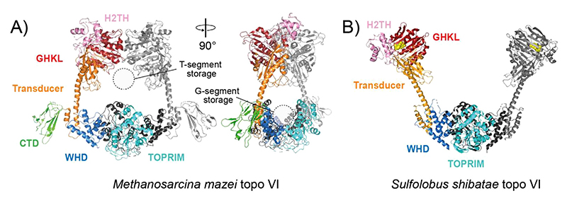
Figure 6. DNA topoisomerase VI (type IIB) structures.
(A) Crystal structure of Methanosarcina mazei topo VI (PDB: 2Q2E).[196] The domains are coloured as labelled in the figure on one TOP6A/Top6B heterodimer, with the second Top6A and Top6B coloured black and grey, respectively. (B) Crystal structure of Sulfolobus shibatae topo VI bound to radicicol (PDB: 2ZBK).[197] Colour coding is the same as in panel A except GHKL-bound radicicol is coloured yellow

