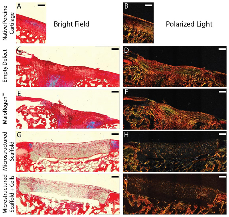
Fig. 4. Optical microscopy of picrosirius red and Alcian blue stained sections of the defect site after 6 months.
(A,C,E,G,I) Bright field images of the stained sections demonstrate the spatial composition of the repair sites. (B,D,F,H,J) Polarized light was used to detect collagen alignment due to its orientation dependent refraction (birefringence). Scale bars = 500 μm. (For interpretation of the references to color in this figure legend, the reader is referred to the Web version of this article.)

