Abstract
Purpose
To evaluate the association between the contact areas of primary molar teeth and approximal caries after three years of observation.
Methods
This prospective cohort study included 1,119 caries-free children, aged three to four years, from Puducherry, India. At baseline, 4,476 contacts were assessed using the OXIS criteria, which denotes O for open contact and X, I, and S for closed contacts. X contact represents a point contact, I is a straight contact, and S is a curved contact. Two calibrated dentists measured dental caries at one-year intervals following the International Caries Detection and Assessment System for three years. Poisson regression analysis with a multilevel approach was used to determine the association between contact type and approximal caries.
Results
Of 3,848 contacts observed at the end of three years, 499 (13 percent) were carious. The adjusted analysis revealed a significant association between contact type and approximal caries (P<0.05). The risk ratios for the development of approximal caries were 2.3 for X-type contacts (0.5 to 11.4, P=0.307), 12.7 for I-type (4.1 to 39.6, P<0.05), and 22.5 for S-type (7.2 to 70.6, P<0.05) when compared with O-type.
Conclusions
Compelling evidence suggests that variations in contacts between teeth are significant in the development of approximal caries. The S-type contact is the most susceptible to approximal caries, followed by the I-type.
Keywords: Approximal Caries, Contact Types, Primary Teeth, Preschool Children
Dental caries is a major global public health problem and the most widespread non-communicable disease. It is the most prevalent condition, per the 2015 Global Burden of Disease Study, with the decay of permanent teeth (2.3 billion people) and primary teeth (560 million children) ranking first and 12th, respectively.1 Among the different types of caries in primary teeth, approximal caries is significant because of its propensity for rapid progression. This may be explained by some of the characteristics of primary teeth, such as thinner enamel and dentin layers,2 a lower degree of mineralization,3 wider dentinal tubules compared to those of the permanent teeth,3 and a broad proximal contact area.2 Among all the factors mentioned, an ideal contact area that prevents plaque accumulation between teeth is the most significant factor for the prevention of approximal caries.4,5 Since 2018, the contact areas of primary molars have been categorized using the OXIS classification. It denotes four different categories of contact areas: open (O); point (X); straight (I); and curved (S).6,7
Multiple studies have reported that closed contact points increase the risk for approximal caries in primary dentition when compared to open contacts.8–10 The predictive power of the approximal surfaces between the primary molars has been evaluated, and when both the approximal surfaces are concave, the risk of caries development is reportedly higher than that associated with any other combination.11 Another study classified contacts as open, light, and substantial, concluding that substantial and/or light contacts between primary molars showed higher caries experience in the approximal surfaces when compared to that associated with open contacts.12 Further, the presence of interproximal caries in one quadrant is a good indicator of caries in the other quadrants as reported by Cho and co-workers.13 A retrospective cohort study reported a significant association between OXIS contacts and approximal caries.14
Understanding how variations in contact areas, namely OXIS, contribute to approximal caries development is crucial. OXIS contact appears to be a significant and reliable risk predictor for approximal caries from the retrospective study mentioned earlier.14 However, a well-designed, prospective, longitudinal study is imperative to elucidate the vulnerability of OXIS contacts to approximal caries over time.
Therefore, the purpose of this study was to evaluate the approximal caries susceptibility of variation in the contact areas of primary teeth among a cohort of 1,119 children. The results, after three years of observation, are presented in the present manuscript.
Methods
Study design and participants
The sample for the present prospective cohort study included preschool children aged three to seven years who had ancestral nativity to Puducherry, a Union Territory in India. The sample was recruited from all five zones of Puducherry, using a two-stage simple random sampling method. The sampling method and selection criteria were described elsewhere.7 Finally, a cohort of 1,119 children (4,476 contacts) aged three to four years was recruited at the baseline. The children were then followed-up longitudinally with annual dental examinations up to six to seven years of age. This study was approved by the Institutional Ethics Committee of (IEC-NI/16/AUG/55/54). Prior to the start of the study, official permission to conduct the study was acquired from the Chief Educational Officer, Puducherry, and principals of the respective schools. Written informed consent was obtained from the parents of all participants for the examination of dental caries. The study was conducted in accordance with the STROBE guidelines for observational studies.15
Assessment of baseline data (independent variables)
All 4,476 contacts of the 1,119 caries-free children were assessed for the type of contact between the mesial surface of the primary second molar and the distal surface of the primary first molar in accordance with OXIS criteria.7 Sectional maxillary and mandibular impressions were obtained on the day of clinical examination for future reference. All recruited children were provided oral hygiene instructions regarding the frequency and method of brushing.
Questionnaire data collection (control variables)
Information concerning infant feeding, diet, and oral hygiene practices was collected using a validated questionnaire16 from every recruited child’s parent during each follow-up visit at 12, 24, and 36 months of age. In the case of a school visit, the questionnaire was given to classroom teachers, who communicated with the parents regarding its completion and return of the questionnaire by the child within 24 to 48 hours. In addition, the parents’ socioeconomic status was assessed using the modified Kuppuswamy scale.17
Annual assessment of dental caries (dependent variable)
The outcome of the present study was the presence of caries on the approximal surfaces (the distal surface of the primary first molar and/or the mesial surface of the primary second molar) at the 12, 24, and 36-month follow-up examinations. The outcome assessment was performed independently by two calibrated examiners who were blinded to baseline data.
Calibration of the examiners
Two dentists were trained to diagnose dental caries clinically and radiographically using the International Caries Detection and Assessment System (ICDAS) prior to the start of this investigation. The training method was carried out according to recommendations in the 2014 ICDAS criteria manual. Training of examiners started with a two-week intensive review and examination, followed by inter- and intra-examiner reliability assessments. Intra-examiner reliability was assessed by examining and re-examining (after two weeks) 25 children not included in the main study. The kappa values for the two examiners were 0.89 and 0.91, respectively. The inter-examiner reproducibility was calculated as 0.93.
Data collection (September 2021 to October 2021)
Data collection for the present study comprised clinical examination, radiographic examination, and questionnaire data collection. Annual dental checkups for the recruited children were planned at the school. However, owing to the COVID-19 pandemic, data were collected through a combination of school, private clinic, and home visits according to the patient’s preference with appropriate safety measures. The screening process was divided into morning, afternoon, and evening. School visits were completed in the morning, and home visits were planned in the afternoon. All evening sessions were dedicated to clinic visits, where the children were screened on an appointment basis. Data were collected through clinical and radiographic examinations. The examiners used plain dental mirrors, sterile wet gauze/cotton, and a CPITN probe (Aggarwal Dental Supply Co., Jalandhar, Punjab, India) for caries assessment. The tip of the CPITN probe has a 0.5-mm ball at the tip and millimeter markings at 3.5, 8.5, and 11.5 mm, and the color codings are 3.5 to 5.5 mm. Each dental surface was scored according to ICDAS criteria.18 Plaque was removed with a piece of wet gauze. The teeth were examined wet and air-dried with a three-in-one syringe in a clinic setting for five seconds, whereas a chip blower was used for the same purpose in the home or school setup. The examiners assessed all surfaces of each tooth and recorded the findings on a custom-made assessment form. However, only the presence of caries on the approximal surfaces was considered for the assessment. In addition to clinical examination, bitewing radiographs were obtained when visual inspection of approximal surfaces was impossible19 or when the ICDAS score was four (underlying dark shadow from dentin). The radiographs were obtained using a portable dental X-Ray unit (Vatech EZRay™ Air Plus Portable X-Ray Machine, Vatech, New Delhi, India) and a digital scanner (SOREDEX™ DIGORA™ Optime, Brea, Calif., USA), which were brought to the schools/home/clinic where the examinations took place. In the present study, the parents of all caries-affected children were contacted and informed regarding their children’s dental health status.
Statistical analysis
Baseline characteristics were described using means with standard deviation for continuous variables and frequencies and proportions for categorical variables. The number of approximal caries was considered as count data. The data had a hierarchical level, which was the tooth and child level. A multilevel Poisson regression analysis was performed. The risk ratio is presented along with a 95 percent confidence interval (95% CI). The Kaplan-Meier method was used to analyze “time-to-event” data. The event of interest was the presence of approximal caries, and time was measured as the number of days. The log-rank test was used to determine whether the difference in survival times between the types of contact was significant. The Cox proportional hazard model was used to test the association between the survival time of carious teeth and the type of contact. The authors used transitional probability computations by dividing each tooth into three stages: sound (S; ICDAS 0), non-cavitated (NC; ICDAS 1,2), and cavitated (C; ICDAS 3-6) lesions. Calculations were performed for each type of contact. Furthermore, the transition probability was calculated overall as well as for the type of contact using the transition frequencies. The statistical significance cutoff threshold was set at 0.05. STATA 16 software (StataCorp LLC®, College Station, Texas), a statistical software that enables users to analyze, manage, and produce graphical visualizations of data, was used for all analyses.
Results
Characteristics of the participants
Of the 1,119 children (4,476 contacts) recruited at baseline, 157 children (with 628 contacts) were lost to follow-up at the end of three years. Therefore, the proportion of dropouts at the end of 36 months was 14 percent. The dropouts were primarily due to a change in address or failure to achieve contact through telephone after three attempts. Finally, 962 (86 percent) subjects with 3,848 contacts remained in the study after completion of both clinical examinations and questionnaires. The sex distribution of the cohort at the three-year follow-up consisted of 402 (41.8 percent) male and 560 (58.2 percent) female children. The children were between six and seven years of age (501 were six years old, and 461 were seven years old) at the final examination. The numbers of children screened via school, home, and clinic visits were 445, 210, and 307, respectively.
Prevalence and frequency of contacts at baseline
The observed prevalence of O, X, I, and S types was 261 (5.8 percent), 148 (3.3 percent), 3,381 (75.5 percent), and 686 (15.3 percent), respectively, among the 4,476 contacts. The most common contact observed was I, followed by S, O, and X.
Assessment of the outcome
Among the 3,848 contacts, 499 contacts (13 percent) were carious at the approximal surfaces (distal surface of primary first molar and/or mesial surface of primary second molar) during the 36-month follow-up visit. The remaining 3,349 contacts were sound. Among the 499 caries-affected contacts, 487 were cavitated lesions and 12 were non-cavitated lesions. Of the caries-affected contacts, 125 on both approximal surfaces were caries-affected (mesial surface of the primary second molar and distal surface of the primary first molar). Among the remaining 374 caries-affected contacts, 102 were mesial (primary second molar) and 272 were distal surfaces (primary first molar), indicating approximal caries. Of the total contacts, 159 required bitewing radiography to confirm the presence of approximal caries. Among them, 132 lesions were cavitated and 27 were sound. Of the total number of carious contacts, I and S contacts accounted for 65.7 percent and 33 percent, respectively, comprising 98.8 percent of all carious contacts. The distribution of approximal caries prevalence at 12, 24, and 36 months is provided as Supplemental Electronic Data—sTable 1.
Of the 962 children, 255 children had the presence of approximal caries in at least one of the contact areas. Of 255 children with dental caries, 174 parents provided consent for treatment (overall, 81 parents did not provide consent or did not report to the private center for dental treatment). In total, 216 carious contacts in 174 children with 216 teeth (carious contact only on one tooth either the mesial surface of the primary second molar or the distal surface of the primary first molar) were provided with appropriate dental treatment. The treatment ranged from 175 glass ionomer restorations, 11 pulpectomies, and 41 stainless steel crowns.
Univariate and multivariate Poisson regression analysis
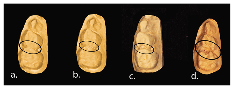
Supplemental Electronic Data—sTable 2 depicts the results of the univariate analysis by Poisson regression with a multilevel approach for the incidence of approximal caries and independent variables. Among the tooth variables, the type of arch and contact were significantly associated with the incidence of dental caries. Regarding the child variables, socioeconomic status, parental supervision of toothbrushing, use of toothpaste, intake of sugary snacks, and frequency of toothbrushing were statistically associated with the incidence of approximal caries. Table 1 displays the multilevel adjusted Poisson regression models with a multilevel approach after adjusting for the cluster effect at the tooth and child level. Model one represents the naive model, model two represents multilevel Poisson regression with dental variables (level one), and model three represents multi-level Poisson regression with dental (level one) and child variables (level two). Model three indicates that the S-type contact showed a risk ratio of 22.5 times greater incidence of approximal caries than that associated with the O-type contact (P<0.001). Furthermore, contacts in the right and maxillary left quadrants showed a 2.2 times greater incidence of approximal caries than those in the mandibular left quadrant (P<0.001). Among the child variables, frequency of toothbrushing (P=0.036) and use of toothpaste (P=0.015) were associated with an increase in approximal caries development after adjusting for the other variables. Figure 1 depicts the models of S contacts at baseline and 12, 24, and 36 months of follow-up. Supplemental Electronic Data—sFigure represents the models of O, X, and I contacts at baseline, and 12, 24, and 36 months of follow-up.
Table 1. Multilevel Adjusted Poisson Regression Analysis of Incidence of Approximal Caries on Primary Teeth Considering Child and Tooth Variables*.
| Variable | Total contacts at 36 months (N=3,848) | Model 1: naive model | Model 2: RR (95% CI) | P-value | Model 3: RR (95% CI) | P-value | |
|---|---|---|---|---|---|---|---|
| Absence of caries | Presence of caries | ||||||
| Intercept | 0.13 (0.12-0.14) | 0.005 (0.002-0.017) | 0.007 (0.002-0.023) | ||||
| Type of contact | |||||||
| O (257) | 254 (98.8) | 3 (1.2) | 1 | 1 | |||
| X (148) | 145 (98.0) | 3 (2.0) | 2.2 (0.4-10.7) | 0.345 | 2.3 (0.5-11.4) | 0.307 | |
| I (2778) | 2450 (88.2) | 328 (11.8) | 13.4 (4.3-41.7) | <0.001 | 12.7 (4.1-39.6) | <0.001 | |
| S (665) | 501 (75.2) | 165 (24.8) | 23.0 (7.4-72.2) | <0.001 | 22.5 (7.2-70.6) | <0.001 | |
| Type of quadrant | |||||||
| Maxillary right (962) | 811 (84.3) | 151 (15.7) | 2.2 (1.6-2.9) | <0.001 | 2.1 (1.6-2.9) | <0.001 | |
| Maxillary left (962) | 807 (83.4) | 155 (16.6) | 2.2 (1.7-2.9) | <0.001 | 2.2 (1.7-2.9) | <0.001 | |
| Mandibular right (962) | 839 (87.2) | 123 (12.8) | 1.8 (1.3-2.4) | <0.001 | 1.8 (1.3-2.4) | <0.001 | |
| Mandibular left (962) | 892 (92.7) | 70 (7.3) | 1 | ||||
| Parental supervision while tooth toothbrushing | |||||||
| Child alone (2,240) | 1919 (85.6) | 321 (14.4) | 1 | ||||
| Parent alone (628) | 556 (88.5) | 72 (11.5) | 1.0 (0.8-1.4) | 0.831 | |||
| Sometimes child, sometimes parent (980) | 874 (89.2) | 106 (10.8) | 0.8 (0.6-1.0) | 0.105 | |||
| Use of toothpaste | |||||||
| Yes (3,248) | 2845 (87.6) | 403 (12.4) | 1 | ||||
| Don’t know (540) | 459 (85.0) | 81 (15.0) | 1.0 (0.8-1.3) | 0.897 | |||
| No (60) | 45 (75.0) | 15 (25.0) | 2.0 (1.1-3.4) | 0.015 | |||
| Frequency of toothbrushing | |||||||
| After every meal (284) | 235 (82.7) | 49 (17.3) | 1 | ||||
| Never (1,456) | 1308 (89.9) | 148 (10.1) | 1.2 (0.7-1.7) | 0.987 | |||
| Occasionally (336) | 305 (90.7) | 31 (9.3) | 1.1 (0.7-1.5) | 0.585 | |||
| Once daily (1,088) | 930 (85.5) | 158 (14.5) | 0.6 (0.4-0.98) | 0.040 | |||
| Twice daily (672) | 561 (83.3) | 111 (16.7) | 0.7 (0.5-0.98) | 0.036 | |||
| SES | |||||||
| Upper middle (232) | 217 (93.5) | 15 (6.5) | 0.6 (0.3-01.0) | 0.064 | |||
| Lower middle (1,068) | 932 (87.2) | 136 (12.8) | 0.9 (0.7-1.1) | 0.271 | |||
| Upper lower (988) | 832 (84.2) | 156 (15.8) | 1.2 (0.9-1.5) | 0.199 | |||
| Lower (1,560) | 1368 (87.7) | 192 (12.3) | 1 | ||||
| Deviance (-2 log likelihood)† | 3,036.62 | 2,876.68 | 2,826.78 | ||||
Abbreviations used in this table: RR=relative risk; CI=confidence interval; O=open contact; X=point contact; I=straight contact; S=curved contact; SES=socioeconomic status.
Deviance (-2 log likelihood)=measure of how well the estimated model fits the likelihood. The model with the lower deviance is the better model. Therefore model 3 is better than model 1 & 2.
Figure 1.
Schematic representation of sectional die models of S contacts at baseline and 12, 24, and 36 months of follow-up, represented as a, b, c, and d, respectively. The S contact became carious at the end of 24 months (c) and progressed to pulpal involvement at the end of 36 months (d).
Survival analysis
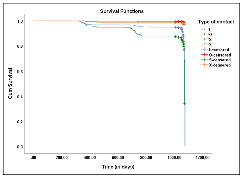
Figure 2 depicts the Kaplan-Meier curve, which explains the plot of the cumulative survival functions for the different groups of the between-subjects factor (i.e., O, X, I, and S types of contacts). The cumulative survival proportion against time for each type of contact is labeled in the survival functions plot. The “event” of interest was considered to be the presence of approximal caries, and others were censored. The survival time is measured in days. The Kaplan-Meier method was used to check whether there are statistically significant differences in the survival distributions between the type of contacts between subjects factor using the log-rank test, Breslow test, and Tarone-Ware test. The result explains that the survival distribution for the four types of contacts was statistically significant (χ2 equals 98.253; P<0.001). The mean and median with a 95% CI for survival time are provided in Supplemental Electronic Data—sTable 3. The estimated mean time until the event of interest (caries yes) is 1066.718 days for the O-type contact, 1061.893 days for the X-type contact, 1042.146 days for the I-type contact, and 1012.262 days for the S-type contact. The median time for caries is 1073.000 days for the I contacts versus 1071.00 days for S contacts. The survival probability is lower for S contacts at all time points as they are more vulnerable to approximal caries. Considering that the P-value (0.001) is less than 0.05, there is significant evidence of a difference in survival times for the type of contacts.
Figure 2. Survival Kaplan-Meier curve of OXIS contacts with approximal caries as the event.
The X-axis represents the survival time of OXIS contacts in days, and the Y-axis represents the cumulative survival function of the event which is approximal caries.
Cox’s regression analysis was performed with the proportional hazards assumption that the hazard ratio between the groups remains constant over time. Cox’s proportional hazards regression is used to relate several risk factors or exposures (considered simultaneously) to survival time. Table 2 shows that the O-type contact was considered as the reference category. The hazard ratio of S-type contact is 25.9 times more risk to approximal caries when compared to the O-type contact (P<0.001). The hazard ratio of I-type contact is 15.3 times more risk to approximal caries when compared to the O-type contact (P<0.001).
Table 2. Multivariable Results of Factors Associated With Time to Having Approximal Caries Using Cox PH Modeling*.
| Types of contact | Total contacts at 36 months (N=3,848) | Hazard ratio (95% CI) | P-value | |
|---|---|---|---|---|
| Absence of caries N (%) | Presence of caries N (%) | |||
| O (257) | 254 (98.8) | 3 (1.2) | 1 | |
| X (148) | 145 (98.0) | 3 (2.0) | 2.5 (0.5-12.6) | 0.253 |
| I (2778) | 2450 (88.2) | 328 (11.8) | 15.3 (4.9-47.5) | <0.001 |
| S (665) | 501 (75.2) | 165 (24.8) | 25.9 (8.3-81.2) | <0.001 |
Abbreviations used in this table: O=open contact; X=point contact; I=straight contact; S=curved contact; CI=confidence interval.
Transitional probability
The percentage of carious contacts at the end of 12, 24, and 36 months were 3.2 percent (NC equals 2.6 percent; C equals 0.6 percent), 2.85 percent (NC equals 0.51 percent; C equals 2.34 percent), and 7.4 percent (NC equals 0.3 percent; C equals 7.1 percent), respectively. Overall, the S-type contact had the maximum transition from the caries-free state (S) to the NC state (1.36 percent) and C state (7.67 percent). Whenever an NC state was observed with an I- or S-type contact in a follow-up examination, it transitioned to a C state in the subsequent year.
Discussion
The results suggest that variations in tooth contacts, namely OXIS-type contacts, are strongly associated with the incidence of approximal caries. Furthermore, teeth with broad contacts (I or S) are at greater risk for approximal caries than teeth with open or narrow contacts (O or X), while also considering variables related to the child. This finding is in agreement with that of a previous prospective cohort study20 conducted in the same field. However, the follow-up period was only for 12 months and the overall prevalence of approximal caries in this study was low (3.3 percent). For outcomes with a low prevalence, a long follow-up period is recommended. Therefore, the present study aimed to investigate the association between OXIS contacts and approximal caries with a follow-up period of 36 months.
Of the total number of carious contacts, I and S contacts accounted for 98.8 percent of all carious contacts. This finding closely agrees with those of previous prospective17 and retrospective cohort14 studies. However, among the closed-type contacts (X, I, and S), the X type is less vulnerable compared to the I and S types. This is because the surface in contact is a point contact and not broad. Hence, this type of contact is more accessible for the removal of plaque using mechanical methods such as toothbrushing from both the buccal and lingual sides of the contact. However, the broad and flat contacts (I and S) are less accessible for mechanical cleansing methods. Therefore, they have proved to be more prone to caries risk than the O and X contacts. It has also been established previously that broad and flat contacts are more prone to approximal caries risk when compared to open contacts.8,9,10 Although the majority of subjects fall in a high-risk category (I and S type) the assessment of OXIS contacts continues to be important owing to the high relative risk of the S (R equals 22.5); I (12.7) contacts. Also, the assessment of contact type is easy and does not require special costly tests.
Children with carious contacts were offered immediate free dental treatment at a private dental center. However, the treatment was provided for the child only when the parents gave their consent and reported for treatment. In the case that consent was not provided by the child’s parent, there was a possibility that the caries contacts of that child progressed to a severe stage when observed during the next follow-up.
Several important results of the longitudinal analysis were noted. Of the caries-affected contacts (499), 125 on both proximal surfaces were caries-affected (mesial surfaces of the primary second molars and distal surfaces of the primary first molars). Among the remaining 374 carious contacts, 272 distal surfaces (primary first molars) and 102 mesial surfaces (primary second molars) revealed approximal caries. This was in accordance with other studies where the most commonly affected surface was the distal surface of primary first molars when compared to the mesial surface of primary second molars.11,14 Second, the S-type contact was the most vulnerable to approximal caries (relative risk [RR] equals 22.5; 95% CI equals 7.2 to 70.6), owing to its complex concave-convex curvature. This was followed by the I-type contact (RR equals 12.7; 95% CI equals 4.1 to 39.6) when compared with the O-type. This result is in agreement with that of a previous retrospective cohort study14 conducted in the same field. Another study reported that concave approximal surfaces between primary molars significantly increased the risk of approximal caries.9 The concave-concave surface could be considered equivalent to the S contact in the present study based on morphology. Substantial and/or light contacts between the primary molars showed higher caries experience in the approximal surfaces when compared to those of teeth with open contacts.10 These may be considered synonymous with the broad contact types (S and I). The rationale for the S contact to pose the maximum risk for approximal caries may be its complex “concave followed by convex” design. The S-type contact would lead to maximum plaque retention between the primary molars since maintenance of oral hygiene by routine mechanical cleansing methods would be difficult in these areas.
Additionally, it was found that, among the child variables, brushing twice daily was protective against approximal caries and the absence of toothpaste use was associated with an incidence of approximal caries. Owing to the lack of studies in this area, this finding could not be compared.
The major strengths of this study are its large sample size, long follow-up period, and low dropout rate. The authors were able to adjust for a diverse set of confounding factors, which makes the association between OXIS contact and approximal caries identified in this study even more robust. The study’s limitations include deviations in the methodology due to the unexpected pandemic situation. Further, there could have been an underestimation of approximal caries, especially the non-cavitated lesions (ICDAS 1 and 2), since bitewing radiographs were not made for all the closed contacts.
Currently, the change in contact type over time is being assessed and prepared for publication submission where the changes in the different contact types from baseline to three years are being evaluated. Based on the earlier prospective and retrospective cohort studies (in context with OXIS contacts), a significant association is observed with OXIS contacts and an occurrence of approximal caries. If similar results are obtained from other studies conducted in different parts of the world, consideration should be given to measure this variable (variations in inter-proximal contacts) during routine caries risk assessment in children. Such measures might potentially modify the International Caries Risk Assessment and Management Protocols of the American Academy of Pediatric Dentistry.
Conclusions
Based on this study’s results, the following conclusions can be made:
Variations in tooth contact, namely OXIS (O equals open, X equals point, I equals straight, and S equals curved) contact types, are strongly associated with the development of approximal caries.
The order of susceptibility of OXIS contacts to approximal caries is S (RR equals 22.5) is greater than I (RR equals 12.7) is greater than X (RR equals 2.3) is greater than O.
The scoring of OXIS contacts is a useful risk factor to detect approximal caries, especially in populations similar to the present study, where 98.8 percent of the contacts belong to the high-risk category (S and I contacts). Scoring of contacts during routine clinical examination using OXIS classification needs further research from different populations.
Supplementary Material
Acknowledgments
The authors wish to thank Mrs. Jothilakshmi, MSc Statistics, Schizophrenia Research Foundation, and Chennai for their assistance with the statistical analysis. This study was funded by the DBT/Wellcome Trust India Alliance (IA/CPHE/17/1/503352). Authors Dr. Kirthiga and Dr. Muthu contributed equally to this research work.
Footnotes
Disclaimer. The authors have no conflicts of interest to declare.
References
- 1.Anderson M, Dahllof G, Warnqvist A, Grindefjord M. Development of dental caries and risk factors between 1 and 7 years of age in areas of high risk for dental caries in Stockholm, Sweden. Eur Arch Paediatr Dent. 2021;22(5):947–57. doi: 10.1007/s40368-021-00642-1. [DOI] [PMC free article] [PubMed] [Google Scholar]
- 2.Mortimer KV. The relationship of deciduous enamel structure to dental disease. Caries Res. 1970;4(3):206–23. doi: 10.1159/000259643. [DOI] [PubMed] [Google Scholar]
- 3.Wilson PR, Beynon AD. Mineralization differences between human deciduous and permanent enamel measured by quantitative microradiography. Arch Oral Biol. 1989;34(2):85–8. doi: 10.1016/0003-9969(89)90130-1. [DOI] [PubMed] [Google Scholar]
- 4.Murray JJ, Majid ZA. The prevalence and progression of approximal caries in the deciduous dentition in British children. Br Dent. 1978;145(6):161–4. doi: 10.1038/sj.bdj.4804135. [DOI] [PubMed] [Google Scholar]
- 5.Pitts NB, Rimmer PA. An in vivo comparison of radiographic and directly assessed clinical caries status of posterior approximal surfaces in primary and permanent teeth. Caries Res. 1992;26(2):146–52. doi: 10.1159/000261500. [DOI] [PubMed] [Google Scholar]
- 6.Kirthiga M, Muthu MS, Kayalvizhi G, Krithika C. Proposed classification for interproximal contacts of primary molars using CBCT: A pilot study. Wellcome Open Res. 2018;3:98. doi: 10.12688/wellcomeopenres.14713.2. [DOI] [PMC free article] [PubMed] [Google Scholar]
- 7.Muthu MS, Kirthiga M, Kayalvizhi G, Mathur VP. OXIS classification of interproximal contacts of primary molars and its prevalence in three-to four-year-olds. Pediatr Dent. 2020;42(3):197–202. [PubMed] [Google Scholar]
- 8.Allison PJ, Schwartz S. Interproximal contact points and proximal caries in posterior primary teeth. Pediatr Dent. 2003;25(4):334–40. [PubMed] [Google Scholar]
- 9.Warren JJ, Slayton RL, Yonezu T, et al. Interdental spacing and caries in the primary dentition. Pediatr Dent. 2003;25(2):109–13. [PubMed] [Google Scholar]
- 10.Subramaniam P, Babu Kl G, Nagarathna J. Interdental spacing and dental caries in the primary dentition of 4- to 6-year-old children. J Dent (Tehran) 2012;9(3):207–14. [PMC free article] [PubMed] [Google Scholar]
- 11.Cortes A, Martignon S, Qvist V, Ekstrand KR. Approximal morphology as predictor of approximal caries in primary molar teeth. Clin Oral Investig. 2018;22(2):951–9. doi: 10.1007/s00784-017-2174-3. [DOI] [PubMed] [Google Scholar]
- 12.Cho VY, King NM, Anthonappa RP. Role of marginal ridge shape and contact extent in approximal caries between primary molars. J Clin Pediatr Dent. 2021;45(2):98–103. doi: 10.17796/1053-4625-45.2.5. [DOI] [PubMed] [Google Scholar]
- 13.Cho V, King N, Anthonappa R. Presence of interproximal carious lesions in primary molars. Pediatr Dent. 2021;43(1):28–33. [PubMed] [Google Scholar]
- 14.Muthu MS, Kirthiga M, Lee JC, et al. OXIS contacts as a risk factor for approximal caries: A retrospective cohort study. Pediatr Dent. 2021;43(4):296–300. [PMC free article] [PubMed] [Google Scholar]
- 15.Von Elm E, Altman DG, Egger M, Pocock SJ, Gøtzsche PC, Vandenbroucke JP, STROBE Initiative The Strengthening the Reporting of Observational Studies in Epidemiology (STROBE) statement: Guidelines for reporting observational studies. J Clin Epidemiol. 2008;61(4):344–9. doi: 10.1016/j.jclinepi.2007.11.008. [DOI] [PubMed] [Google Scholar]
- 16.Folayan MO, Kolawole KA, Oziegbe EO, et al. Prevalence and early childhood caries risk indicators in preschool children in suburban Nigeria. BMC Oral Health. 2015;15:72. doi: 10.1186/s12903-015-0058-y. [DOI] [PMC free article] [PubMed] [Google Scholar]
- 17.Bairwa M, Rajput M, Sachdeva S. Modified Kuppuswamy’s socioeconomic scale: Social researcher should include updated income criteria, 2012. Indian J Community Med. 2013;38(3):185–6. doi: 10.4103/0970-0218.116358. [DOI] [PMC free article] [PubMed] [Google Scholar]
- 18.Ismail AI, Sohn W, Tellez M, et al. The International Caries Detection and Assessment System (ICDAS): An integrated system for measuring dental caries. Community Dent Oral Epidemiol. 2007;35(3):170–8. doi: 10.1111/j.1600-0528.2007.00347.x. [DOI] [PubMed] [Google Scholar]
- 19.American Academy of Pediatric Dentistry. The Reference Manual of Pediatric Dentistry. American Academy of Pediatric Dentistry; Chicago, Ill: 2022. Prescribing dental radiographs for infants, children, adolescents, and individuals with special health care needs; pp. 273–6. [Google Scholar]
- 20.Kirthiga M, Muthu MS, Kayalvizhi G, Mathur VP, Jayakumar N, Praveen R. OXIS contacts and approximal caries in preschool children: A prospective cohort study. Caries Res. 2023;57(2):133–140. doi: 10.1159/000529160. [DOI] [PMC free article] [PubMed] [Google Scholar]
Associated Data
This section collects any data citations, data availability statements, or supplementary materials included in this article.


