Limbus is formed by the junction of the corneal and conjunctival epithelia, where the epithelium gradually becomes thicker toward the sclera.[1] It is a 1–2-mm-wide anatomical ring-shaped transition zone between the opaque sclera and the clear cornea.[2] Anatomically, the limbus contains important features related to eye functions including fibrovascular ridges radially oriented known as palisades of Vogt that host corneal stem cells for epithelial turnover.[1] Scanning electron microscopy (SEM) is a technique for obtaining high-resolution images of biological and nonbiological specimens.
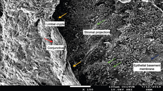
Our aim was to show the ultrastructure of the corneal limbus using SEM [Fig. 1].[3]
Figure 1.
(×150): Red arrow: conjunctiva; yellow arrows: limbal cryptx; green arrows: stromal projections; blue arrow: epithelial basement membrane. Superficial stromal projections around the limbal crypts providing a niche for stem and epithelial cells. There is a transitional zone form peripheral projections to a smoother epithelial basement membrane centrally
Financial support and sponsorship
Nil.
Conflicts of interest
There are no conflicts of interest.
References
- 1.Consejo A, Llorens-Quintana C, Radhakrishnan H, Iskander DR. Mean shape of the human limbus. J Cataract Refract Surg. 2017;43:667–72. doi: 10.1016/j.jcrs.2017.02.027. [DOI] [PubMed] [Google Scholar]
- 2.Spaniol K, Witt J, Mertsch S, Borrelli M, Geerling G, Schrader S. Generation and characterisation of decellularised human corneal limbus. Graefes Arch Clin Exp Ophthalmol. 2018;256:547–57. doi: 10.1007/s00417-018-3904-1. [DOI] [PubMed] [Google Scholar]
- 3.Versura P, Bonvicini F, Caramazza R, Laschi R. Scanning electron microscopy study of human cornea and conjunctiva in normal and various pathological conditions. Scan Electron Microsc. 1985:1695–708. [PubMed] [Google Scholar]


