Abstract
Diplomonad parasites of the genus Giardia have adapted to colonizing different hosts, most notably the intestinal tract of mammals. The human-pathogenic Giardia species, Giardia intestinalis, has been extensively studied at the genome and gene expression level, but no such information is available for other Giardia species. Comparative data would be particularly valuable for Giardia muris, which colonizes mice and is commonly used as a prototypic in vivo model for investigating host responses to intestinal parasitic infection. Here we report the draft-genome of G. muris. We discovered a highly streamlined genome, amongst the most densely encoded ever described for a nuclear eukaryotic genome. G. muris and G. intestinalis share many known or predicted virulence factors, including cysteine proteases and a large repertoire of cysteine-rich surface proteins involved in antigenic variation. Different to G. intestinalis, G. muris maintains tandem arrays of pseudogenized surface antigens at the telomeres, whereas intact surface antigens are present centrally in the chromosomes. The two classes of surface antigens engage in genetic exchange. Reconstruction of metabolic pathways from the G. muris genome suggest significant metabolic differences to G. intestinalis. Additionally, G. muris encodes proteins that might be used to modulate the prokaryotic microbiota. The responsible genes have been introduced in the Giardia genus via lateral gene transfer from prokaryotic sources. Our findings point to important evolutionary steps in the Giardia genus as it adapted to different hosts and it provides a powerful foundation for mechanistic exploration of host–pathogen interaction in the G. muris–mouse pathosystem.
Keywords: parasite, diplomonad, Giardia, streamlined, antigenic variation, lateral gene transfer
Data Summary
Raw DNA and RNA sequence reads are archived at NCBI Sequence Read Archive (SRA) under accession numbers SRR8858297–SRR8858305.
This Whole Genome Shotgun project has been deposited at DDBJ/EMBL/GenBank under the accession number PRJNA524057. The version described in this paper is version VDLU00000000.1. The genome sequence and annotations generated in this project have been integrated into GiardiaDB (https://giardiadb.org/giardiadb/).
Impact Statement.
The Giardia genus comprises eukaryotic single-celled parasites that infect many animals. The Giardia intestinalis species complex, which can colonize and cause diarrheal disease in humans and different animal hosts has been extensively explored at the genomic and cell biologic levels. Other Giardia species, such as the mouse parasite Giardia muris, have remained uncharacterized at the genomic level, hampering our understanding of in vivo host–pathogen interactions and the impact of host dependence on the evolution of the Giardia genus. We discovered that the G. muris genome encodes many of the same virulence factors as G. intestinalis. The G. muris genome has undergone genome contraction, potentially in response to a more defined infective niche in the murine host. We describe differences in metabolic and microbiome modulatory gene repertoire, mediated mainly by lateral gene transfer, that could be important for understanding infective success and host specificity across the Giardia genus. Our findings provide new insights for the use of G. muris as a powerful model for exploring host–pathogen interactions in giardiasis.
Background
Many eukaryotes have evolved from free-living to parasitic lifestyles over evolutionary time, yet parasitism has developed independently in different taxonomic groups and is therefore characterized by many unique features [1, 2]. Comparative genomics provides an opportunity to investigate the factors of parasitism such as loss of morphological, metabolic and genomic complexity, and consequently reduced evolutionary potential for a free-living lifestyle [2]. It can also identify the drivers and consequences of a parasitic lifestyle and generate new testable ecological and evolutionary hypotheses [3].
Giardia is a protozoan parasite that non-invasively colonizes the intestinal tract of many vertebrates. The human pathogen, Giardia intestinalis, is estimated to cause 300 million cases of giardiasis in the world each year, being a major cause of diarrheal disease [4]. Giardiasis is also a problem in domestic animals, and the zoonotic potential of Giardia has been highlighted in recent years [5]. In vitro models of the interaction of G. intestinalis with human cells have helped to unravel clues to how Giardia causes disease [6–8], such as the importance of, the adhesive disc for attachment [9], flagella for motility [10, 11], secreted cysteine proteases for interference with host defenses [12–16], interactions with the intestinal microbiota [6, 17], differentiation into cysts for transmission [4, 18] and interference with nitric oxide (NO) production [19, 20]. Despite this progress it remains uncertain whether these in vitro models are representative of the natural infection, particularly because animal models of G. intestinalis infection have significant limitations. For example, infection of mice, the most commonly used laboratory animals, with human G. intestinalis isolates is unreliable and requires manipulations such as antibiotic conditioning [6]. Giardia muris, one of six recognized species of Giardia [21], has been used as a mouse model since the 1960s for exploring the pathogenesis and immunological responses of the mammalian host to infection [22]. The availability of knock-out mice and other host-related resources makes G. muris a powerful model to investigate host–pathogen interactions [6]. The life cycle and infective process of G. muris is closely related to infection by G. intestinalis [23]. Major findings in Giardia biology such as flagellar and disc function, cellular differentiation [22, 24, 25] and immunity [23, 26–29] have been pioneered with G. muris, and later been shown to be transferable to human G. intestinalis infections [29, 30]. Unfortunately, research on G. muris has been hampered by the lack of genome information and gene-expression data [5].
Here we describe the draft genome of G. muris, representing the first genome of any Giardia outside of the G. intestinalis species complex. We performed comparative genomics with free-living (Kipferlia bialata [31]) and parasitic (G. intestinalis [32–34] and S. salmonicida [35]) relatives to G. muris to determine how G. muris may have evolved into an intestinal pathogen of rodents.
Results
Genome assembly
We extracted DNA from freshly excysted G. muris cysts purified from the faeces of infected mice (Fig. S1a, available in the online version of this article) and assembled a high-quality draft genome using sequences obtained by PacBio and Illumina technologies. In addition, we generated RNA-Seq data for gene prediction and gene-expression analyses with total RNA extracted from cysts, recently excysted cells (excyzoites) and trophozoites isolated from the small intestine of infected mice (Fig. S1a).
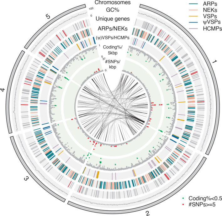
The G. muris draft genome consists of 59 contigs spanning 9.8 Mbp, which is notably smaller than the G. intestinalis WB genome (12.6 Mbp, Table 1). Most of the genome (9.0 Mbp, 92 %) is found on five contigs (>1 Mbp). None of these are terminated by telomeric repeats, but among the remaining short contigs (<30 kbp), ten are terminated in telomeric repeats (TAGGG), suggesting they represent the terminal points of five chromosomes. The karyotype of G. muris was previously shown to consist of four separable chromosomes [36]. We hypothesize that our five major contigs represent a total of five chromosomes in G. muris, two of which are so close in size (1.290 and 1.297 Mbp) that they were not readily resolved using pulsed-field gels, and are named accordingly from 1 to 5 from largest to smallest in size (Fig. 1). Overall, 42 of the 44 small contigs contain ribosomal DNA (rDNA) clusters that encode 28S, 18S and 5.8S rRNAs. In fact, rDNA clusters make up 2.0 % of the total genome, and account for 91.6 % of the identified repeats (Methods S1). Half of the contigs terminated by telomeric repeats have adjacent rDNA clusters (Fig. S1b), suggesting that multiple of the G. muris chromosomes [36], like those in G. intestinalis [37], have long repeats of rDNAs close to the telomeres. In contrast to G. intestinalis chromosomes [37], no retrotransposon sequences were found in the telomeric regions and overall very few retrotransposon sequences were detected in the G. muris genome.
Table 1.
Comparison of genome content between G. muris, G. intestinalis, S. salmonicida and K. bialata
|
Species |
G. muris |
G. intestinalis |
S. salmonicida |
K. bialata |
|---|---|---|---|---|
|
Genome size (Mbp) |
9.8 |
12.6 |
12.9 |
51.0 |
|
Chromosomes (scaffolds) |
5 (59) |
5 (35) |
9 (233) |
ND (11,564) |
|
G+C % |
54.7 |
46.3 |
33.4 |
49.4 |
|
No. of protein encoding genes |
4653 |
4963 |
8067 |
17 389 |
|
Mean/median protein size (aa) |
578/428 |
635/457 |
373 |
333 |
|
Mean/median intergenic size (bp)* |
264/37 |
470/81 |
421 |
597 |
|
Coding density %† |
84.5/88.6 |
81.5/84.7 |
72.1 |
ND |
|
No. of introns |
3 cis, 5 trans |
8 cis, 5 trans |
3 cis |
124 912 |
|
tRNA genes |
68 |
65 |
145 |
ND |
|
ASH % |
0.016 |
0.028 |
0.15 |
ND |
|
References |
This study |
Ref [38] |
Ref [35] |
Ref [31] |
*Mean/Median intergenic distance is based on all RNAs (mRNAs, tRNAs, rRNAs), but not pseudogenized genes.
†Coding density: First value is based on all RNAs (mRNAs, tRNAs, rRNAs), but not pseudogenized genes; Second value is based on all RNAs including ψVSP.
Fig. 1.
Circular representation of the G. muris chromosomes. From outside inward: five chromosomes, GC percent, unique genes (grey) including unique metabolic genes in Table 4 (red), ARPs (greenblue) / NEKs (pink), VSPs (orange) / ψVSPs (blue) / HCMPs (purple), Coding percent / 5 kbp (green if <=0.5), # SNPs / kbp >=5 (red), blastn matches with >95 % identity and >1000 bp in size. The circular plot was drawn with circlize [94].
Allelic sequence heterozygosity (ASH) in the assembly was estimated to be 0.016 % (Table 1), equivalent to the low level found in the G. intestinalis WB genome (0.026 %, Table 1). Distribution of ASH along chromosomes showed only weak clustering in certain areas, particularly at the ends of chromosomes (Fig. 1).
Genome streamlining and synteny
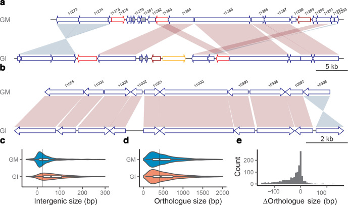
Gene prediction and manually curated annotation identified 4653 protein coding genes in G. muris (Table 1). This makes 84.5 % of the genome coding, counting also the tRNAs and rRNAs (Table 1). Thus, the G. muris genome is an example of a very compact eukaryotic genome. Consistent with that, the average intergenic size is 264 bp (Table 1), with a prominent skew towards shorter intergenic regions for a high proportion of genes (median size at 37 bp, Fig. 2c). The compactness of the genome is also illustrated by multiple instances of overlapping genes, with 441 genes (9.5 % of all genes) showing an average overlapping size of 21 bp (spanning 1–327 bp) with neighbouring genes.
Fig. 2.
Examples of synteny between G. muris and G. intestinalis. (a) A 50 kbp region on chromosome 3, which share synteny to a 58 kbp region on chromosome 5 in WB. Synteny plot was plotted using genoplotR [95]. Shades of red and blue represent forward and inverted matches between orthologues. Genes are drawn as arrows in blue. ARPs in red, NEKs in dark red and VSPs in orange. Dark grey filled genes are unique genes to that the genome in comparison to the other. (b) A 14 kbp region on chromosome 1, which shares synteny to a 16 kbp region on chromosome 5 in G. intestinalis. It uses the same colour scheme as in (a). (c) Violin plots of intergenic sizes of neighbouring positional orthologues of G. muris and G. intestinalis, and the grey vertical line represents the median intergenic size of G. muris. (d) Violin plots of positional orthologue sizes of G. muris and G. intestinalis, and the grey vertical line represents the median orthologue size of G. muris. (e) Histogram of the positional orthologue size difference between G. muris and G. intestinalis.
The G. muris and the new improved G. intestinalis WB genome [38] do not maintain clear chromosomal synteny even though both are assembled as five near-complete chromosomes (Fig. S2). However, local synteny (Fig. 2a, b) was retained among 3043 one-to-one orthologues (Table S1a) (an average amino acid similarity of 44.7 %) shared by the two genomes. Comparing local synteny, it becomes obvious that G. muris keeps shorter orthologous gene and intergenic region sizes (Fig. 2b–e, paired t-test significant with P-value of 4.0e-50 and 6.5e-53, respectively). We found that the average number of domains in G. muris orthologues was not significantly different from G. intestinalis (paired t-test, P-value 0.25), indicating that protein domain loss is not the cause of protein shortening.
Gene regulation
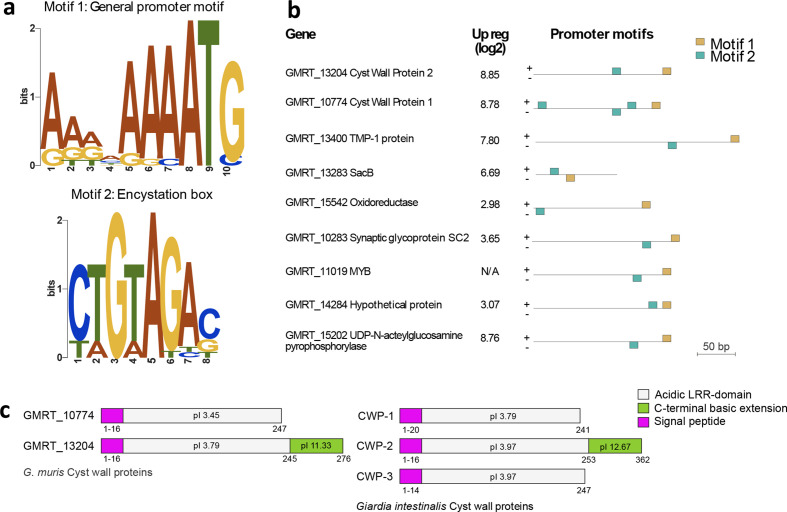
We could not identify any universal, conserved promoter motifs shared by all G. muris genes except for an enrichment of A residues around the start codon (Fig. S3a, one proportion z test against 25 %, shows the significance of A residue with P-value <0.05, up to 13 bases upstream of the start codon), which resembles observations in G. intestinalis [39]. The streamlining of the G. muris genome was also apparent at the 3′ end of genes where the putative polyadenylation signal, which is similar to the one described in G. intestinalis [39], is overlapping with the stop codon for most genes (Fig. S3b). Genes up-regulated early during encystation in G. intestinalis have specific promoter elements [40, 41]. Most of these genes were also identified in G. muris, with one notable exception of cyst-wall protein 3 (Fig. 3c). Encystation-related genes in G. muris share promoter motifs (Fig. 3a), similar to the Myb binding sites found in G. intestinalis [41], suggesting a similar type of regulation. We also noted that the encystation-related genes are among the most highly expressed genes in G. muris trophozoites in vivo (Fig. S3c, Table S1b), similar to G. intestinalis infection in mice [42, 43].
Fig. 3.
Gene regulation and organization of VSPs in G. muris. (a) Promoter motifs shared by encystation-related genes. Motif 1 [gold in (b)] represents the general promotor motif positioned directly adjacent to the start codon. Motif 2 [teal in (b)] resembles the encystation-regulated promoter previously identified in G. intestinalis (52). (b) The distribution and position of motif 1 (gold) and motif 2 (teal) in chosen genes regulated during encystation. (c) Giardia cyst wall proteins. Cyst wall protein 3 is missing in G. muris. Signal peptide (pink). Acidic LRR-domain (grey). C-terminal basic extension (green).
Very few genes in G. intestinalis contain introns, with only eight known cis-spliced and four trans-spliced genes (five trans-introns) [44–46]. Similarly, only three cis- and no trans-introns were identified in the parasitic diplomonad S. salmonicida [35], whereas the free-living fornicate K. bialata has on average seven cis-introns per protein encoding gene [31]. G. muris maintains homologues to the eight cis-spliced G. intestinalis genes, but has only three retained introns (Fig. S4a). All four trans-spliced genes in G. intestinalis have homologues in G. muris with conserved splicing motifs (Fig. S4). Mining genes with similar motifs did not reveal additional intron-containing genes in G. muris. Similar to G. intestinalis [44] all the trans-spliced genes in G. muris preserve a similar cleavage motif TCCTTTACTCAA (Fig. S4c) as the RNA processing sequence motif [44]. Thus, we observe a reduction of introns in G. muris, and cis-introns seem to be easier to lose than trans-introns.
VSPs and antigenic variation in G. muris
Variant specific-surface proteins (VSPs) in G. intestinalis are characterized as cysteine-rich proteins with frequent CXXC motifs and a conserved C-terminal transmembrane (TM) domain followed by a cytoplasmic pentapeptide (CRGKA, Fig. S5A). We identified 265 VSP homologues in G. muris. Their C-terminal pentapeptide (GCRGK, Fig. S5a, Table S1c) differed slightly from that in G. intestinalis. However, the cysteine and arginine residues in the pentapeptide, which are known to be post-translationally modified in G. intestinalis, are conserved [47, 48]. In addition, the preceding 24 aa of the G. muris VSP TM domain show conservation to the TM domain of G. intestinalis VSPs (Fig. S5a). Most G. muris VSPs contain the conserved GGCY motif present in most G. intestinalis VSPs (Table S1c). Since bona fide VSPs need signal peptides (SPs) at the N-terminus to guide VSPs to the parasite surface, we divided this group into two subgroups; proteins with predicted SPs are called VSPs, whereas VSP proteins without SPs are referred to as pseudogenized VSPs (ψVSPs). The 26 complete VSP genes (16 unique at 98 % identity to each other) are mostly located chromosome-centrally (Fig. 1, Table 2). Seven pairs of VSP genes were identified with identical sequences arranged either as head-to-head (two pairs) or tail-to-tail (five pairs) (Table 3). The intervening sequence between the different VSP pairs have coding sequences or truncated pseudogenes with homology to NimA (never in mitosis gene a)-related kinase (NEKs), ankyrin repeat proteins (ARPs) and zinc-finger domains (Table 3). There are also four copies of identical VSPs clustering close to the 3′ end of chromosome 2 (Fig. 2) interspersed with tandem repeats, NEKs and zinc-finger domain proteins (Table 3).
Table 2.
Summaries of gene families within Giardia
|
G. intestinalis* |
G. muris* |
|
|---|---|---|
|
NEK |
184 (26) |
216 (23) |
|
ARP-1 |
269 (5) |
298 (6) |
|
ARP-2 |
33 |
86 (16) |
|
ARP-3 |
8 |
33 |
|
VSP |
133 |
26 |
|
ψVSP |
208 |
239 |
*Values in () indicate the number of pseudogenized copies.
Table 3.
Arrangement of VSP genes in the G. muris genome
|
Chr |
Geneid1 |
Geneid2 |
Arrangement |
Gene size (aa) |
Distance (bp) |
Genes in between |
|---|---|---|---|---|---|---|
|
1 |
20 512 |
13 275 |
-> <- |
596 |
2025 |
ψARP-2 |
|
1 |
21 145 |
21 149 |
-> <- |
594 |
1658 |
ψARP-2 |
|
1 |
13 124 |
21 374 |
-> <- |
620 |
8400 |
ψARP-2, alpha-tubulin |
|
2 |
16 008 |
21 957 |
<- -> |
624 |
8794 |
NEK, ARP-2 |
|
2 |
22 301 |
22 304 |
-> <- |
523 |
1656 |
ψARP-2 |
|
2 |
12 920 |
22 758 (22764, 22769) |
<- -> |
515 |
10 612 (6824, 8428) |
NEK, TR, Zinc |
|
4 |
24 220 |
24 228 |
-> <- |
623 |
8303 |
ψARP-2, alpha-tubulin |
|
5 |
24 787 |
24 792 |
<- -> |
619 |
7158 |
NEK, Zinc, ARP-2 |
In contrast to complete VSPs, most ψVSPs (183/239) are found in linear arrays (n=17) in G. muris, herein defined as having >3 ψVSPs genes (Fig. S5b, c). Strikingly, nine out of the ten ends of the main contigs have a ψVSP array at or close to the ends of the chromosomes (telomere-adjacent) containing a total of 131 genes (Table S1c). The only main contig that ends without an array has a cluster of two ψVSPs close to the chromosome terminus. We found a single ψVSP array consisting of 12 genes in a chromosome position that was non-telomere adjacent (on chromosome 5) (Fig. 1). The ψVSP arrays vary in copy numbers (5–23 genes) and the terminal part of the ψVSP array is always arranged with the tail-end towards the chromosome terminus. The tandem arrangement of the gene arrays suggested that they were generated by gene duplication. Two of the terminal arrays are scrambled and have a shift in the ψVSP array directionality at the site of an intact VSP (Fig. S5b).
We constructed a phylogeny of all the VSPs and ψVSPs in G. muris to investigate their evolutionary dynamics. The phylogeny revealed relaxed clustering of ψVSP genes originating in each linear array (Fig. S5c). However, the internal linear array on chromosome 5 represents a noteworthy exception showcasing a very recent gene duplication event. Interestingly, the great majority of full-length VSPs (23/28) are clustered in the phylogeny, including the VSPs in the scrambled ψVSP arrays, despite these genes being distributed in physically separate chromosomal locations across all five primary scaffolds (Fig. S5c). The few non-clustered VSP genes in the phylogeny that are not part of pairs or clusters are found directly adjacent to ψVSPs genes. The relaxed clustering of VSPs and ψVSPs suggested that these genes might be undergoing periodical recombination or gene conversion.
The VSPs and ψVSPs showed distinct expression patterns. Essentially all the ψVSP genes were non-transcribed in the three surveyed life-stages (Fig. S5d). VSPs, on the other hand, showed on average higher expression with one or a few loci displaying dominant expression in the different life-stages (Fig. S5d).
There is also another, less characterized VSP-related cysteine-rich protein family in G. intestinalis, high cysteine membrane proteins (HCMPs) [49], with 104 members [38]. Many are highly up-regulated during interaction with intestinal epithelial cells [50]. The HCMPs have several CXXC and CXC motifs, one VSP-like transmembrane domain but with longer C-terminals than in the VSPs [50]. The 34 genes matching these criteria in the G. muris genome were named HCMPs after the corresponding gene family in G. intestinalis. They are found spread-out on the five chromosomes (Fig. 1).
Multigene families in G. muris
The largest multigene families in G. intestinalis outside the VSPs and HCMPs are the NEKs [51] and ankyrin repeat containing proteins (Protein 21.1) [32]. There are 230 NEKs in G. muris (Fig. 1, Table 3), making up 71 % of its kinome, slightly more than what was found in G. intestinalis [51]. We classified ankyrin repeat containing proteins further into three groups. The ankyrin repeat protein-1 (ARP-1) with only ankyrin repeats, ARP-2 with ankyrin repeats plus zinc finger domains, and ARP-3 with both ankyrin repeats plus domains other than zinc finger domains. The NEKs and the different classes of ARPs are scattered throughout the chromosomes without obvious clustering (Fig. 1). A phylogenetic analysis revealed that 79 of the NEKs are conserved as 1 : 1 orthologues between G. muris and G. intestinalis (Fig. S6a). Among the NEK orthologues, we found that 65 % of them consists of NEKs that in G. intestinalis are predicted to be catalytically ‘dead’ (as defined in [51]). This proportion of ‘dead’ NEKs is only marginally smaller than the G. intestinalis average (71 %) [51] indicating a high level of functional conservation in this protein group. Each species has one massively expanded cluster of NEKs with G. muris having the largest with 104 members and the one in G. intestinalis having 79 members. ARPs show a similar evolutionary stability with 132 conserved 1 : 1 orthologues between species (Fig. S6b). G. muris shows a major species-specific expansion of 91 genes whereas the largest expanded clusters in G. intestinalis amounts to two groups of 15 genes each. The partly shared domain-structure of NEKs and ARPs prompted us to investigate their relationship by a network analysis employing reciprocal blastp (1e-05 cutoff). The two groups are not recovered as clearly separated clusters but form partially overlapping networks (Fig. S6c). The larger numbers of genes in these multigene families in G. muris compared to G. intestinalis and their evolutionary dynamics are intriguing given the otherwise streamlined features of the G. muris genome, perhaps suggesting that they have unique roles in adaptation to their murine hosts.
Virulence factors in Giardia
G. intestinalis is not known to possess classical virulence factors, such as enterotoxins, but several genes are important for colonization of the host and thus for pathogenesis. These include genes for motility [10], the adhesive disc for attachment [9], secreted cysteine proteases that can degrade host defensive factors [12, 13], and cysteine-rich surface protein like the VSPs [52] and the HCMPs [49] that undergo antigenic variation. The cytoskeletal protein repertoire in G. muris is very similar to G. intestinalis apart from several fragmented alpha-tubulins (three complete genes with homologues in G. intestinalis and nine incomplete gene fragments). The adhesive disc is a unique cytoskeletal structure of Giardia parasites essential for attachment of the trophozoite in the small intestine, but is missing in other fornicates [9]. The first detailed studies of the adhesive disc were performed on G. muris trophozoites [24], but more recent work has mostly focused on G. intestinalis. The vast majority (82 of 85) of G. intestinalis disc proteins [9] were also identified in G. muris (Table S1d); 12 were NEK kinases and 27 were ARP-1 proteins. Two of the three G. intestinalis disc proteins not found in G. muris (Table S1d) localize to a structure in the G. intestinalis disc on top of the ventral groove, but this structure is missing in the G. muris disc [22], suggesting functional disc differences. Many of the disc proteins that are immunodominant during G. intestinalis infections (e.g. alpha-giardins, beta-giardin, SALP-1, alpha- and beta-tubulin [53] are highly expressed (here defined as >500 FPKM) in G. muris trophozoites in the small intestine (Table S1b).
Proteases are important virulence factors in many pathogens and cysteine-protease activities have been suggested to play a role in Giardia virulence [4, 13]. We identified 81 proteins classified as proteases in the G. muris genome, compared to 96 proteins identified in an identical search in G. intestinalis WB (Table S1e). The largest family of proteases in G. muris, with 15 members, are papain-like cysteine proteases (C1A family). This protein family is also the largest protease group in G. intestinalis with 21 members [54, 55]. Several conserved groups of proteases were found to have been present in the ancestor to Giardia and Spironucleus, although we also found evidence for lineage-specific gene loss and expansion in G. muris (Fig. S7). The most highly expressed cysteine protease of G. muris in vivo is the closest homologue to the highest expressed protease in G. intestinalis WB [55].
Metabolic pathways in G. muris
Our metabolic reconstruction identified 95 metabolic pathways in G. muris compared to 98 pathways detailed in G. intestinalis WB; four pathways were unique in G. muris and eight in G. intestinalis WB. Even though the overall metabolism is highly similar between G. muris and G. intestinalis WB, the genes for several specific enzymes and their putative reactions show distinct differences. Thus, 18 unique reactions (14 enzymes) were predicted in G. muris and 25 in G. intestinalis WB (10 enzymes) (Table 4). Several of these unique proteins showed moderate-high identity to prokaryotic proteins (Table S1f). Five of these prokaryote-like genes are present in the genome of assemblage B strain G. intestinalis GS.
Table 4.
Lateral gene transfers in G. muris (Gm), G. intestinalis (Gi WB, Gi GS, Gi P15), S. salmonicida (Ss), Trepomonas spp. (Trep), K. bialata (Kb) and T. vaginalis (Tv)
|
Species |
Gm |
Gi WB |
Gi GS |
Gi P15 |
Ss |
Trep |
Kb |
Tv |
|---|---|---|---|---|---|---|---|---|
|
2.5-diketo-d-gluconic acid reductase |
X |
X |
X |
X |
||||
|
Arginase |
X |
X |
X |
|||||
|
Carboxymuconolactone decarboxylase |
X |
X |
||||||
|
Ferritin-like |
X |
X |
||||||
|
Fructokinase |
X |
|||||||
|
Ketosteroid isomerase |
X |
|||||||
|
l-ascorbate-6-phosphate lactonase |
X |
X |
||||||
|
Maltose-O-acetyltransferase |
X |
X |
X |
|||||
|
Mannose-6-phosphate isomerase |
X |
X |
||||||
|
Phosphopantothenate-cysteine ligase |
X |
X |
||||||
|
Quorum-quenching N-acyl-homoserine lactonase |
X |
X |
||||||
|
Ribonuclease 3 |
X |
X |
X |
|||||
|
Tae4 |
X |
|||||||
|
Tryptophanase |
X |
X |
X |
|||||
|
β-phosphoglucomutase |
|
X |
X |
X |
X |
X |
||
|
Extracellular nuclease |
|
X |
X |
X |
X |
X |
X |
X |
|
Flavohemoprotein |
|
X |
X |
X |
||||
|
Glycerol kinase |
|
X |
X |
X |
X |
X |
||
|
Inositol-3-phosphate synthase |
|
X |
X |
X |
||||
|
Methyltransferase |
|
X |
X |
X |
||||
|
NADPH-ferrihemoprotein |
|
X |
X |
X |
X |
X |
||
|
Purine nucleoside phosphorylase |
|
X |
X |
X |
X |
X |
X |
X |
|
Pyruvate phosphate dikinase |
|
X |
X |
X |
X |
|||
|
Sugar/H+ symporter |
|
X |
X |
X |
||||
|
Threonine dehydratase |
|
X |
X |
X |
X |
X |
The potential utilization of carbohydrate sources for glycolysis is different in G. muris compared to G. intestinalis. Fructokinase and mannose 6-phosphate isomerase enable G. muris to use fructose and mannose 6-phosphate unlike G. intestinalis. In both cases, the enzymes were acquired from bacteria via lateral gene transfer (Table S1f, Fig. S8a, b). Curiously, the G. muris gene for mannose 6-phosphate isomerase is found next to a bacterial transcriptional regulator/sugar kinase gene. Phylogenetic analyses show that G. muris mannose 6-phosphate isomerase and the transcriptional regulator/sugar kinase gene clusters deep in the Bacteroidetes group. This gene arrangement is observed in bacteria of the genus Alistipes , whose genomes harbour the most similar homologues, supporting the notion that a single event of lateral gene transfer best explains the origin of these genes in G. muris (Fig. S8b, c). Both genes have the A-rich initiator that precedes the start codon in most G. muris genes and are expressed in G. muris trophozoites (Table S1b).
The utilization of glycerol for ATP synthesis via glycerol kinase has been suggested in G. intestinalis upon depletion of primary carbon sources [56]. This enzyme is found in both Spironucleus and Trichomonas but has been lost in G. muris. Another notable metabolic difference to G. intestinalis WB is the lack of pyrophosphate-dependent pyruvate phosphate dikinase (PPDK) that leaves a single, less energy efficient, route from phosphoenol pyruvate to pyruvate via pyruvate kinase in G. muris.
G. muris is predicted to synthesize coenzyme A from pantothenate, employing a bifunctional phosphopantothenoyl decarboxylase-phosphopantothenate synthase. The same pathway is described in S. salmonicida [35], but the complete pathway is missing in G. intestinalis (Table 4).
As in all other studied metamonads, G. muris encodes the arginine dihydrolase (ADH) pathway that enables the use of arginine as an energy source and at the same time, reduces the available free arginine in the environment, preventing nitric oxide (NO) production in host cells [57]. NO efficiently kills G. intestinalis trophozoites and the main scavenger enzyme for NO in G. intestinalis is Flavohemoprotein [58], which is lacking in G. muris (Table 4). Arginine is an important modulator of virulence in many infectious organisms since it interferes with NO production [59]. G. muris encodes arginases, which converts arginine directly to ornithine and urea. Arginases are present in G. intestinalis GS and T. vaginalis, representing an ancestral acquisition in Metamonada followed by subsequent losses in Spironucleus and G. intestinalis assemblage A and E (Table 4, Fig. S8d).
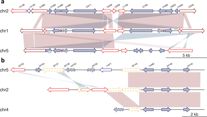
We noticed that several of the bacterial derived genes that are shared with G. intestinalis GS are clustered together on the chromosomes in G. muris in highly dynamic genomic regions. For example, arginase genes are found in a four-gene genomic region that is present in two adjacent copies in chromosome 2, two on chromosome 1 and one on chromosome 5 (dark grey filled arrows, Fig. 4a). Intact arginase genes are only found on chromosome 2 adjacent to the genes encoding 2,5-diketo-d-gluconic acid reductase. All the duplication events have occurred between ARPs (red arrows) and NEKs (dark red arrows). The homologous regions on the three chromosomes likely originated via two duplication events.
Fig. 4.
Synteny plot of two duplicated regions in the genome. (a, b) Shades of red and blue represent forward and inverted matches between neighbouring sequences. ARPs are drawn in red, NEKs in dark red and VSPs in orange. Pseudogenized genes are drawn in dashed lines. Dark grey filled genes are unique genes to G. muris in comparison with G. intestinalis. Point mutation in arginase is marked by the red asterisk. Homologous sequences that are not annotated in the genome are drawn in a dashed box on sides of the backbone grey line. Genes discussed in the paper: 21 985 and 21 992 encode the intact arginase genes, 21 986 and 21 992 encode the enzyme 2,5-diketo-d-gluconic acid reductase, whereas 21 984 and 21 994 are hypothetical proteins in (a); 14 480 encodes 2,5-diketo-d-gluconic acid reductase, 24 746 encodes CMD and 24 745 encodes ketosteroid isomerase-like protein in (b).
Nucleotide substitutions have accumulated since the duplication events and in-frame stop codons (marked by red asterisk in Fig. 4a) have rendered three of the arginase genes pseudogenized. The small ORFs sitting at the other side of arginase are hypothetical proteins and have similar homologues in other parts of the genome, but the sequences of their duplicated homologues in those regions are pseudogenized (dotted light grey block in Fig. 4a). Both genes have their closest relatives in bacteria, even though the genes can be found in other eukaryotes (Fig. S13e). Phylogenetic analyses indicate the genes might have been transferred from different bacterial donors multiple times into different eukaryotic lineages (Fig. S13e).
A second, distantly related 2,5-diketo-d-gluconic acid reductase gene copy (Fig. 4b, and S8f) in the genome is present in three different genomic locations together with two other enzymes, carboxymuconolactone decarboxylase (CMD) and ketosteroid isomerase-like protein. All three genes, constitute putative lateral gene transfers (Fig. S8g, h). This three-gene region is close to ψVSPs (orange arrows) and ARPs.
Interaction with the intestinal microbiota
Giardia trophozoites colonize the intestinal lumen where they can potentially interact with other intestinal microbes. Although little is functionally known about such interactions and their consequences for parasite survival, four proteins encoded in the G. muris genome could play a role in these interactions.
Bactericidal/permeability-increasing (BPI) proteins are innate immune defense proteins that bind to lipopolysaccharide and display potent killing activity against Gram-negative bacteria by increasing membrane permeability. Beyond this basic function BPI proteins might also act as effectors in controlling mutualistic symbioses [60]. Homologues of BPI proteins are found in G. muris and G. intestinalis [61], but it remains to be determined if they have anti-microbial activity.
G. muris encodes tryptophanase, an enzyme that metabolizes tryptophan to pyruvate with concomitant release of indole and ammonia. While pyruvate can be utilized in energy metabolism, indole and its metabolites have been shown to affect gut microbiota composition, possibly by interfering with quorum-sensing systems, and might be able to influence host health [62]. Phylogenetic analysis of this protein showed that this enzyme represents an ancestral acquisition in diplomonads with subsequent loss in G. intestinalis (Fig. S8i).
Two more proteins with potential importance for microbiota interactions are encoded in G. muris: Tae4 and quorum-quenching N-acyl-homoserine lactonase. The Tae4 proteins are wide-spread amidases that were first described in association with the T6SS system effector Tae4 in Salmonella Typhimurium [63]. The Tae4 proteins degrade bacterial peptidoglycan by hydrolysing the amide bond, γ-d-glutamyl-mDAP (dl-bond) of Gram-negative bacteria [63], and is required for interbacterial antagonism and successful gut colonization by S. Typhimurium [64]. Quorum-quenching N-acyl-homoserine lactonase degrades N-acyl-homoserine lactone, a molecule used by both Gram-positive and Gram-negative bacteria, for quorum sensing [65]. Our phylogenetic analysis supports the lateral acquisition of both genes (Fig. S8j, k). While Tae4 has been a recent acquisition in G. muris, quorum-quenching N-acyl-homoserine lactonase was present in the common ancestor of G. muris and G. intestinalis and lost in G. intestinalis WB and P15 (Fig. S8k).
Discussion
Our data shows that G. muris has an even more compact genome than G. intestinalis, whose genome is already known to be highly streamlined [32]. Genome compaction via reduction of mobile or repetitive elements have been seen in other eukaryotic parasites [1, 66]. G. muris appears to fall into this category as it encodes no known classes of mobile elements and repetitive elements are mostly confined to telomeric contexts. The shortness of intergenic regions in G. muris ranks among the most extreme recorded for any eukaryote, even shorter than Microsporidia, which are known as the most compact and reduced eukaryotic genomes [67]. The global synteny map of G. muris to G. intestinalis indicates many frequent small-scale genome rearrangements that often favours a more efficient gene packing in G. muris, thus allowing shorter intergenic regions. This evidence of gene shuffling and the fact that there is very little evidence of genome degradation would argue for optimization of growth as the driving force of G. muris genome streamlining.
G. muris trophozoites have not been grown axenically in vitro, which has hampered exploration of its genome, gene regulation and metabolism [5, 21], and has limited the use of G. muris as an in vitro model system for the human parasite G. intestinalis and other intestinal protozoan parasites [5]. We identified several metabolic differences between G. muris and G. intestinalis that might indicate avenues to successful strain axenization. Most of these differences are represented by instances of lateral gene transfer of metabolic genes or losses thereof in either G. intestinalis or G. muris. The lack of metabolic genes such as glycerol kinase and PPDK might shift the relative emphasis of metabolic pathways compared to G. intestinalis. The possible utilization of additional carbohydrates (such as mannose and fructose) and differences in amino acid utilization (via tryptophanase and arginase) and their impact on growth in vitro are interesting avenues to investigate in any future axenization efforts.
G. intestinalis isolates are typically poor at infecting mice. Despite this, the assemblage B isolate GS, which shares more metabolic enzymes with G. muris than the assemblage A isolate WB, is better able to establish infection in mice than WB. This suggests that the shared metabolic capacity of G. muris and G. intestinalis GS enables survival in the murine intestinal tract. Additionally, G. muris might be able to interact or interfere with intestinal Gram-positive and Gram-negative bacteria and this could be a key to establish successful infections. G. intestinalis, which lacks some of the putative microbiome modulators, such as Tryptophanase and Tae4, is dependent on reduction of the small intestinal microbiota in order to efficiently infect mice [68]. G. muris is cleared from the murine host by secretory IgA [26], whereas the role of IgA in anti-giardial defense is less clear for G. intestinalis GS [26, 69]. We speculate that G. muris is more resistant to elimination by innate factors such as competition with the normal microbiota, or host production of reactive oxygen species and/or NO, whereas GS is more sensitive to innate factors and eliminated much faster within 1–3 weeks (while G. muris clearance requires 4–8 weeks). Future insights into the importance of innate factors in G. muris infection should be facilitated by the availability of the complete genome sequence.
Sub-telomeric regions in parasitic protozoa often contain arrays of expanded gene families that are under positive selection by the immune system [66]. The relaxed evolutionary pressure offered by keeping pseudogenized copies of surface antigens might be an advantage for G. muris that allows genetic drift and recombination to drive rapid and stealthy diversification, thus avoiding elimination by adaptive immune defenses. It was previously reported that G. muris is capable of antigenic variation and encodes VSP genes with high similarity and conserved structural features (CRGKA pentapeptide) to those G. intestinalis [70]. We failed to identify close homologues to the previously sequenced G. muris VSPs in our G. muris genome. Instead, these VSP genes show much greater similarity and in one case almost absolute identity to G. intestinalis VSPs of assemblage B. Importantly, our finding that G. muris VSPs have distinctive structural features (GCRGK cytoplasmic tail) not reported in G. intestinalis VSPs to date indicates that the previously reported G. muris VSPs are likely due to contamination from a G. intestinalis assemblage B isolate.
The linear ψVSP arrays in G. muris have previously been described in G. intestinalis [71]. Our phylogenetic analyses of G. muris VSPs and ψVSPs revealed evidence of recombination or segmental gene conversion, as previously demonstrated in G. intestinalis [71]. However, we recognized two clear differences in the VSP repertoire in G. muris and G. intestinalis. First, G. muris encodes a low number of intact VSP loci that are located internally on the chromosomes. Second, the ψVSP arrays are almost exclusively telomere adjacent, as opposed to G. intestinalis where this tendency is not apparent [71]. These aspects of the G. muris VSP repertoire resemble the antigenic variation systems of Pneumocystis spp. and Trypanosoma brucei [72, 73]. Despite clear mechanistic differences, all these systems have converged on having large reservoirs of mostly telomeric positioned, arrayed genes that are transcriptionally silent and are sources for recombination and gene conversion into expression sites.
The function of ankyrin repeat proteins and NEK kinases remains mostly unknown. They represent, together with VSPs, the most dynamic protein families in the Giardia genomes [51]. The Giardia NEK kinases lack transmembrane domains and have been suggested to target and localize to different intracellular structures with their ankyrin repeats [51] and many of the G. intestinalis NEK kinases localize to cytoskeletal structures, including the flagella and adhesive disc [9]. Rearrangements and duplications in the G. muris genome are frequently associated with these large gene families (Figs. 2a and 4), indicating they might serve as anchoring-points for recombination.
Lateral gene transfer is an important shaping factor in the evolution of metabolism in protists [74]. The origins of laterally transferred genes in G. muris are here inferred to be by prokaryotic sources that are members of the gastrointestinal flora, in agreement to previous observations [75]. Most of the putative differences in metabolic potential in the Giardia genomes are attributable to lateral gene transfers, either by lineage-specific gene gain or loss. For example, the ability to utilize mannose has been introduced from bacteria of the genus Alistipes via lateral gene transfer. This event is supported by phylogenetic reconstruction, shows a high degree of sequence conservation (>70 % at the amino acid level) and displays maintained gene order to the one seen in the closest related bacterial lineages (Fig. S8b). Interestingly, several lateral gene transfers were found clustered in amplified areas of the G. muris chromosome. Curiously, G. intestinalis assemblage B also maintains clustered copies of arginase and 2,5-diketo-d-gluconic acid reductase, while these genes have both been lost in the G. intestinalis assemblage A and E lineages.
Anti-microbial peptides of several classes, such as defensins and trefoil-factor 3, are up-regulated in the small intestine of G. muris infected mice [29]. Secreted cysteine proteases from G. intestinalis have been shown to be able to degrade defensins [12]. We detected prominent expression of several cysteine proteases in G. muris. The protease with the highest expression is suggested to have a role in encystation and excystation [55]. Its G. intestinalis homologue is up-regulated and secreted during interactions with human intestinal epithelial cells [8] and it cleaves chemokines, tight junction proteins and defensins [12, 76]. Thus, this is most likely also an important virulence factor in G. muris.
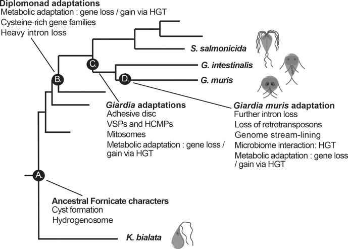
Our results from this study are summarized in a model of the evolution of Giardia’s virulence traits in Fig. 5. A number of characters important for Giardia’s ability to infect the intestine of mammals are pre-parasitic inventions (such as modified mitochondria and differentiation into transmissive cysts, Fig. 5) and some are found in all diplomonad parasites (e.g. loss of metabolic functions, streamlined microtubular cytoskeleton, expansion of gene families like ankyrins and cysteine proteases and loss of introns, Fig. 5). Giardia-specific innovations include the adhesive disc for attachment, VSPs and HCMPs for antigenic variation and mitosomes involved in Fe-S complex synthesis (Fig. 5). Whereas some are only found in G. muris (e.g. metabolic genes involved in microbiota interactions, Fig. 5), suggesting adaptation to the intestinal environment of mice. Our data shows that the environment in the host’s intestine, most of all the immune system and the microbiota, apply selective pressure for changes in the genome, metabolic potential and the parasite surface proteome.
Fig. 5.
Model of the evolution of virulence traits in Giardia parasites. A set of important diplomonad evolutionary innovations and their chronology is depicted at relevant phylogenetic nodes. (a) Free-living fornicate ancestor. (b) Diplomonad ancestor. (c) Giardia ancestor. (d) Giardia muris.
Methods
Cell preparation and nucleic acid extraction
In total, 4.5×10⁷ muris trophozoites (day 7 post-infection) were collected from small intestines of three C57 mice, washed once in PBS and pellet frozen at −80 °C (Biosample SAMN11231832). Viable cysts of G. muris isolate Roberts–Thomson passaged through mice were obtained from Waterborne. These cysts had been purified from faecal material using Percoll and sucrose gradients (Biosample SAMN11231833). DNA and RNA were extracted from 1×10⁷ cysts using standard methods.
Then, 1×10⁸ cysts were excysted according to the procedure in Feely et al. [77] (Biosample SAMN11231834). RNA was purified from cell material equivalent to 1×10⁷ cysts. DNA for long-read sequencing was prepared from the remaining cysts as described in Methods S1.
Sequencing, assembly, contamination removal and annotation
Total genomic DNA was sequenced using both Illumina MiSeq and PacBio RS II sequencers. The stranded transcriptome mRNA and the miRNA libraries were sequenced with Illumina HiSeq 2000 system. The RNA samples extracted from excysted cells and cysts prior excystation were prepared using the TruSeq stranded mRNA sample preparation kit and sequenced by HiSeq 2500.
PacBio long reads were assembled de novo using the SMRT Analysis (v2.3.0) pipeline.
Reads were assembled with HGAP followed by consensus sequence calling with Quiver. The resulting assembly contained 317 contigs. A blastn (e-value<=0.1) of the contigs against the NT database revealed contamination, mostly from fungi, and the contaminated contigs were removed from the final assembly, resulting in 59 final contigs.
The Illumina MiSeq reads were mapped to the PacBio assembly using BWA v0.7.12-r1039 [78] and Nesoni v0.130 (http://bioinformatics.net.au/software.nesoni.shtml) was used to correct mostly the indels that we have observed to cause frame-shifts in certain genes. Overall, 8 deletion, 46 insertion and 16 SNPs were corrected by Nesoni with setting –majority 0.75 based on the mapped bam file.
Structural annotation was made from a union of Prodigal (v2.60) [79] and GlimmerHMM (v3.0.1) [80] predicted genes. Functional annotation consisted of a combination of information from blastp results against NR database as well as HMMER (v3.0) search results of domain information against Pfam (v27.0). All genes were then manually examined, with RNA-Seq reads mapped (using BWA [78]) as a guideline for manual structural annotation. Introns were manually curated based on sequence similarity to G. intestinalis introns.
A detailed description of genome sequencing, annotation, protein families and synteny analyses is available in Methods S1. This Whole Genome Shotgun project has been deposited at DDBJ/EMBL/GenBank under the accession number PRJNA524057. The version described in this paper is version VDLU00000000.1.
RNA-Seq expression analysis
BAM files were generated from mapping the RNA-Seq reads to the reference genome using BWA (v0.7.12) [78]. Cufflinks (v104700) [78] was used to calculate the FPKM values from the BAM files, by FPKM definition: # read pairs in genes / # total reads / 1,000,000 / size of the gene.
Pathway analysis
The metabolic pathways of G. muris and G. intestinalis WB were predicted with a combination of BlastKOALA [79] implemented in KEGG [80], Pathway Tools v21.5 [81] and GiardiaDB [82]. The different predictions were combined and manually curated under Pathway Tools [83]. Pathway Tools function pathway hole filler [83] was used to further complete the pathway, and transport inference parser [84] was used to infer transport reaction(s) for transporters, which were then verified with Conserved Domain databases [85].
Phylogenetic analysis
G. muris sequences were used as queries to retrieve at least 5000 hits with e-value <0.001, using blastp against the nr database and the organism-specific proteomes. The datasets were aligned in the forward and reverse orientation using MAFFT v6.603b [86] and PROBCONS v1.12 [87]. The four resulting alignments were combined with T-COFFEE [88] and trimmed by BMGE v1.12 [89]. Maximum-likelihood (ML) trees were computed using IQtree v1.6.5 [90] under LG4X substitution model [91]. Branch supports were assessed using ultrafast bootstrap approximation (UFboot) [92] with 1000 bootstrap replicates and 1000 replicates for SH-like approximate likelihood ratio test (SH-aLRT) [93]. A detailed description of the phylogenetic analyses is available in Methods S1.
Supplementary Data
Funding information
The project was supported by funding from Vetenskapsrådet-M (2017–02918) to SGS, and the San Diego Digestive Diseases Research Center (NIH grant P30 DK120515).
Acknowledgements
Elaine Hanson is acknowledged for technical support performing trophozoite purifications from infected mice. Phylogenetic analyses were performed on resources provided by the Swedish National Infrastructure for Computing (SNIC) at Uppsala Multidisciplinary Center for Advanced Computational Science (UPPMAX).
Author contributions
S.G.S., L.E., J.O.A. and J.J.H. conceived and supervised the study. J.J.H. and E.E. developed methodology and performed experiments. F.X. developed software and performed genome assembly. F.X., A.J.G., J.J.H., A.A., D.P., J.O.A., S.G.S. and E.E. curated the genome annotations. F.X., J.J.H., A.J.G. and S.G.S. performed formal analysis. L.E. and S.G.S. acquired funding. J.O.A., L.E. and S.G.S. provided resources. J.J.H., F.X. and A.J.G. visualized the data. J.J.H., F.X., A.J.G., E.E., L.E., J.O.A. and S.G.S. drafted the manuscript. All authors edited the manuscript.
Conflicts of interest
The authors declare that there are no conflicts of interest.
Ethical statement
Animal care and use for this study was approved by the University of California, San Diego Institutional Animal Care and Use Committee under protocol number S00205 and United States Public Health Service assurance numbers A3033-1 and D16-00020. Animal use adhered to the guidelines in the most recent edition of the Guide for the Care and Use of Laboratory Animals of the National Research Council of the United States National Academies and the Guidelines on Euthanasia by the American Veterinary Medical Association.
Footnotes
Abbreviations: ADH, arginine dihydrolase; ARP, ankyrin repeat protein; ASH, allelic sequence heterozygosity; BPI, bactericidal/permeability-increasing; HCMP, high cysteine membrane protein; HGT, horizontal gene transfer; NEK, NimA (never in mitosis gene a)-related kinase; NO, nitric oxide; SNP, single-nucleotide polymorphism; SP, signal peptide; TM, transmembrane; VSP, Variant specific-surface protein; ψVSP, pseudogenized variant specific-surface protein.
All supporting data, code and protocols have been provided within the article or through supplementary data files. One supplementary table and eight supplementary figures are available with the online version of this article.
References
- 1.Poulin R, Randhawa HS. Evolution of parasitism along convergent lines: from ecology to genomics. Parasitology. 2015;142:S6–S15. doi: 10.1017/S0031182013001674. [DOI] [PMC free article] [PubMed] [Google Scholar]
- 2.Hupalo DN, Bradic M, Carlton JM. The impact of genomics on population genetics of parasitic diseases. Curr Opin Microbiol. 2015;23:49–54. doi: 10.1016/j.mib.2014.11.001. [DOI] [PMC free article] [PubMed] [Google Scholar]
- 3.Jackson AP, Otto TD, Aslett M, Armstrong SD, Bringaud F, et al. Kinetoplastid phylogenomics reveals the evolutionary innovations associated with the origins of parasitism. Current Biology. 2016;26:161–172. doi: 10.1016/j.cub.2015.11.055. [DOI] [PMC free article] [PubMed] [Google Scholar]
- 4.Einarsson E, Ma’ayeh S, Svärd SG. An up-date on Giardia and giardiasis. Curr Opin Microbiol. 2016;34:47–52. doi: 10.1016/j.mib.2016.07.019. [DOI] [PubMed] [Google Scholar]
- 5.Cacciò SM, Lalle M, Svärd SG. Host specificity in the Giardia duodenalis species complex. Infect. Genet. Evol. 2018;66:335–345. doi: 10.1016/j.meegid.2017.12.001. [DOI] [PubMed] [Google Scholar]
- 6.Fink MY, Singer SM. The intersection of immune responses, microbiota, and pathogenesis in giardiasis. Trends in Parasitology. 2017;33:901–913. doi: 10.1016/j.pt.2017.08.001. [DOI] [PMC free article] [PubMed] [Google Scholar]
- 7.Ma’ayeh SY, Knörr L, Sköld K, Granham A, Ansell BRE, et al. Responses of the differentiated intestinal epithelial cell line Caco-2 to infection with the Giardia intestinalis Gs isolate. Front Cell Infect Microbiol. 2018;8:244. doi: 10.3389/fcimb.2018.00244. [DOI] [PMC free article] [PubMed] [Google Scholar]
- 8.Ma’ayeh SY, Liu J, Peirasmaki D, Hörnaeus K, Bergström Lind S, et al. Characterization of the Giardia intestinalis secretome during interaction with human intestinal epithelial cells: the impact on host cells. PLOS Neglected Tropical Diseases. 2017;11:e0006120. doi: 10.1371/journal.pntd.0006120. [DOI] [PMC free article] [PubMed] [Google Scholar]
- 9.Nosala C, Hagen KD, Dawson SC. ‘Disc-o-Fever’: getting down with giardia’s groovy microtubule organelle. Trends in Cell Biology. 2018;28:99–112. doi: 10.1016/j.tcb.2017.10.007. [DOI] [PMC free article] [PubMed] [Google Scholar]
- 10.Nosala C, Dawson SC. The critical role of the cytoskeleton in the pathogenesis of Giardia. Clin Microbiol Rev Report. 2015;2:155–162. doi: 10.1007/s40588-015-0026-y. [DOI] [PMC free article] [PubMed] [Google Scholar]
- 11.McInally SG, Dawson SC. Eight unique basal bodies in the multi-flagellated diplomonad Giardia lamblia. Cilia. 2016;5 doi: 10.1186/s13630-016-0042-4. [DOI] [PMC free article] [PubMed] [Google Scholar]
- 12.Liu J, Ma’ayeh S, Peirasmaki D, Lundström-Stadelmann B, Hellman L, et al. Secreted Giardia intestinalis cysteine proteases disrupt intestinal epithelial cell junctional complexes and degrade chemokines. Virulence. 2018;9:879–894. doi: 10.1080/21505594.2018.1451284. [DOI] [PMC free article] [PubMed] [Google Scholar]
- 13.Ortega-Pierres G, Argüello-García R, Laredo-Cisneros MS, Fonseca-Linán R, Gómez-Mondragón M, et al. Giardipain-1, a protease secreted by Giardia duodenalis trophozoites, causes junctional, barrier and apoptotic damage in epithelial cell monolayers. Int J Parasitol. 2018;48:621–639. doi: 10.1016/j.ijpara.2018.01.006. [DOI] [PubMed] [Google Scholar]
- 14.Amat CB, Motta J-P, Fekete E, Moreau F, Chadee K, et al. Cysteine Protease–Dependent mucous disruptions and differential mucin gene expression in Giardia duodenalis infection. The American Journal of Pathology. 2017;187:2486–2498. doi: 10.1016/j.ajpath.2017.07.009. [DOI] [PubMed] [Google Scholar]
- 15.Cotton JA, Bhargava A, Ferraz JG, Yates RM, Beck PL, et al. Giardia duodenalis cathepsin B proteases degrade intestinal epithelial interleukin-8 and attenuate interleukin-8-induced neutrophil chemotaxis. Infection and Immunity. 2014;82:2772–2787. doi: 10.1128/IAI.01771-14. [DOI] [PMC free article] [PubMed] [Google Scholar]
- 16.Cotton JA, Motta J-P, Schenck LP, Hirota SA, Beck PL, et al. Giardia duodenalis infection reduces granulocyte infiltration in an in vivo model of bacterial toxin-induced colitis and attenuates inflammation in human intestinal tissue. PLoS ONE. 2014;9:e109087. doi: 10.1371/journal.pone.0109087. [DOI] [PMC free article] [PubMed] [Google Scholar]
- 17.Allain T, Amat CB, Motta J-P, Manko A, Buret AG. Interactions of Giardia sp. with the intestinal barrier: epithelium, mucus, and microbiota. Tissue Barriers. 2017;5:e1274354. doi: 10.1080/21688370.2016.1274354. [DOI] [PMC free article] [PubMed] [Google Scholar]
- 18.Mendez TL, De Chatterjee A, Duarte T, De Leon J, Robles-Martinez L, et al. And giardial encystation: the show must go on. Curr Trop Med Rep. 2015;2:136–143. doi: 10.1007/s40475-015-0052-0. [DOI] [PMC free article] [PubMed] [Google Scholar]
- 19.Stadelmann B, Merino MC, Persson L, Svärd SG. Arginine consumption by the intestinal parasite Giardia intestinalis reduces proliferation of intestinal epithelial cells. PLoS ONE. 2012;7 doi: 10.1371/journal.pone.0045325. [DOI] [PMC free article] [PubMed] [Google Scholar]
- 20.Eckmann L, Laurent F, Langford TD, Hetsko ML, Smith JR, et al. Nitric oxide production by human intestinal epithelial cells and competition for arginine as potential determinants of host defense against the Lumen-Dwelling pathogen Giardia lamblia. J Immunol. 2000;164:1478–1487. doi: 10.4049/jimmunol.164.3.1478. [DOI] [PubMed] [Google Scholar]
- 21.Helmy YA, Spierling NG, Schmidt S, Rosenfeld UM, Reil D, et al. Occurrence and distribution of Giardia species in wild rodents in Germany. Parasites & Vectors. 2018;11 doi: 10.1186/s13071-018-2802-z. [DOI] [PMC free article] [PubMed] [Google Scholar]
- 22.Friend DS. The fine structure of Giardia muris. J Cell Biol. 1966;29:317–332. doi: 10.1083/jcb.29.2.317. [DOI] [PMC free article] [PubMed] [Google Scholar]
- 23.Dann SM, Le CHY HEM, Ross MC, Eckmann L. Giardia infection of the small intestine induces chronic colitis in genetically susceptible hosts. J Immunol. 2018;201:548–559. doi: 10.4049/jimmunol.1700824. [DOI] [PMC free article] [PubMed] [Google Scholar]
- 24.Holberton DV. Fine structure of the ventral disk apparatus and the mechanism of attachment in the flagellate Giardia muris. J Cell Sci. 1973;13:11–41. doi: 10.1242/jcs.13.1.11. [DOI] [PubMed] [Google Scholar]
- 25.Schaefer FW, Rice EW, Hoff JC. Factors promoting in vitro excystation of Giardia muris cysts. Trans R Soc Trop Med Hyg. 1984;78:795–800. doi: 10.1016/0035-9203(84)90024-5. [DOI] [PubMed] [Google Scholar]
- 26.Langford TD, Housley MP, Boes M, Chen J, Kagnoff MF, et al. Central importance of immunoglobulin A in host defense against Giardia spp. Infect Immun. 2002;70:11–18. doi: 10.1128/IAI.70.1.11-18.2002. [DOI] [PMC free article] [PubMed] [Google Scholar]
- 27.Davids BJ, Palm JED, Housley MP, Smith JR, Andersen YS, et al. Polymeric immunoglobulin receptor in intestinal immune defense against the lumen-dwelling protozoan parasite Giardia. J Immunol. 2006;177:6281–6290. doi: 10.4049/jimmunol.177.9.6281. [DOI] [PubMed] [Google Scholar]
- 28.Dreesen L, De Bosscher K, Grit G, Staels B, Lubberts E, et al. Giardia muris infection in mice is associated with a protective interleukin 17A response and induction of peroxisome proliferator-activated receptor alpha. Infect Immun. 2014;82:3333–3340. doi: 10.1128/IAI.01536-14. [DOI] [PMC free article] [PubMed] [Google Scholar]
- 29.Manko A, Motta J-P, Cotton JA, Feener T, Oyeyemi A, et al. Giardia co-infection promotes the secretion of antimicrobial peptides beta-defensin 2 and trefoil factor 3 and attenuates attaching and effacing bacteria-induced intestinal disease. Plos One. 2017;12:e0178647. doi: 10.1371/journal.pone.0178647. [DOI] [PMC free article] [PubMed] [Google Scholar]
- 30.Saghaug CS, Sørnes S, Peirasmaki D, Svärd S, Langeland N, et al. Human memory CD4+ T cell immune responses against Giardia lamblia. Clinical and Vaccine Immunology. 2016;23:11–18. doi: 10.1128/CVI.00419-15. [DOI] [PMC free article] [PubMed] [Google Scholar]
- 31.Tanifuji G, Takabayashi S, Kume K, Takagi M, Nakayama T, et al. The draft genome of Kipferlia bialata reveals reductive genome evolution in fornicate parasites. Plos One. 2018;13:e0194487. doi: 10.1371/journal.pone.0194487. [DOI] [PMC free article] [PubMed] [Google Scholar]
- 32.Morrison HG, McArthur AG, Gillin FD, Aley SB, Adam RD, et al. Genomic minimalism in the early diverging intestinal parasite Giardia lamblia. Science. 2007;317:1921–1926. doi: 10.1126/science.1143837. [DOI] [PubMed] [Google Scholar]
- 33.Franzén O, Jerlström-Hultqvist J, Castro E, Sherwood E, Ankarklev J, et al. Draft genome sequencing of Giardia intestinalis assemblage B isolate Gs: is human giardiasis caused by two different species? PLoS Pathog. 2009;5:e1000560. doi: 10.1371/journal.ppat.1000560. [DOI] [PMC free article] [PubMed] [Google Scholar]
- 34.Jerlström-Hultqvist J, Franzén O, Ankarklev J, Xu F, Nohýnková E, et al. Genome analysis and comparative genomics of a Giardia intestinalis assemblage E isolate. BMC Genomics. 2010;11:543. doi: 10.1186/1471-2164-11-543. [DOI] [PMC free article] [PubMed] [Google Scholar]
- 35.Xu F, Jerlström-Hultqvist J, Einarsson E, Á Ástvaldsson, Svärd SG, et al. The genome of Spironucleus salmonicida highlights a fish pathogen adapted to fluctuating environments. PLoS Genetics. 2014;10 doi: 10.1371/journal.pgen.1004053. [DOI] [PMC free article] [PubMed] [Google Scholar]
- 36.Campbell SR, van Keulen H, Erlandsen SL, Senturia JB, Jarroll EL. Giardia sp.: comparison of electrophoretic karyotypes. Experimental Parasitology. 1990;71:470–482. doi: 10.1016/0014-4894(90)90073-l. [DOI] [PubMed] [Google Scholar]
- 37.Prabhu A, Morrison HG, Martinez CR, Adam RD. Characterisation of the subtelomeric regions of Giardia lamblia genome isolate WBC6. Int. J. Parasitol. 2007;37:503–513. doi: 10.1016/j.ijpara.2006.12.011. [DOI] [PubMed] [Google Scholar]
- 38.Xu F, Jex A, Svärd SG. A chromosome-scale reference genome for Giardia intestinalis WB. Sci Data. 2020;7:38. doi: 10.1038/s41597-020-0377-y. [DOI] [PMC free article] [PubMed] [Google Scholar]
- 39.Franzén O, Jerlström-Hultqvist J, Einarsson E, Ankarklev J, Ferella M, et al. Transcriptome profiling of Giardia intestinalis using strand-specific RNA-Seq. PLoS Computational Biology. 2013;9:e1003000. doi: 10.1371/journal.pcbi.1003000. [DOI] [PMC free article] [PubMed] [Google Scholar]
- 40.Einarsson E, Troell K, Hoeppner MP, Grabherr M, Ribacke U, et al. Coordinated changes in gene expression throughout encystation of Giardia intestinalis. PLOS Neglected Tropical Diseases. 2016;10:e0004571. doi: 10.1371/journal.pntd.0004571. [DOI] [PMC free article] [PubMed] [Google Scholar]
- 41.Morf L, Spycher C, Rehrauer H, Fournier CA, Morrison HG, et al. The transcriptional response to encystation stimuli in Giardia lamblia is restricted to a small set of genes. Eukaryotic Cell. 2010;9:1566–1576. doi: 10.1128/EC.00100-10. [DOI] [PMC free article] [PubMed] [Google Scholar]
- 42.Barash NR, Nosala C, Pham JK, McInally SG, Gourguechon S, et al. Giardia Colonizes and Encysts in high-density foci in the murine small intestine. mSphere. 2017;2 doi: 10.1128/mSphere.00343-16. [DOI] [PMC free article] [PubMed] [Google Scholar]
- 43.Pham JK, Nosala C, Scott EY, Nguyen KF, Hagen KD, et al. Transcriptomic profiling of high-density Giardia foci encysting in the murine proximal intestine. Front Cell Infect Mi. 2017;7 doi: 10.3389/fcimb.2017.00227. [DOI] [PMC free article] [PubMed] [Google Scholar]
- 44.Hudson AJ, Moore AN, Elniski D, Joseph J, Yee J, et al. Evolutionarily divergent spliceosomal snRNAs and a conserved non-coding RNA processing motif in Giardia lamblia. Nucleic Acids Res. 2012;40:10995–11008. doi: 10.1093/nar/gks887. [DOI] [PMC free article] [PubMed] [Google Scholar]
- 45.Kamikawa R, Inagaki Y, Tokoro M, Roger AJ, Hashimoto T. Split introns in the genome of Giardia intestinalis are excised by spliceosome-mediated trans-splicing. Current Biology. 2011;21:311–315. doi: 10.1016/j.cub.2011.01.025. [DOI] [PubMed] [Google Scholar]
- 46.William Roy S. Transcriptomic analysis of diplomonad parasites reveals a trans-spliced intron in a helicase gene in Giardia. Peer J. 2017;5:e2861. doi: 10.7717/peerj.2861. [DOI] [PMC free article] [PubMed] [Google Scholar]
- 47.Touz MC, Conrad JT, Nash TE. A novel palmitoyl acyl transferase controls surface protein palmitoylation and cytotoxicity in Giardia lamblia VSP palmitoylation. Mol Microbiol. 2005;58:999–1011. doi: 10.1111/j.1365-2958.2005.04891.x. [DOI] [PubMed] [Google Scholar]
- 48.Touz MC, Ropolo AS, Rivero MR, Vranych CV, Conrad JT, et al. Arginine deiminase has multiple regulatory roles in the biology of Giardia lamblia. Journal of Cell Science. 2008;121:2930–2938. doi: 10.1242/jcs.026963. [DOI] [PMC free article] [PubMed] [Google Scholar]
- 49.Davids BJ, Reiner DS, Birkeland SR, Preheim SP, Cipriano MJ, et al. A new family of giardial cysteine-rich non-VSP protein genes and a novel cyst protein. PLoS ONE. 2006;1:e44. doi: 10.1371/journal.pone.0000044. [DOI] [PMC free article] [PubMed] [Google Scholar]
- 50.Ringqvist E, Avesson L, Söderbom F, Svärd SG. Transcriptional changes in Giardia during host-parasite interactions. Int J Parasitol. 2011;41:277–285. doi: 10.1016/j.ijpara.2010.09.011. [DOI] [PubMed] [Google Scholar]
- 51.Manning G, Reiner DS, Lauwaet T, Dacre M, Smith A, et al. The minimal kinome of Giardia lamblia illuminates early kinase evolution and unique parasite biology. Gen Bio. 2011;12:R66. doi: 10.1186/gb-2011-12-7-r66. [DOI] [PMC free article] [PubMed] [Google Scholar]
- 52.Gargantini PR, Serradell M del C, Ríos DN, Tenaglia AH, Luján HD. Antigenic variation in the intestinal parasite Giardia lamblia. Curr Opin Microbiol. 2016;32:52–58. doi: 10.1016/j.mib.2016.04.017. [DOI] [PubMed] [Google Scholar]
- 53.Palm JED, Weiland ME-L, Griffiths WJ, Ljungström I, Svärd SG. Identification of immunoreactive proteins during acute human giardiasis. J. Infect. Dis. 2003;187:1849–1859. doi: 10.1086/375356. [DOI] [PubMed] [Google Scholar]
- 54.Liu J, Svärd SG, Klotz C. Giardia intestinalis cystatin is a potent inhibitor of papain, parasite cysteine proteases and, to a lesser extent, human cathepsin B. FEBS Letters. 2019;593:1313–1325. doi: 10.1002/1873-3468.13433. [DOI] [PubMed] [Google Scholar]
- 55.DuBois KN, Abodeely M, Sakanari J, Craik CS, Lee M, et al. Identification of the major cysteine protease of Giardia and its role in encystation. J. Biol. Chem. 2008;283:18024–18031. doi: 10.1074/jbc.M802133200. [DOI] [PMC free article] [PubMed] [Google Scholar]
- 56.Ansell BRE, McConville MJ, Baker L, Korhonen PK, Young ND, et al. Time-Dependent transcriptional changes in axenic Giardia duodenalis trophozoites. PLoS Neglected Tropical Diseases. 2015;9:1–24. doi: 10.1371/journal.pntd.0004261. [DOI] [PMC free article] [PubMed] [Google Scholar]
- 57.Stadelmann B, Merino MC, Persson L, Svärd SG. Arginine consumption by the intestinal parasite Giardia intestinalis reduces proliferation of intestinal epithelial cells. PLoS ONE. 2012;7:e45325. doi: 10.1371/journal.pone.0045325. [DOI] [PMC free article] [PubMed] [Google Scholar]
- 58.Mastronicola D, Testa F, Forte E, Bordi E, Pucillo LP, et al. Flavohemoglobin and nitric oxide detoxification in the human protozoan parasite Giardia intestinalis. Biochem Biophys Res Commun. 2010;399:654–658. doi: 10.1016/j.bbrc.2010.07.137. [DOI] [PubMed] [Google Scholar]
- 59.Das P, Lahiri A, Lahiri A, Chakravortty D. Modulation of the arginase pathway in the context of microbial pathogenesis: a metabolic enzyme moonlighting as an immune modulator. PLoS Pathogens. 2010;6 doi: 10.1371/journal.ppat.1000899. [DOI] [PMC free article] [PubMed] [Google Scholar]
- 60.Krasity BC, Troll JV, Weiss JP, McFall-Ngai MJ. LBP/BPI proteins and their relatives: conservation over evolution and roles in mutualism. Biochem Soc Trans. 2011;39:1039–1044. doi: 10.1042/BST0391039. [DOI] [PMC free article] [PubMed] [Google Scholar]
- 61.Ankarklev J, Franzén O, Peirasmaki D, Jerlström-Hultqvist J, Lebbad M, et al. Comparative genomic analyses of freshly isolated Giardia intestinalis assemblage a isolates. BMC Genomics. 2015;16 doi: 10.1186/s12864-015-1893-6. [DOI] [PMC free article] [PubMed] [Google Scholar]
- 62.Lee J-H, Wood TK. Lee J. Roles of indole as an interspecies and Interkingdom signaling molecule. Trends in Microbiology. 2015;23:707–718. doi: 10.1016/j.tim.2015.08.001. [DOI] [PubMed] [Google Scholar]
- 63.Russell AB, Singh P, Brittnacher M, Bui NK, Hood RD, et al. A widespread bacterial type VI secretion effector superfamily identified using a heuristic approach. Cell Host & Microbe. 2012;11:538–549. doi: 10.1016/j.chom.2012.04.007. [DOI] [PMC free article] [PubMed] [Google Scholar]
- 64.Sana TG, Flaugnatti N, Lugo KA, Lam LH, Jacobson A, et al. Salmonella typhimurium utilizes a T6SS-mediated antibacterial weapon to establish in the host gut. Proceedings of the National Academy of Sciences. 2016;113:E5044–E5051. doi: 10.1073/pnas.1608858113. [DOI] [PMC free article] [PubMed] [Google Scholar]
- 65.Kim MH, Choi W-C, Kang HO, Lee JS, Kang BS, et al. The molecular structure and catalytic mechanism of a quorum-quenching N-acyl-L-homoserine lactone hydrolase. Proceedings of the National Academy of Sciences. 2005;102:17606–17611. doi: 10.1073/pnas.0504996102. [DOI] [PMC free article] [PubMed] [Google Scholar]
- 66.Leckenby A, Hall N. Genomic changes during evolution of animal parasitism in eukaryotes. Curr Opin Genet Dev. 2015;35:86–92. doi: 10.1016/j.gde.2015.11.001. [DOI] [PubMed] [Google Scholar]
- 67.Peyretaillade E, Boucher D, Parisot N, Gasc C, Butler R, et al. Exploiting the architecture and the features of the microsporidian genomes to investigate diversity and impact of these parasites on ecosystems. Heredity. 2015;114:441–449. doi: 10.1038/hdy.2014.78. [DOI] [PMC free article] [PubMed] [Google Scholar]
- 68.Singer SM, Nash TE. The Role of Normal Flora in Giardia lamblia Infections in Mice. Int J Infect Dis. 2000;181:1510–1512. doi: 10.1086/315409. [DOI] [PubMed] [Google Scholar]
- 69.Singer SM, Nash TE. T-Cell-Dependent control of acute Giardia lamblia infections in mice. Infect Immun. 2000;68:170–175. doi: 10.1128/iai.68.1.170-175.2000. [DOI] [PMC free article] [PubMed] [Google Scholar]
- 70.Ropolo AS, Saura A, Carranza PG, Lujan HD. Identification of variant-specific surface proteins in Giardia muris trophozoites. Infect Immun. 2005;73:5208–5211. doi: 10.1128/IAI.73.8.5208-5211.2005. [DOI] [PMC free article] [PubMed] [Google Scholar]
- 71.Adam RD, Nigam A, Seshadri V, Martens CA, Farneth GA, et al. The Giardia lamblia VSP gene repertoire: characteristics, genomic organization, and evolution. BMC Genomics. 2010;11:424. doi: 10.1186/1471-2164-11-424. [DOI] [PMC free article] [PubMed] [Google Scholar]
- 72.Schmid-Siegert E, Richard S, Luraschi A, Mühlethaler K, Pagni M, et al. Mechanisms of surface antigenic variation in the human pathogenic fungus Pneumocystis jirovecii. mBio. 2017;8 doi: 10.1128/mBio.01470-17. [DOI] [PMC free article] [PubMed] [Google Scholar]
- 73.Mugnier MR, Stebbins CE, Papavasiliou FN. Masters of disguise: antigenic variation and the VSG coat in Trypanosoma brucei. PLoS Pathog. 2016;12:e1005784. doi: 10.1371/journal.ppat.1005784. [DOI] [PMC free article] [PubMed] [Google Scholar]
- 74.Husnik F, McCutcheon JP. Functional horizontal gene transfer from bacteria to eukaryotes. Nat Rev Microbiol. 2018;16:67–79. doi: 10.1038/nrmicro.2017.137. [DOI] [PubMed] [Google Scholar]
- 75.Alsmark C, Foster PG, Sicheritz-Ponten T, Nakjang S, Martin Embley T, et al. Patterns of prokaryotic lateral gene transfers affecting parasitic microbial eukaryotes. Genome Biology. 2013;14:R19. doi: 10.1186/gb-2013-14-2-r19. [DOI] [PMC free article] [PubMed] [Google Scholar]
- 76.Liu J, Fu Z, Hellman L, Svärd SG. Cleavage specificity of recombinant Giardia intestinalis cysteine proteases: degradation of immunoglobulins and defensins. Molecular and Biochemical Parasitology. 2019;227:29–38. doi: 10.1016/j.molbiopara.2018.10.004. [DOI] [PubMed] [Google Scholar]
- 77.Feely DE, Gardner MD, Hardin EL. Excystation of Giardia muris induced by a phosphate-bicarbonate medium: localization of acid phosphatase. J. Parasitol. 1991;77:441–448. [PubMed] [Google Scholar]
- 78.Trapnell C, Williams BA, Pertea G, Mortazavi A, Kwan G, et al. Transcript assembly and quantification by RNA-seq reveals unannotated transcripts and isoform switching during cell differentiation. Nat Biotechnol. 2010;28:511–515. doi: 10.1038/nbt.1621. [DOI] [PMC free article] [PubMed] [Google Scholar]
- 79.Kanehisa M, Sato Y, Morishima K. BlastKOALA and GhostKOALA: KEGG tools for functional characterization of genome and metagenome sequences. J Mol Bio. 2016;428:726–731. doi: 10.1016/j.jmb.2015.11.006. [DOI] [PubMed] [Google Scholar]
- 80.Kanehisa M, Furumichi M, Tanabe M, Sato Y, Morishima K. KEGG: new perspectives on genomes, pathways, diseases and drugs. Nucleic Acids Res. 2017;45:D353–D361. doi: 10.1093/nar/gkw1092. [DOI] [PMC free article] [PubMed] [Google Scholar]
- 81.Karp PD, Latendresse M, Paley SM, Krummenacker M, Ong QD, et al. Pathway tools version 19.0 update: software for pathway/genome informatics and systems biology. Brief. Bioinformatics. 2016;17:877–890. doi: 10.1093/bib/bbv079. [DOI] [PMC free article] [PubMed] [Google Scholar]
- 82.Aurrecoechea C, Brestelli J, Brunk BP, Carlton JM, Dommer J, et al. GiardiaDB and TrichDB: integrated genomic resources for the eukaryotic protist pathogens Giardia lamblia and Trichomonas vaginalis. Nucleic Acids Res. 2009;37:D526–530. doi: 10.1093/nar/gkn631. [DOI] [PMC free article] [PubMed] [Google Scholar]
- 83.Green ML, Karp PD. A Bayesian method for identifying missing enzymes in predicted metabolic pathway databases. BMC Bioinformatics. 2004;5:76. doi: 10.1186/1471-2105-5-76. [DOI] [PMC free article] [PubMed] [Google Scholar]
- 84.Lee TJ, Paulsen I, Karp P. Annotation-based inference of transporter function. Bioinformatics. 2008;24:i259–267. doi: 10.1093/bioinformatics/btn180. [DOI] [PMC free article] [PubMed] [Google Scholar]
- 85.Marchler-Bauer A, Bryant SH. CD-Search: protein domain annotations on the fly. Nucleic Acids Res. 2004;32:W327–331. doi: 10.1093/nar/gkh454. [DOI] [PMC free article] [PubMed] [Google Scholar]
- 86.Katoh K, Misawa K, Kuma K, Miyata T. MAFFT: a novel method for rapid multiple sequence alignment based on fast Fourier transform. Nucleic acids research. 2002;30:3059–3066. doi: 10.1093/nar/gkf436. [DOI] [PMC free article] [PubMed] [Google Scholar]
- 87.CB D, Mahabhashyam MSP, Brudno M, Batzoglou S. ProbCons: probabilistic consistency-based multiple sequence alignment. Genome research. 2005;15:330–340. doi: 10.1101/gr.2821705. [DOI] [PMC free article] [PubMed] [Google Scholar]
- 88.Notredame C, Higgins DG, Heringa J. T-Coffee: a novel method for fast and accurate multiple sequence alignment. J. Mol. Biol. 2000;302:205–217. doi: 10.1006/jmbi.2000.4042. [DOI] [PubMed] [Google Scholar]
- 89.Criscuolo A, BMGE GS. Block mapping and Gathering with Entropy): a new software for selection of phylogenetic informative regions from multiple sequence alignments. BMC Evol Biol. 2010;10:210. doi: 10.1186/1471-2148-10-210. [DOI] [PMC free article] [PubMed] [Google Scholar]
- 90.Nguyen L-T, Schmidt HA, von Haeseler A, Minh BQ. IQ-TREE: a fast and effective stochastic algorithm for estimating maximum-likelihood phylogenies. Mol. Biol. Evol. 2015;32:268–274. doi: 10.1093/molbev/msu300. [DOI] [PMC free article] [PubMed] [Google Scholar]
- 91.Kalyaanamoorthy S, Minh BQ, Wong TKF, von Haeseler A, Jermiin LS. ModelFinder: fast model selection for accurate phylogenetic estimates. Nat. Methods. 2017;14:587–589. doi: 10.1038/nmeth.4285. [DOI] [PMC free article] [PubMed] [Google Scholar]
- 92.Hoang DT, Chernomor O, von Haeseler A, Minh BQ, Vinh LS. UFBoot2: improving the ultrafast bootstrap approximation. Mol. Biol. Evol. 2018;35:518–522. doi: 10.1093/molbev/msx281. [DOI] [PMC free article] [PubMed] [Google Scholar]
- 93.Guindon S, Dufayard J-F, Lefort V, Anisimova M, Hordijk W, et al. New algorithms and methods to estimate maximum-likelihood phylogenies: assessing the performance of PhyML 3.0. Syst. Biol. 2010;59:307–321. doi: 10.1093/sysbio/syq010. [DOI] [PubMed] [Google Scholar]
- 94.Gu Z, Gu L, Eils R, Schlesner M, Brors B. circlize implements and enhances circular visualization in R. Bioinformatics. 2014;30:2811–2812. doi: 10.1093/bioinformatics/btu393. [DOI] [PubMed] [Google Scholar]
- 95.Guy L, Kultima JR, Andersson SGE. genoPlotR: comparative gene and genome visualization in R. Bioinformatics. 2010;26:2334–2335. doi: 10.1093/bioinformatics/btq413. [DOI] [PMC free article] [PubMed] [Google Scholar]
Associated Data
This section collects any data citations, data availability statements, or supplementary materials included in this article.


