Abstract
Most model bacteria have been domesticated in laboratory conditions. Yet, the tempo with which a natural isolate diverges from its ancestral phenotype under domestication to a novel laboratory environment is poorly understood. Such knowledge, however is essential to understanding the rate of evolution, the time scale over which a natural isolate can be propagated without loss of its natural adaptive traits, and the reliability of experimental results across labs. Using experimental evolution, phenotypic assays, and whole-genome sequencing, we show that within a week of propagation in a common laboratory environment, a natural isolate of Bacillus subtilis acquires mutations that cause changes in a multitude of traits. A single adaptive mutational step in the gene coding for the transcriptional regulator DegU impairs a DegU-dependent positive autoregulatory loop and leads to loss of robust biofilm architecture, impaired swarming motility, reduced secretion of exoproteases, and to changes in the dynamics of sporulation across environments. Importantly, domestication also resulted in improved survival when the bacteria face pressure from cells of the innate immune system. These results show that degU is a target for mutations during domestication and underscores the importance of performing careful and extremely short-term propagations of natural isolates to conserve the traits encoded in their original genomes.
Subject terms: Experimental evolution, Bacteria, Biofilms, Microbial genetics
Introduction
Most bacteria grown in the laboratory face environmental conditions that are distinct from those in their natural habitat. In its natural environment, hardly anywhere a bacterium would find a niche with plentiful nutrients and optimal aeration. Therefore, when sampled from the diverse natural world and subsequently cultured in the lab, bacteria can rapidly adapt and modify their original phenotypes. Such evolutionary domestication can result in increased fitness in the lab at the cost of losing previous adaptations1. Studies in Escherichia coli, Bacillus subtilis, Caulobacter crescentus, and Saccharomyces cerevisiae have shown that adaptation to laboratory environments occurs via diverse genotypic paths, while some evolutionary parallelism (e.g. mutations in the same gene and changes in colony morphology) has been reported2–5. The tempo and mode of evolution under domestication remains poorly understood. Yet, it is important to characterize and quantify the extent to which current model bacteria have changed as the result of undetermined domestication paths and to determine how fast genetic changes can arise and rise to fixation in populations.
A well-studied strain of the model organism for spore-forming bacteria, B. subtilis strain 168, is known to have lost a plasmid and acquired mutations in sfp, epsC, swrA, and degQ throughout its laboratory life6. These genetic changes lead to the loss of traits likely to be important to the B. subtilis natural life cycle in the soil, root plants, or the gastrointestinal tract of various organisms. Robust biofilm formation is one of such traits. Yet, some 168 strains and derivatives remain able to form complex biofilms7. In addition, a study using genetic engineering to correct the mutated genes and to re-introduce the lost plasmid showed that it was possible to restore biofilm phenotypes exhibited by the parental, less domesticated strain NCIB 36106. Although strain NCIB 3610 is commonly used as a model for biofilm development, it carries two separate mutations, in rapP and in dtd8, which impair biofilm formation9,10. Thus, this strain seems to be less than an ideal model for biofilm formation by B. subtilis.
The ability to produce endospores (spores for simplicity) is another important trait of the life cycle of B. subtilis. Spores are highly resistant to external stress and are produced as a response to extreme nutrient depletion. Under laboratory conditions, sporulation is triggered at the onset of the stationary phase of growth11. A recently characterized natural isolate of B. subtilis, BSP1, starts the process of spore formation during growth, unlike its domesticated relatives, and reaches a higher spore titer12. This occurs as a result of the main activator of the sporulation process, Spo0A ~ P, reaching higher levels per cell and in a larger fraction of the population during exponential growth. Spo0A is activated by phosphorylation via a phosphorelay that integrates multiple environmental, cell cycle, and nutritional cues11,12. The precocious and increased sporulation of BSP1 is due to the lack of two genes coding for Rap phosphatases, able to drain phosphoryl groups from the phosphorelay, through the dephosphorylation of Spo0F, a phosphorelay component12. Interestingly, continuous evolution under laboratory conditions leads to a decrease and loss of sporulating ability13–16.
The large number of traits that can potentially be lost during the continuous growth of B. subtilis in classical laboratory environments and the lack of knowledge regarding the first steps of adaptation makes it imperative to understand when and how the domestication occurs. Experimental evolution offers a powerful methodology to study the dynamics of the repeatability of evolutionary change under the same laboratory conditions and to study domestication13,17,18. When coupled with genome sequencing, it allows a real-time assessment of the tempo and genetic basis of adaptation to novel environments, as well as the order with which adaptive mutations fix in evolving populations15,19–23.
Here we use experimental evolution to follow the evolutionary path taken by a natural isolate of B. subtilis during domestication to a common laboratory environment. We find that within 1 week (eight passages), mutations in degU, coding for a global transcriptional regulator24–26, spread and cause a rapid change in several social traits of B. subtilis, which are likely to be important for its fitness in the wild27. We show that one of the degU mutations causes attenuation of swarming motility, reduction of biofilm production and alteration of its architecture, increased resistance to bacteriophage SPP1 infection, and reduced exoprotease secretion. Importantly, the initial process of domestication also changes the sporulation dynamics across environments and allows increased survival when the bacteria face pressure from the innate immune system. DegU thus emerges as a central mutational target during domestication. Overall, our results indicate that the propagation of natural isolates in the laboratory should be performed with extreme care as domestication can lead to rampant loss of traits that while important for the B. subtilis natural life cycle are largely dispensable in laboratory conditions.
Results
Emergence of a new adaptive colony morphology during B. subtilis domestication.
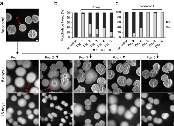
Five populations derived from a B. subtilis natural isolate (BSP1) hereinafter named Ancestral, were passaged daily for 16 days, via dilution in a rich medium with agitation and aeration. Samples were frozen every 2 days so that evolutionary steps during short-term domestication could be followed through whole-genome sequencing. Daily plating revealed that, within the first week of the experiment, two new colony morphotypes emerged (Fig. 1a). The ancestral type, a, dominated the initial populations, while a new type b, characterized by a flat colony morphotype, and a type c, an intermediate morphotype, reached appreciable frequencies rapidly. Type b was detected in all populations, while type c was only observed in three out of five populations (Fig. 1b). As type b achieved the highest frequency, reaching fixation after 16 days in population 1 (Fig. 1c), we conducted a detailed phenotypic and genotypic characterization of one clone from this population. The rapid spread of type b, as well as its emergence in all evolved populations, suggests that it carries an advantage when growing in the laboratory environment. To test this hypothesis, we selected a type b colony from population 1 on day eight, hereinafter named Evolved, and characterized its growth in LB. Indeed both the maximum growth rate per hour and the carrying capacity after 7 h of growth of the Evolved were significantly higher than those of the Ancestral (see Supplementary Fig. S1a,b online). This suggests that Evolved has increased growth traits in the laboratory environment.
Figure 1.
Changes in colony morphology with domestication. (a) Representative image of the Ancestral colony morphology and the three different types of colony morphology, a, b and c, observed at the eighth and sixteenth days of the domestication experiment in the five evolved populations; (b) frequency of each morphotype in the five populations at day 8 (Ancestral, n = 163; Population 1, n = 407; Population 2, n = 140; Population 3, n = 110; Population 4, n = 132; Population 5, n = 162); (c) frequency of morphotype b in population 1 over time (Ancestral, n = 163; day 2, n = 200; day 4, n = 189; day 6, n = 238; day 8, n = 407). The scale bar represents 1 cm and applies to all panels. For panel b and c, n stands for the number of colonies. This figure was generated with Microsoft Excel 2019 MSO (version 16.0.10366.20016; https://www.microsoft.com) and Microsoft PowerPoint 2019 MSO (version 16.0.10366.20016; https://www.microsoft.com).
DegU as a target for the adaptation to the laboratory environment
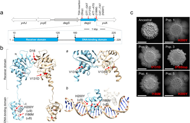
To determine the genetic basis of the adaptive morphotype we performed whole-genome sequencing of the Ancestral and the Evolved clone. A single mutation was observed, in the degU gene, coding for the response regulator DegU28. This non-synonymous mutation is a T-to-G transversion causing the substitution of isoleucine 186 by a methionine (I186M) in the helix-turn-helix motif (HTH), within the DNA-binding domain of DegU28 (Fig. 2a). degU is part of the degS-degU operon, coding for a two-component system that controls social behaviors in B. subtilis24,25,29,30. Interestingly, the laboratory strain 168 is known to have a mutation in degQ that leads to a decreased phosphorylation of DegU29,31,32. In the laboratory strain of B. subtilis, the unphosphorylated form of DegU activates genes involved in the ability to uptake external DNA during competence development33. During growth, the intracellular concentration of the phosphorylated form of DegU (DegU~P), increases, leading to the progressive activation of genes required for swarming motility, biofilm formation, and the secretion of extracellular enzymes29,34. Given the central importance of DegU in processes that could be costly in the laboratory environment we tested whether additional clones of the natural isolate BSP1, which had evolved independently, also carried mutations in degU. Sanger sequencing revealed that all the five clones isolated from each population had mutations in degU: in addition to the mutation causing the I186M substitution (Evolved, population 1; see above), the same mutation was identified in a clone from population 4; a mutation causing the substitution of histidine 200 by a tyrosine (H200Y) was identified in two populations (population 1 and population 5), and another, causing the replacement of valine 131 by an aspartate (V131D) was identified in one (population 2) (Fig. 2a,b). Finally, a mutation caused the insertion of a TGA codon leading to premature translational arrest after codon D18 (Fig. 2a,c; population 3). These populations exhibited distinct colony morphologies but all appeared less structured than the colonies formed by Ancestral (Fig. 2c). Although these colony morphologies seem to slightly differ depending on the mutation found in degU, we cannot exclude the contribution of other mutations that might have arisen during the experimental evolution. The V131D substitution lies within the linker region that separates the receiver and DNA-binding domains of DegU, while H200Y (as for I186M; above) is located in the HTH motif (Fig. 2b). All of the missense mutations affect amino acid residues that are conserved among DegU orthologs (see Supplementary Fig. S2a,b online) and are thus likely to be functionally important. In particular, the I186M and H200Y substitutions are likely to affect DNA binding (Fig. 2a; see also Supplementary Results and Discussion online for a discussion of the effect of the various substitutions). These results suggest a high degree of evolutionary parallelism at the gene level and implicate DegU as the first main target of domestication to the laboratory for the natural isolate BSP1.
Figure 2.
Domestication is accompanied by mutations in degU. (a) degU region of the B. subtilis chromosome (top) and domain organization of the DegU protein (bottom). The position of the various mutations detected and the corresponding amino acid substitution is indicated. (b) Model of the full-length DegU protein of B. subtilis obtained by comparative modeling and using the crystal structure of the LiaR protein from Enterococcus faecalis as the template (PDB code: 5hev). The protein is thought to form a dimer and the two monomers are represented in blue and light brown, with the position of the receiver and DNA-binding domains indicated. The red arrows indicate the location of the single amino acid substitutions found in DegU. a and b show a magnification of the regions encompassing the V131D (a) and the I186M and H200Y (b) substitutions. In b, the region of the helix-turn-helix (HTH) motif is modeled with DNA, to highlight the likely involvement of residues I186 and H200 in DNA binding. The HTH motif was independently modeled using the crystal structure of the LiaR DNA-binding domain as the template (PDB code: 4wuh). (c) Representative images showing the complex biofilm morphology of Ancestral and clones representative of each population after 16 days of domestication. The mutations in DegU present in each clone are indicated in red. All strains were incubated in MSgg for 96 h at 28 °C. Scale bar 1 cm. This figure was generated with Microsoft PowerPoint 2019 MSO (version 16.0.10366.20016; https://www.microsoft.com).
The I186M substitution in DegU is responsible for the colony phenotype of evolved
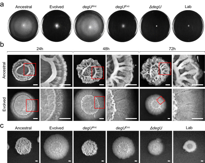
We proceeded with a detailed characterization of the Evolved from population 1 which carried the I186M substitution in DegU. To test whether this substitution caused the alteration in colony morphology, the wildtype degU allele (or degUAnc, for Ancestral) and the allele coding for DegUI186M (found in population 1, or degUEvo) were introduced ectopically at the non-essential amyE gene in a strain bearing a degU knockout constructed in the background of Ancestral (see Supplementary Fig. S3; see also Supplementary Results and Discussion online). The resulting strains, termed degUAnc and degUEvo, had a colony morphology indistinguishable from Ancestral and Evolved / degUEvo respectively (Fig. 3c). We infer that the degUEvo allele is responsible for the colony phenotype of Evolved.
Figure 3.
degUEvo is responsible for the alteration in swarming motility and colony architecture. (a) Swarming motility assay of Ancestral, Evolved, degUAnc, degUEvo, ΔdegU, and Lab. LB plates fortified with 0.7% of agar were inoculated incubated for 16 h at 28 °C. Swarm expansion, resulting from bacterial growth, appears in white, whereas uncolonized agar appears in black. (b) Representative images showing the complex colony architecture development along with the indicated time points of the Ancestral and Evolved. The strains were grown in MSgg medium at 28 °C. (c) Representative images showing the complex colony architecture of the indicated strains on MSgg agar plates incubated for 96 h at 28 °C. Scale bars 1 cm. For all panels, the assays were repeated a minimum of three times. This figure was generated with Microsoft PowerPoint 2019 MSO (version 16.0.10366.20016; https://www.microsoft.com).
degUI186M causes loss of major social traits in BSP1
In commonly used laboratory strains of B. subtilis, such as 168 and NCIB 3610, several social traits are regulated by the unphosphorylated form as well as by low, medium, or high levels of DegU~P25,26. DegU functions as a “rheostat”, sensing environmental signals and allowing the expression of competence, social motility (or swarming), biofilm development, and exoprotease production along a gradient in the cellular accumulation of Deg~P25. Competence development is positively regulated by the unphosphorylated form of DegU33,35,36. ComK is a regulatory protein required for competence development that drives transcription of the genes coding for the DNA uptake and integration machinery but also stimulates transcription of its own gene37. Unphosphorylated DegU functions as a priming protein in competence development by binding to the comK promoter and facilitating ComK stimulation of comK transcription at low ComK concentrations33,37,38. Given this, we proceeded to test the effect of the I186M substitution in Evolved, and whether the phenotypes described above and observed in laboratory strains were also regulated by DegU in the natural isolate strain BSP1. We found no differences in the development of competence between Ancestral or degUAnc and Evolved or degUEvo (see Supplementary Fig. S4 online). This suggests that the I186M substitution does not affect the function of unphosphorylated DegU in promoting competence development and thus, that degUEvo is not a loss-of-function allele. Low levels of DegU~P, however, activate transcription of genes involved in social motility, or swarming24,29,39,40. Swarming motility assays revealed that while the Ancestral and degUAnc have the ability to swarm, the Evolved and degUEvo show poor swarming ability (Fig. 3a). A widely used laboratory strain, PY79, hereinafter termed Lab, as well as other laboratory strains, carry mutations that prevent swarming motility41–43. In our assay, Lab as well as a degU insertional mutant, are deficient in swarming motility (Fig. 3a). Thus, the I186M substitution leads to limited pleiotropic effects, i.e., decreased swarming motility without affecting competence.
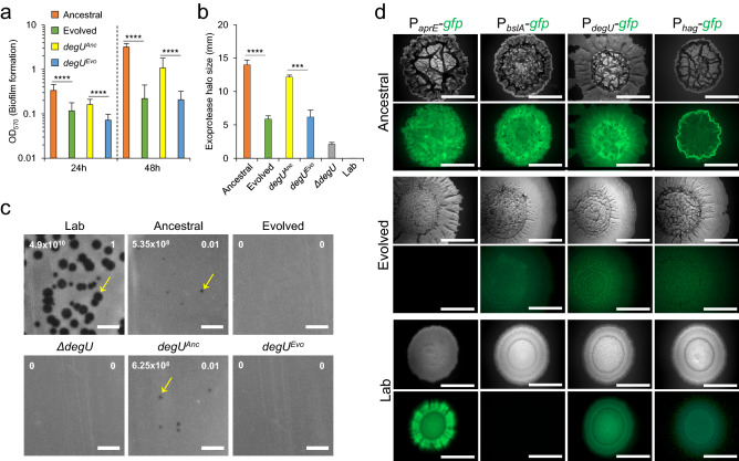
The production of proteins responsible for biofilm formation is another trait under the control of DegU~P24,29,31,44,45. To query whether biofilm architecture and robustness were affected by the I186M substitution, we examined both the colony (a biofilm formed at the solid medium/air interface) architecture over time, as well as the formation of biofilms at the liquid/air interface in liquid cultures. Both Ancestral and degUAnc showed a complex colony architecture characterized by many wrinkles after 24 h of incubation, and the complexity of the colony architecture increased with time (Fig. 3b). In contrast, Evolved and degUEvo formed colonies that were flatter and had fewer wrinkles (Fig. 3b,c). As a control, colonies formed by a degU insertional mutant show extremely low complexity and, when compared to Ancestral, Lab also formed colonies with a simpler architecture (Fig. 3c). These results are consistent with the importance of DegU for the formation of a biofilm at a solid medium/air interface44,45. Similarly, in liquid cultures, both Evolved and degUEvo formed a biofilm at the liquid/air interface which was less robust than that of the Ancestral as determined through quantification of the pellicle formed using cristal violet (Fig. 4a; see Supplementary Fig. S5 online). Thus, degUEvo affects the ability of B. subtilis to form complex, robust biofilms.
Figure 4.
degUEvo is responsible for the alteration in biofilm complexity, exoprotease secretion, phage resistance, and the pattern of gene expression during biofilm formation. (a) Quantification by the crystal violet method of the biofilms formed by the Ancestral (n = 18 for 24 h; n = 15 for 48 h), Evolved (n = 22 for 24 h; n = 15 for 48 h), degUAnc (n = 18 for 24 h, n = 19 for 48 h), and degUEvo (n = 19 for 24 h; n = 18 for 48 h) in MSgg broth incubated at 25 °C for the indicated time points. Mann–Whitney U tests were used. ****p < 0.0001. (b) Dimension of the halos produced by the Ancestral (n = 4), Evolved (n = 4), degUAnc (n = 3), degUEvo (n = 3), and ΔdegU (n = 4) in LB fortified with 1.5% agar and supplemented with 2% of skimmed milk incubated at 37 °C for 48 h. Unpaired t test with Welch’s correction were used. ****p < 0.0001 and ***p = 0.007. The error bar represents the standard deviation. (c) Efficiency of Plating (EOP) shown in white numbers in the superior right corner for the Ancestral, Evolved, degUAnc, degUEvo, ΔdegU using as a reference the indicator strain Lab (PY79), which is phage sensitive. The number of plaque-forming units (PFU’s) is shown in the superior left corner in white numbers. The yellow arrows indicate SPP1 phage plaques. Note that Ancestral is sensitive to SPP1 but the plaque size is reduced when compared to the Lab strain, while the Evolved is resistant. Scale bars 1 cm. (d) Representative images of the expression of transcriptional fusions between the aprE, bslA, hag, and degU promoter regions and gfp in Ancestral, Evolved, and Lab after 96 h of incubation in MSgg at 28 °C. Scale bar 1 cm. In panel a and b the error bars represent the standard deviation. This figure was generated with Microsoft Excel 2019 MSO (version 16.0.10366.20016; https://www.microsoft.com) and Microsoft PowerPoint 2019 MSO (version 16.0.10366.20016; https://www.microsoft.com).
Other processes important for the natural B. subitlis social lifestyle dependent on high levels of DegU~P include the secretion of exoproteases and survival under bacteriophage predation25,39,46,47. We found decreased exoprotease secretion in Evolved and the degUEvo strain, as compared to Ancestral and degUAnc (Fig. 4b). For reference, exoprotease secretion was severely impaired in both the degU insertional mutant and in Lab (Fig. 4b). Lastly, when infected with the SPP1 bacteriophage, both Evolved and degUEvo showed increased resistance to phage infection, as compared to Ancestral and degUAnc or Lab (Fig. 4c). Overall, these results show that in the natural isolate BSP1, as in the laboratory strain, DegU is a key regulator of social straits. They also reinforce the view that the degUEvo mutation is pleiotropic, albeit but not fully, as it affects the phenotypes regulated by DegU~P, including social mobility, biofilm formation, exoprotease production, and resistance to phage infection, while does not impair the function of unphosphorylated DegU in priming competence development.
Decreased transcription of DegU~P-target genes after domestication
The phenotypic assays performed showed that in the natural isolate BSP1 the I186M substitution in DegU changed traits regulated by DegU~P in the first steps of domestication. The location of the I186M substitution in the HTH motif of DegU raised the possibility that the transcription of genes regulated by DegU~P could be impaired in Evolved relative to Ancestral. To test this, we constructed transcriptional fusions between the promoters for the hag, bslA, and aprE genes and the gfp gene. hag codes for flagellin, the main component of the flagellum and is required for social motility; moreover, the expression of hag indirectly requires low levels of DegU~P24,48. bslA codes for a self-assembling hydrophobin that forms a hydrophobic coat at the surface of biofilms; as shown for strain NC3610, BslA is required for the formation of structurally complex colonies and biofilms49–52. Lastly, aprE codes for subtilisin, a major alkaline exoprotease28,53. Transcription of both the bslA and aprE genes is subject to a logic AND gate, in that it requires both derepression of both promoters under the control of Spo0A~P and in addition, DegU~P24,28,49,54.
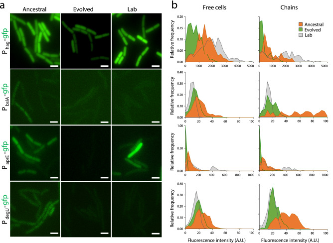
We examined the transcription of these genes at the colony (biofilms) and single-cell (stationary phase in LB) levels. In Ancestral, the PaprE-, PbslA- and PdegU-gfp fusions were expressed throughout the architecturally complex colonies, whereas expression of Phag-gfp was expressed mostly at the colony edge (Fig. 4d). Expression of PaprE-, PdegU- and Phag-gfp was also detected in Lab, but at lower levels, and expression of PbslA-gfp was not detected, which is consistent with the simpler colony morphology (Fig. 4d). Interestingly, in spite of the maintenance of a complex colony architecture, expression of PbslA-, PdegU- and Phag-gfp was detected at very low levels in Evolved, and expression of PaprE-gfp was not detected (Fig. 4d). These results suggest that DegUI186M reduces the expression of DegU target genes during biofilm development.
During planktonic growth, transcription of hag decreases markedly in the absence of DegU~P29. Moreover, the expression of hag is heterogeneous, with free cells showing higher expression levels than chained cells55. Accordingly, in Ancestral, the free cells showed higher fluorescence intensity from the Phag-gfp fusion than chained cells (p < 0.0001, Kruskal–Wallis test with Dunn’s test of multiple comparisons; Fig. 5a,b). In contrast, Evolved showed a decrease in the GFP signal from Phag-gfp both in free cells (~ 2.2 fold; p < 0.0001, Kruskal–Wallis test with Dunn’s test of multiple comparisons; NB: unless otherwise stated, all p values below were determined in the same manner) and chains (~ 2.8 fold; p < 0.0001), relative to the Ancestral (Fig. 5b). Although Lab does not exhibit swarming motility, as previously reported55 it showed increased expression of Phag-gfp in both free cells (p < 0.0001) and chains (p < 0.0001) when compared to Ancestral, which exhibits swarming motility (Fig. 5b). Expression of bslA is heterogeneous between free cells and chains in the Ancestral, as also found for hag (Fig. 5a; see also above), and was ~ 2.8 fold lower in free cells when compared to chained cells (p < 0.0001; Fig. 5b). Strikingly, no heterogeneous expression between free cells and chains of bslA was observed in the Evolved clone (Fig. 5b). Moreover, the level of bslA expression was similar in free cells and chains (p = 0.09), although bslA expression was ~ 1.5 fold lower (p < 0.0001) in the free cells when compared to Ancestral (Fig. 5b). In the Lab strain, which does not produce robust biofilms, transcription of bslA is markedly reduced (Fig. 5b). In laboratory strains, DegU~P is a direct positive regulator of aprE, and expression of aprE was shown to be bi-stable in a laboratory strain54. Consistent with this finding, low and high levels of aprE expression were detected for Lab, both in free cells and chains (Fig. 5). In contrast, Ancestral did not express high levels of aprE, although it showed heterogeneity between free cells and chains in aprE expression (Fig. 5). Lastly, Evolved showed greatly reduced expression of aprE, both in free cells (p < 0.0001, Kruskal–Wallis test with Dunn’s test of multiple comparisons) and chains (p < 0.0001) (Fig. 5). Thus, domestication in the natural isolate BSP1 is accompanied by a sharp decrease in the expression of aprE.
Figure 5.
degUEvo alters the pattern of gene expression at the single-cell level. (a) Representative images of the expression of hag-, bslA-, aprE- and degU-gfp transcriptional fusions in Ancestral, Evolved, and Lab one hour after the onset of stationary phase in LB. The cultures were grown with agitation at 37 °C. Scale bar 1 µm. (b) Relative frequency of expression of transcriptional fusions of the indicated promoters to gfp in the same conditions as above. For the relative frequency of expression of transcriptional fusions in free cells, a total of 371 (hag-gfp), 453 (bslA-gfp), 713 (aprE-gfp), and 561 (degU-gfp) cells from Ancestral, Evolved and Lab were analyzed. For the relative frequency of expression of transcriptional fusions in chains, a total of 150 (hag-gfp), 79 (bslA-gfp), 168 (aprE-gfp), and 191 (degU-gfp) cells from Ancestral, Evolved and Lab were analyzed. This figure was generated with GraphPad Prism 7 software for Windows (version 7.04; https://www.graphpad.com/scientific-software/prism/) and Microsoft PowerPoint 2019 MSO (version 16.0.10366.20016; https://www.microsoft.com).
Overall, these results show that in the natural isolate BSP1, the I186M substitution in DegU reduces the expression of DegU~P regulated genes during biofilm development and planktonic growth.
Domestication impairs a degU positive auto-regulatory loop
In its high-level phosphorylated state, DegU~P activates transcription of degU itself56,57 by binding to a site in the degU regulatory region34,58. Importantly, this positive auto-regulatory loop contributes to the heterogeneous expression of degU and the DegU~P-dependent genes in laboratory strains54. Since Evolved shows reduced expression of DegU~P-target genes, we wanted to test whether DegUI186M further impaired expression of degU, reducing the levels of DegU. We found expression of PdegU-gfp to be heterogeneous between free cells and chains in Ancestral, with chains showing a ~ 1.6 fold higher expression relative to free cells (p < 0.0001; Fig. 5)55. In Evolved and Lab strains, expression of degU was reduced when compared to Ancestral, both in free cells (p < 0.0001) and chains (p < 0.0001) (Fig. 5).
Together, these results suggest that I186M impairs the ability of DegU~P to activate transcription of degU itself. Impaired activation of the DegU auto-regulatory loop, in turn, reduces transcription of degU itself, suggesting an explanation as to why the expression of genes directly or indirectly regulated by DegU~P in Evolved is reduced.
Domestication leads to increased survival in the presence of cells of the immune system
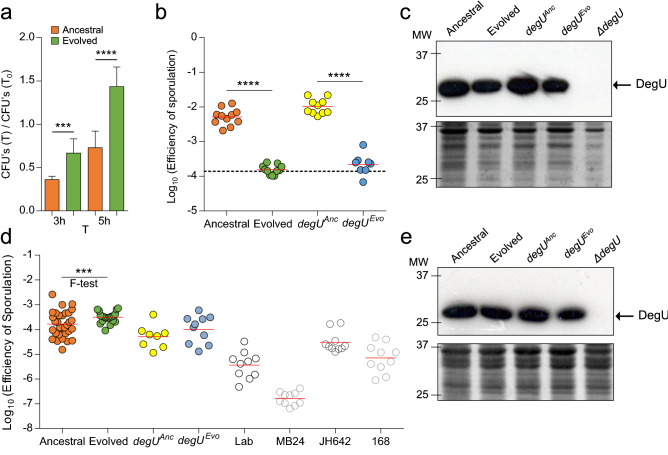
Bacillus subtilis has been isolated from the gastrointestinal tract of several animals including humans59,60 and has been found to grow, sporulate and persist in the murine gut61,62. Thus, it can experience selective pressures inside a host. To determine if domestication could impair the ability of B. subtilis to withstand a hostile host environment, we measured the survival of Evolved and Ancestral in the presence of cells of the innate immune system—macrophages. Interestingly, the Evolved strain showed an increased survival over the Ancestral in the presence of macrophages, both at 3 h (p = 0.001, Unpaired t test with Welch’s correction) and 5 h (p < 0.0001, Unpaired t test with Welch’s correction) (Fig. 6a). This result illustrates that the domestication to the laboratory environment coincidently leads to a survival advantage for B. subtilis when facing cells of the host immune system.
Figure 6.
degUEvo increases survival in the presence of macrophages and changes sporulation efficiency in an environment-dependent manner. (a) Macrophages were infected with Ancestral (n = 9) and Evolved (n = 9) and colony-forming units of both the intracellular and extracellular bacteria obtained by plating at the indicated time points. Unpaired t test with Welch’s correction were used, where ***corresponds to p = 0.001 and **** to p < 0.0001. The error bar represents the standard deviation. (b) Comparison of the sporulation efficiency in RPMI medium between Ancestral (n = 11), Evolved (n = 11), degUAnc (n = 10) and degUEvo (n = 8). The efficiency of sporulation was calculated as the ratio between the heat resistant spore counts and total (viable) cells. The dashed line indicates the average sporulation efficiency for the Ancestral in LB. ANOVA with Tukey’s multiple comparison tests were used, where **** corresponds to p < 0.0001. (c) Accumulation of DegU in Ancestral, Evolved, degUAnc, degUEvo, and the degU insertional mutant in RPMI. (d) Comparison of the sporulation efficiency and variance in LB between Ancestral (n = 31), Evolved (n = 21), degUAnc (n = 8), degUEvo (n = 10), Lab (n = 10) and three other commonly used laboratory strains (MB24, n = 10, JH642, n = 10, and 168, n = 10). For the mean sporulation efficiency, an ANOVA and Tukey’s multiple comparison test was used. For the variance the F test was used. ***p = 0.0002. (e) The levels of DegU are similar between Ancestral and Evolved in LB. Accumulation of DegU in Ancestral, Evolved, degUAnc, degUEvo, and the degU insertional mutant. In (c,e), the cells were collected after growth in RPMI (c) or LB (e) and whole-cell lysates prepared (see “Methods”). Proteins (20 µg) in whole-cell lysates were resolved by SDS-PAGE and subject to immunoblot analysis with an anti-DegU antibody. The arrow shows the position of DegU; the red arrows indicate slightly higher levels of DegU. The panel below the immunoblot shows part of a Coomassie-stained gel, run in parallel, as a loading control. The position of molecular weight markers (in kDa) is shown on the left side of the panels. In panels (b,d) the red line indicates the mean. The full-length blots and full-length Coomassie-stained gels are presented in Supplementary Figure S6. This figure was generated with GraphPad Prism 7 software for Windows (version 7.04; https://www.graphpad.com/scientific-software/prism/) and Microsoft PowerPoint 2019 MSO (version 16.0.10366.20016; https://www.microsoft.com).
Sporulation efficiency changes across environments
High levels of DegU~P promote sporulation by increasing the levels of Spo0A~P30. Since the I186M substitution reduced transcription of degU, we reasoned that changes in the frequency of sporulation could have occurred during domestication. We first tested this phenotype in a host-related environment (RPMI medium) and found the sporulation efficiency of Ancestral (and of degUAnc) to be ~ 1.5 Log10-fold higher than that of Evolved (or degUEvo) (Fig. 6b). The levels of DegU, as assessed by immunoblot analysis with an anti-DegU antibody of established specificity63, are slightly higher in RPMI for both Ancestral and degUAnc when compared to Evolved or degUEvo (Fig. 6c).
We further tested the ability of our strains to sporulate in the environment where the domestication process occurred (LB medium). Most laboratory strains sporulate at very low levels in LB (about 104 spores/ml of culture as compared to 108 spores/ml in a medium such as DSM that support efficient sporulation)12. We found no significant difference in the mean efficiency of sporulation between Evolved (and degUEvo) and Ancestral (or degUAnc) in LB (Fig. 6d). Interestingly, the variance was reduced in the Evolved when compared to the Ancestral (F test, p = 0.0002). In addition, all of the laboratory strains tested sporulated in LB at efficiencies lower than that of the Ancestral, although one strain, JH642, sporulated better than the other laboratory strains tested under these conditions (Fig. 6d). This is consistent with the initial description of Ancestral, which enters sporulation during growth and reaches a higher titer of spores than any of the laboratory strains tested in DSM12. The steady-state levels of DegU in whole-cell lysates obtained 1 h after the onset of the stationary phase in LB revealed very similar levels of DegU for all the strains (Fig. 6e).
These results suggest that the I186M mutation can affect the intracellular levels of DegU in a manner that depends on the environment and illustrates that the influence of DegU on sporulation exhibits antagonistic pleiotropy.
Discussion
BSP1 is a gastrointestinal isolate of B. subtilis which initiates sporulation during growth. This happens because BSP1 lacks three rap genes, coding for phosphatases that normally drain phosphoryl groups from the phosphorelay, thus negatively regulating the activation of Spo0A. As such, more cells in the population have Spo0A active above a threshold level required to induce sporulation. Several other gastrointestinal isolates of B. subtilis, including from the human gut, also lack combinations of the rap genes and exhibit enhanced sporulation12,62. B. subtilis completes its entire life cycle in the gut62, and it seems likely that sporulation is important for survival and/or propagation in the gut ecosystem, as described for other spore-formers64,65. Sporulation is also important for the efficient dispersal of spore-formers through the environment and among hosts66–68. Sporulation is, however, a time and energetically costly process, requiring the differential expression of over 10% of the genome over a period of 7–8 h12,69. Hence, the propagation of B. subtilis in the laboratory in the absence of selection for sporulation results in a reduction in the ability to sporulate13–16. B. subtilis has been used in laboratory conditions for more than 50 years and has accumulated mutations likely to be adaptive in that environment and which, relative to wild strains, lead to the attenuation of phenotypic traits which include swarming motility41,42, poly-γ-glutamate synthesis31, production of antibiotics, the secretion of degradative enzymes70 or the formation of robust biofilms6,31,71. These processes have either become neutral with respect to fitness, or selection favored their loss under laboratory conditions. In addition, repeatedly selecting individual colonies to cultivate and maintain bacteria in laboratory conditions can increase the chances of loss of phenotypes independently of fitness differences72.
Here we traced one example of possible domestication routes for the natural isolate B. subtilis strain BSP1. Rapid changes in colony morphotypes were observed in parallel cultures, leading to complete fixation of a specific colonial morphotype, termed type b, in all the replicate cultures after only 2 weeks (Fig. 1). The adaptive morphotype is characterized by a smooth and flat colony, lacking the complex architectural features of the original strain (Fig. 2c). Similar colony morphology changes were previously observed during the domestication of other B. subtilis strains3,13–15,18. Colonies are biofilms formed at the solid/air interface1. As such, these observations hinted at the attenuation of an important social behavior. Studies in Salmonella enterica, Saccharomyces cerevisiae, Bacillus licheniformis, Aneurinibacillus migulanus, and Myxococcus xanthus have also documented the appearance of smooth colonies within a short period of time5,73–76. This suggests that phenotypic parallelism across species is a broad pattern of adaptation to the laboratory environment.
At the genomic level we detected mutations in the coding region of the degU gene (Fig. 2) in clones isolated from the independently evolved populations. It is known that many bacteria and yeast can evolve very rapidly in laboratory environments77–79 and with considerable level of gene parallelism across the independent replicates21. As such, it is not unexpected that the same gene could be targeted by selection in our domestication experiment. Yet another possible explanation for the emergence of these mutations in the same gene could be that degU is a mutational hotspot. However, a mutation accumulation experiment performed in the undomesticated strain of B. subtilis NCIB 3610 with a normal mutation rate did not detect mutations in degU80. This suggests that the most likely explanation for the high level of convergence observed in this work is due to strong selection for mutations in degU when BSP1 is adapting to the laboratory environment. DegU is the response regulator of the two-component system DegS–DegU and controls social traits such as biofilm formation, swarming motility, and exoprotease secretion24. In its non-phosphorylated state, DegU is responsible for the development of competence while the rise in DegU~P levels sequentially activates swarming, biofilm formation, and exoprotease secretion29. DegU belongs to the NarL/FixJ subfamily of DNA binding proteins81. We characterized in detail the effects of a mutation leading to the I186M substitution in DegU. The I186M substitution occurs in the DNA-recognition helix of the DegU HTH motif, located in the C-terminal domain of the protein (Fig. 2a). Modeling studies indicate that this substitution is likely to affect a contact of the HTH motif with bases in the major groove of DNA as is evident in the crystal structure of NarL, in which I186 is conserved, with DNA82 (Fig. 2; see Supplementary Fig. S2 online; see also Supplementary Results and Discussion online). Moreover, alanine-scanning mutagenesis resulted in decreased transcription of the DegU-controlled genes comG (as a proxy for the activity of ComK, a direct target of DegU) and aprE in a strain producing DegUI186A; the substitution affected the binding of DegU to the comK and aprE target promoters28. I186 is conserved in LuxR, another NarL family member, and its replacement by Ala results in reduced binding to target DNA sequences83. One other substitution found in DegU in our study, H200Y, is likely to impair DNA binding, as suggested by the study of a single Ala substitution in transcription and DNA binding to cognate sites in the promoters of the DegU-responsive genes comK and aprE28. This residue, however, as suggested by the structure of a NArL-DNA co-crystal, contacts the DNA phosphate backbone82 (see also Supplementary Results and Discussion online). Importantly, while the I186M substitution found in the domesticated clone, Evolved, did not cause changes in the efficiency of transformation with exogenous DNA, it impaired processes regulated by DegU~P, such as swarming motility, biofilm formation, SPP1 bacteriophage sensitivity, and exoprotease secretion. In accordance with these observations, the transcription of genes regulated by DegU~P was reduced in the domesticated clone. The transcription of degU itself was similarly reduced (Fig. 5b); since our degU transcriptional reporter fusion includes all promoters known to contribute to the expression of the gene (see Supplementary Fig. S3 online), including the DegU~P-recognized P3 promoter, it suggests that the I186M substitution affects the auto-regulatory loop that controls the production and activity of DegU57. Failure to successfully activate the auto-regulatory loop due to impaired binding to DNA of the I186M substitution could be an explanation for the defective biofilm development, exoprotease production, and phage sensitivity, which require high levels of DegU~P.
Earlier work has shown that the extensive propagation of B. subtilis in a nutrient-rich medium, i.e., under conditions of relaxed selection for sporulation, resulted in the emergence of a strain that accumulated mutations in genes of biosynthetic pathways, sporulation competence, DNA repair, and others14,15. Relative to the ancestral, the resulting strain displayed different cell and colony morphologies, loss of sporulation and competence, but an overall increased fitness under laboratory conditions14,15. Interestingly, our selection, which was also performed in rich medium, LB, did not result in loss of sporulation. Rather, the I186M substitution modulated the efficiency of sporulation across conditions, specifically, in a host-related condition (RPMI medium) and LB (Fig. 6). Importantly, the accumulation of DegU was only slightly higher in Ancestral (and degUAnc) relative to Evolved when sporulation was tested in RPMI, where the sporulation efficiency of Ancestral (and degUAnc) was also higher than that of Evolved. Since high levels of DegU~P control production of Spo0A~P30, it is possible that I186M places DegU~P below a threshold required for the stimulation of sporulation through Spo0A. This is consistent with the decreased transcription of degU by DegUI186M (Fig. 5b). In contrast, no differences in the mean sporulation efficiency of Evolved and Ancestral were found under nutritional conditions (LB medium) that do not support efficient sporulation by most laboratory strains (Fig. 6), and the accumulation of DegU did not differ between Evolved and Ancestral. We note, however, that the accumulation of DegU reflects both transcription/production of the protein and proteolysis; since DegU~P is a preferred substrate for the ClpXP protease, the steady-state levels of the protein will reflect the ratio of DegU/DegU~P levels57. The inability of some domesticated strains of B. subtilis to form robust biofilms results from the accumulation of mutations in four chromosomal genes (sfp, epsC, swrA, and degQ), in addition to the loss of plasmid-borne gene, rapP6. In contrast, under our experimental conditions and using a natural isolate of B. subtilis, the target for mutation during domestication is degU. Our results are consistent with what was observed in the laboratory strain 168, that has a mutation in degQ6, targeting the DegS–DegU system. Considering that targeting the DegS–DegU system is a strategy used by two different strains of B. subtilis, with very different genomes, suggests that this system is a major target of adaptation to the laboratory environment.
It seems possible that our experimental conditions did not cause relaxed selection for sporulation; rather, our selection may have first targeted the costliest phenotypes under the test conditions, at least in the context of the BSP1 genome. Those phenotypes are directly controlled by DegU~P, including the formation of complex colonies and robust biofilms. It is interesting to note that a 2-month culture of the laboratory strain NCIB 3610, resulted in the emergence of strains with different levels of biofilm robustness, as is evident in the colony architecture and the expression of genes required for matrix production3. These phenotypes were the result of mutations in the sinR gene, coding for a master regulator of biofilm development, and arose both on plates as well as in LB cultures3. One conclusion offered was that matrix overproduction can be neutral or advantageous in rich medium3. The difference in the mutations obtained under our experimental conditions and the study of Leiman et al. is in line with the idea that adaptations to a new environment, depend both on the initial genome as well as the culture history of the strain84. In addition, since other phenotypes other than biofilm formation are probably under selection in the laboratory environment, pleiotropic genes as degU may be selected.
Taken together, the results suggest that the I186M mutation impairs the ability of DegU to function as a transcription factor and that this feature confers an advantage to the natural isolate BSP1 when growing under laboratory conditions. Interestingly, arcA in E. coli and rpoS in S. enterica and E. coli, regulators of stationary phase processes, are also common targets of laboratory adaptation73,85. This strengthens the evidence for a general rule that the initial adaptations to a new environment involve changes in genes that act as regulatory hubs of networks that affect the stationary phase of growth.
The accumulation of mutations during adaptation to a laboratory environment over a relatively small number of passages also unraveled a signal of antagonistic pleiotropy: evolved exhibited changes in traits in host-related environments, and showed increased survival in the presence of macrophages (Fig. 6a). This provides support for the coincidental hypothesis86,87 that posits that adaptations to new environments can lead to changes in complex interactions with hosts. Increased survival of the Evolved when facing the cells of the host immune system also implicates DegU potential relevance for the interaction between B. subtilis and its host in its natural environment. Importantly, the genus Bacillus is commonly used as a probiotic88,89, and B. subtilis was shown to stimulate macrophage activity and the host immune response90–93. However, the mechanisms behind Bacillus role as a probiotic are still unclear88. Given that DegU is widely conserved amongst the Bacillus genus (see Supplementary Fig. S2 online), the role of DegU in this interaction should be studied in future work. In addition, our study highlights the importance of performing short-term cultivation of bacterial natural isolates to prevent the loss of traits that may be important for the probiotic activity of B. subtilis.
Methods
General methods
Lysogeny Broth (LB) medium was used for the routine growth of B. subtilis and Escherichia coli. The E. coli strain DH5α was used as the host strain for the construction and maintenance of plasmids and was grown in the presence of 100 µg ml−1 ampicillin when carrying vectors or recombinant plasmids. When appropriate, B. subtilis strains were grown in the presence of antibiotics, used at the following concentrations: 5 µg ml−1 chloramphenicol, 1 µg ml−1 erythromycin, and 1 µg ml−1 neomycin for liquid cultures, and 3 µg ml−1 neomycin on solid media.
Other methods
The construction of all plasmids and strains is described in detail in the Supplementary Methods online. The B. subtilis strains used or constructed in this work are listed in Supplementary Table S1 online. Plasmids are listed in Supplementary Table S2 online and oligonucleotides in Supplementary Table S3 online. Modeling of the DegU structure is also described in Supplementary Results and Discussion online.
Domestication experiments
Five independent populations from five different colonies, all derived from the ancestral natural isolate BSP160, were grown for 16 days in LB with a 1:100 dilution into fresh medium every 24 h. This is a common media for growing B. subtilis in the laboratory environment and may introduce selective pressure against sporulation and biofilm formation. At the point of dilution, an aliquot from each culture was collected and kept frozen at – 80 °C for subsequent analysis.
Whole-genome sequencing
To identify the mutations that emerged after 8 days of evolution we extracted DNA from the Evolved clone from population 1 and the Ancestral. The DNA library construction and sequencing were carried out by the Instituto Gulbenkian de Ciência (IGC) genomics facility. Each sample was pair-end sequenced on an Illumina MiSeq Benchtop Sequencer. Standard procedures produced data sets of Illumina paired-end 250-bp read pairs. The mean coverage per sample was 30 × and 18 × for the Evolved and Ancestral, respectively. Mutations were identified using the BRESEQ pipeline version 0.32.194 with default parameters and using the available BSP1 genome95 as a reference genome. All predicted mutations were manually inspected using the Integrative Genomics Viewer96.
Macrophages culture and infection assay
The murine macrophage cell line RAW 264.7 was cultured in RPMI medium (Sigma), supplemented with 2 mM l-glutamine, 1 mM sodium pyruvate, 10 mM hepes, 50 µM 2-mercaptoethanol solution and 10% heat-inactivated Fetal Bovine Serum in an atmosphere of 5% CO2. For the infection, B. subtilis and the macrophages were grown separately in a 24-well tissue plate containing fresh RPMI media as described above. At 24 h of acclimatization, B. subtilis was diluted 1:100 into fresh RPMI. Macrophages were washed, re-suspended in fresh RPMI, and activated with 2 µg ml−1 CpG for another 24h97. Then, the macrophages were washed to remove the remaining CpG, fresh RPMI media was added, and B. subtilis was added to a 1:8 MOI (multiplicity of infection; about 8 × 10⁶ cells). At the indicated time points of infection, the wells were scraped and the contents centrifuged at 6000g for 10 min at room temperature. After centrifugation, the samples were serially diluted and plated to determine the titer of total viable cells and heat-resistant spores.
Sporulation assays
Sporulation of B. subtilis was usually analyzed in LB and supplemented RPMI. When using LB, B. subtilis cultures were grown overnight, diluted 1:100, and incubated for 24 h at 37 °C. At this time, dilutions of the cultures were plated for total viable counts and treated for 20 min at 80 °C to determine the titer of heat-resistant spores. For supplemented RPMI, the cultures were grown as described above for 48 h and plated for viable cells and spore counts as described above for LB. The sporulation efficiency was defined as the ratio of heat-resistant spores relative to the total viable cell count12.
SPP1 phage lysates and transduction
SPP1 lysates were prepared as described by Yasbin and Young98. Briefly, a dense culture of B. subtilis was infected with different dilutions of SPP1 in a semisolid LB agar (LB containing 0.7% agar). The plate containing near confluent phage plaques was washed with 4 ml of TBT, centrifuged at 5000g for 10 min, treated with 12 µg ml−1 DNase, and filtered through a 0.45 µm syringe filter. The indicator strain PY79 was used for titration of the SPP1 lysates as described by São-José et al.99. SPP1 phage transduction was performed as described42. The recipient strains were grown in LB until the stationary phase after which 1 ml of the culture of the recipient strain was mixed in a glass tube with 10 mM CaCl2 and infected with an MOI of 1 of the donor SPP1 lysate. The transduction mixture was then incubated at 37 °C for 25 min with agitation, centrifuged at 5000g for 10 min, washed with 2 ml of LB, and centrifuged again at 5000g for 10 min. The supernatant was discarded, the pellet was resuspended in 100 µl of LB and plated onto LB plates fortified with 1.5% agar with the appropriate antibiotics and 10 mM of sodium citrate.
Competence assay
The development of competence was performed as described by Baptista et al.47. Briefly, B. subtilis cultures were grown overnight and diluted 1:100 in GM1 at 37 °C. Ninety minutes after the end of the exponential growth, the cultures were diluted 1:10 in GM2 and incubated for 90 min at 37 °C. At this point, a sample of the cultures was serially diluted in LB and plated for determination of total colony forming units (CFU) per milliliter. For transformation, DNA from strain AH7605 or W648 was added to 500 µl of the culture samples, to a concentration of 5 µg ml−1, the mixture incubated for 30 min at 37 °C and finally plated with the appropriate antibiotics. The transformation efficiency is the ratio between the number of transformants and the total number of colonies.
Protease activity assay
Secreted proteases were observed essentially as described by Saran et al.100. The strains were grown until they reached an absorbance of 0.8 at 600 nm. At this time the cultures were diluted to an absorbance of 0.01 at 600 nm. 10 µl of this dilution was spotted in a 2% skimmed milk plate and incubated at 37 °C for 48 h. Then, 6 ml of 10% Tannic Acid was added for the detection of the protease-positive strains. The diameter of the halos observed was measured, and the diameter of the colony was subtracted to obtain the real value of the halo.
Swarming and colony morphology assays
Swarming motility was examined according to the method described by Kearns and Losick42. For colony morphology, the B. subtilis cultures were grown overnight and 3 µl of the culture was spotted onto an MSgg101 plate fortified with 1.5% agar. The plates were incubated at 28 °C or 37 °C. The images were captured at the times indicated in the figures.
Biofilm quantification by crystal violet
The method used for estimating the solid-surface-associated biofilm formation with crystal violet was as described by Morikawa et al102. Briefly, an overnight culture was diluted to an absorbance of 0.03 at 600 nm and mixed 1:100 into 100 µl of MSgg in a 96-well plastic titer plate. The plate was incubated for 48 h at 25 °C. Then, the culture was carefully removed from the wells. After washing two times with distilled water, 150 µl of 1% crystal violet was added to the wells and incubated for 25 min at room temperature. The wells were washed again two times with distilled water and the crystal violet attached to the biofilm matrix was solubilized in 150 µl of DMSO and incubated for 10 min at room temperature. The removed culture was quantified by measuring its absorbance at 600 nm and the biofilm attached to the crystal violet was quantified measuring its absorbance at 570 nm.
Biofilm fluorescence imaging
For biofilm imaging, the B. subtilis cultures were grown overnight and 3 µl of the culture was spotted onto an MSgg plate fortified with 1.5% agar and incubated for 96 h at 28 °C. Images were acquired on a Zeiss Axio Zoom.V16 stereomicroscope equipped with a Zeiss Axiocam 503 mono CCD camera and controlled with the Zeiss Zen 2.1 (blue edition) software, using the 1 × 0.25 NA objective, the fluorescence filter set GFP and the Bright Field optics.
Fluorescence microscopy and image analysis
Cultures were grown in LB until one hour after the end of the exponential phase. The cells were collected by centrifugation (1 min at 2400×g, room temperature), and washed with 1 ml of phosphate-buffered saline (PBS). Finally, the cells were resuspended in 100 μl of PBS and applied to microscopy slides coated with a film of 1.7% agarose. Images were taken with standard phase contrast and GFP filter, using a Leica DM 6000B microscope equipped with an aniXon + EM camera (Andor Technologies) and driven by Metamorph software (Meta Imaging series 7.7, Molecular Devices). For quantification of the GFP signal, 6 × 6 pixel regions were defined in the desired cell and the average pixel intensity was calculated and corrected by subtracting the average pixel intensity of the background, using Metamorph software (Meta Imaging series 7.7, Molecular Devices).
Immunoblot analysis
Immunoblot of DegU was analyzed in LB and supplemented RPMI. When using LB, B. subtilis cultures were grown until one hour after the end of the exponential phase and samples (10 ml) were withdrawn. For supplemented RPMI, the cultures were grown as described above and samples (10 ml) were withdrawn. In both media, the cells were collected by centrifugation (5 min at 15,300 ×g, 4 °C). The cells were resuspended in 1 ml Lysis buffer (50 mM NaH2PO4, 0.5 M NaCl, 10 mM Imidazole, pH 8.0) and whole-cell lysates prepared using a French press cell (19,000 lb/in2). Proteins in the lysates (10 µg) were then separated on 15% SDS-PAGE gels and the gels subject to immunoblot analysis using an anti-DegU antibody of established specificity at a 1:1000 dilution103. Gels run in parallel were stained with Coomassie brilliant blue to be used as loading controls.
Supplementary information
Acknowledgements
We thank Dr. Mitsuo Ogura for the gift of the anti-DegU antibody, Dr. Kazuo Kobayahis for the gift of strains WTF28 and W648, Dr. Jan-Willem Veening for the gift of strains 08G52, 08G57, and 08I09, and Dr. Carlos São José for the gift of the SPP1 lysate and advice on its use. We thank Tanja Ðapa and Anke Konrad for critically reading this manuscript. We also thank Mónica Serrano for helpful discussions. This work was supported by Project LISBOA-01-0145-FEDER-007660 (“Microbiologia Molecular, Estrutural e Celular”) funded by FEDER funds through COMPETE2020-“Programa Operacional Competitividade e Internacionalização” (POCI), by national funds through the FCT (“Fundação para a Ciência e a Tecnologia") grant POCI/BIABCM/60855/2004 to A.O.H. and partially supported by PPBI—Portuguese Platform of BioImaging (PPBI-POCI-01-0145-FEDER-022122) co-funded by national funds from OE—"Orçamento de Estado" and by European funds from FEDER. The research leading to these results also received funding from the European Research Council under the European Community’s Seventh Framework Programme (FP7/2007–2013)/ERC grant agreement no 260421—ECOADAPT to I.G. H.C.B. was the recipient of a doctoral fellowship from the FCT (PD/BD/128429/2017). The funders had no role in study design, data collection and analysis, decision to publish, or preparation of the manuscript.
Author contributions
Conceptualization, H.C.B., A.O.H., and I.G.; data curation, H.C.B., methodology, H.C.B., T.N.C., A.O.H., and I.G.; validation, H.C.B., A.O.H., and I.G.; formal analysis, H.C.B., T.N.C., A.O.H., and I.G.; investigation, H.C.B.; resources, A.O.H., and I.G.; writing—original draft, H.C.B., T.N.C., A.O.H., and I.G.; writing—review and editing, H.C.B., A.O.H., and I.G.; visualization, H.C.B., T.N.C., A.O.H., and I.G.; funding acquisition and supervision, A.O.H. and I.G, project administration, I.G.
Data availability
Genome sequencing data have been deposited with links to BioProject accession number PRJNA592868 in the NCBI BioProject database (https://www.ncbi.nlm.nih.gov/bioproject/).
Competing interests
The authors declare no competing interests.
Footnotes
Publisher's note
Springer Nature remains neutral with regard to jurisdictional claims in published maps and institutional affiliations.
Contributor Information
Adriano O. Henriques, Email: aoh@itqb.unl.pt
Isabel Gordo, Email: igordo@igc.gulbenkian.pt.
Supplementary information
is available for this paper at 10.1038/s41598-020-76017-1.
References
- 1.Vlamakis H, Chai Y, Beauregard P, Losick R, Kolter R. Sticking together: Building a biofilm the Bacillus subtilis way. Nat. Rev. Microbiol. 2013;11:157–168. doi: 10.1038/nrmicro2960. [DOI] [PMC free article] [PubMed] [Google Scholar]
- 2.Eydallin G, Ryall B, Maharjan R, Ferenci T. The nature of laboratory domestication changes in freshly isolated Escherichia coli strains. Environ. Microbiol. 2014;16:813–828. doi: 10.1111/1462-2920.12208. [DOI] [PubMed] [Google Scholar]
- 3.Leiman SA, Arboleda LC, Spina JS, McLoon AL. SinR is a mutational target for fine-tuning biofilm formation in laboratory-evolved strains of Bacillus subtilis. BMC Microbiol. 2014;14:301. doi: 10.1186/s12866-014-0301-8. [DOI] [PMC free article] [PubMed] [Google Scholar]
- 4.Marks ME, et al. The genetic basis of laboratory adaptation in Caulobacter crescentus. J. Bacteriol. 2010;192:3678–3688. doi: 10.1128/JB.00255-10. [DOI] [PMC free article] [PubMed] [Google Scholar]
- 5.Kuthan M, et al. Domestication of wild Saccharomyces cerevisiae is accompanied by changes in gene expression and colony morphology. Mol. Microbiol. 2003;47:745–754. doi: 10.1046/j.1365-2958.2003.03332.x. [DOI] [PubMed] [Google Scholar]
- 6.McLoon AL, Guttenplan SB, Kearns DB, Kolter R, Losick R. Tracing the domestication of a biofilm-forming bacterium. J. Bacteriol. 2011;193:2027–2034. doi: 10.1128/JB.01542-10. [DOI] [PMC free article] [PubMed] [Google Scholar]
- 7.Gallegos-Monterrosa R, Mhatre E, Kovács ÁT. Specific Bacillus subtilis 168 variants form biofilms on nutrient-rich medium. Microbiology (Reading, Engl.) 2016;162:1922–1932. doi: 10.1099/mic.0.000371. [DOI] [PubMed] [Google Scholar]
- 8.Pollak S, Omer Bendori S, Eldar A. A complex path for domestication of B. subtilis sociality. Curr. Genet. 2015;61:493–496. doi: 10.1007/s00294-015-0479-9. [DOI] [PubMed] [Google Scholar]
- 9.Omer Bendori S, Pollak S, Hizi D, Eldar A. The RapP–PhrP quorum-sensing system of Bacillus subtilis strain NCIB3610 affects biofilm formation through multiple targets, due to an atypical signal-insensitive allele of RapP. J. Bacteriol. 2015;197:592–602. doi: 10.1128/JB.02382-14. [DOI] [PMC free article] [PubMed] [Google Scholar]
- 10.Leiman SA, et al. D-amino acids indirectly inhibit biofilm formation in Bacillus subtilis by interfering with protein synthesis. J. Bacteriol. 2013;195:5391–5395. doi: 10.1128/JB.00975-13. [DOI] [PMC free article] [PubMed] [Google Scholar]
- 11.Narula J, et al. Slowdown of growth controls cellular differentiation. Mol. Syst. Biol. 2016;12:871. doi: 10.15252/msb.20156691. [DOI] [PMC free article] [PubMed] [Google Scholar]
- 12.Serra CR, Earl AM, Barbosa TM, Kolter R, Henriques AO. Sporulation during growth in a gut isolate of Bacillus subtilis. J. Bacteriol. 2014;196:4184–4196. doi: 10.1128/JB.01993-14. [DOI] [PMC free article] [PubMed] [Google Scholar]
- 13.Zeigler DR, Nicholson WL. Experimental evolution of Bacillus subtilis. Environ. Microbiol. 2017;19:3415–3422. doi: 10.1111/1462-2920.13831. [DOI] [PubMed] [Google Scholar]
- 14.Maughan H, Nicholson WL. Increased fitness and alteration of metabolic pathways during Bacillus subtilis evolution in the laboratory. Appl. Environ. Microbiol. 2011;77:4105–4118. doi: 10.1128/AEM.00374-11. [DOI] [PMC free article] [PubMed] [Google Scholar]
- 15.Brown CT, et al. Whole-genome sequencing and phenotypic analysis of Bacillus subtilis mutants following evolution under conditions of relaxed selection for sporulation. Appl. Environ. Microbiol. 2011;77:6867–6877. doi: 10.1128/AEM.05272-11. [DOI] [PMC free article] [PubMed] [Google Scholar]
- 16.Maughan H, Masel J, Birky CW, Nicholson WL. The roles of mutation accumulation and selection in loss of sporulation in experimental populations of Bacillus subtilis. Genetics. 2007;177:937–948. doi: 10.1534/genetics.107.075663. [DOI] [PMC free article] [PubMed] [Google Scholar]
- 17.Jerison ER, Desai MM. Genomic investigations of evolutionary dynamics and epistasis in microbial evolution experiments. Curr. Opin. Genet. Dev. 2015;35:33–39. doi: 10.1016/j.gde.2015.08.008. [DOI] [PMC free article] [PubMed] [Google Scholar]
- 18.Kovács ÁT, Dragoš A. Evolved biofilm: Review on the experimental evolution studies of Bacillus subtilis pellicles. J. Mol. Biol. 2019 doi: 10.1016/j.jmb.2019.02.005. [DOI] [PubMed] [Google Scholar]
- 19.Weinreich DM, Delaney NF, Depristo MA, Hartl DL. Darwinian evolution can follow only very few mutational paths to fitter proteins. Science. 2006;312:111–114. doi: 10.1126/science.1123539. [DOI] [PubMed] [Google Scholar]
- 20.Barrick JE, Lenski RE. Genome-wide mutational diversity in an evolving population of Escherichia coli. Cold Spring Harb. Symp. Quant. Biol. 2009;74:119–129. doi: 10.1101/sqb.2009.74.018. [DOI] [PMC free article] [PubMed] [Google Scholar]
- 21.Tenaillon O, et al. The molecular diversity of adaptive convergence. Science. 2012;335:457–461. doi: 10.1126/science.1212986. [DOI] [PubMed] [Google Scholar]
- 22.Herron MD, Rashidi A, Shelton DE, Driscoll WW. Cellular differentiation and individuality in the ‘minor’ multicellular taxa. Biol. Rev. Camb. Philos. Soc. 2013;88:844–861. doi: 10.1111/brv.12031. [DOI] [PMC free article] [PubMed] [Google Scholar]
- 23.Waters SM, Zeigler DR, Nicholson WL. Experimental evolution of enhanced growth by Bacillus subtilis at low atmospheric pressure: Genomic changes revealed by whole-genome sequencing. Appl. Environ. Microbiol. 2015;81:7525–7532. doi: 10.1128/AEM.01690-15. [DOI] [PMC free article] [PubMed] [Google Scholar]
- 24.Verhamme DT, Kiley TB, Stanley-Wall NR. DegU co-ordinates multicellular behaviour exhibited by Bacillus subtilis. Mol. Microbiol. 2007;65:554–568. doi: 10.1111/j.1365-2958.2007.05810.x. [DOI] [PubMed] [Google Scholar]
- 25.Murray EJ, Kiley TB, Stanley-Wall NR. A pivotal role for the response regulator DegU in controlling multicellular behaviour. Microbiology (Reading, Engl.) 2009;155:1–8. doi: 10.1099/mic.0.023903-0. [DOI] [PubMed] [Google Scholar]
- 26.Lopez D, Vlamakis H, Kolter R. Generation of multiple cell types in Bacillus subtilis. FEMS Microbiol. Rev. 2009;33:152–163. doi: 10.1111/j.1574-6976.2008.00148.x. [DOI] [PubMed] [Google Scholar]
- 27.Tarnita CE. The ecology and evolution of social behavior in microbes. J. Exp. Biol. 2017;220:18–24. doi: 10.1242/jeb.145631. [DOI] [PubMed] [Google Scholar]
- 28.Shimane K, Ogura M. Mutational analysis of the helix-turn-helix region of Bacillus subtilis response regulator DegU, and identification of cis-acting sequences for DegU in the aprE and comK promoters. J. Biochem. 2004;136:387–397. doi: 10.1093/jb/mvh127. [DOI] [PubMed] [Google Scholar]
- 29.Kobayashi K. Gradual activation of the response regulator DegU controls serial expression of genes for flagellum formation and biofilm formation in Bacillus subtilis. Mol. Microbiol. 2007;66:395–409. doi: 10.1111/j.1365-2958.2007.05923.x. [DOI] [PubMed] [Google Scholar]
- 30.Marlow VL, et al. Phosphorylated DegU manipulates cell fate differentiation in the Bacillus subtilis biofilm. J. Bacteriol. 2014;196:16–27. doi: 10.1128/JB.00930-13. [DOI] [PMC free article] [PubMed] [Google Scholar]
- 31.Stanley NR, Lazazzera BA. Defining the genetic differences between wild and domestic strains of Bacillus subtilis that affect poly-gamma-dl-glutamic acid production and biofilm formation. Mol. Microbiol. 2005;57:1143–1158. doi: 10.1111/j.1365-2958.2005.04746.x. [DOI] [PubMed] [Google Scholar]
- 32.Miras M, Dubnau D. A DegU-P and DegQ-dependent regulatory pathway for the K-state in Bacillus subtilis. Front Microbiol. 2016;7:1868. doi: 10.3389/fmicb.2016.01868. [DOI] [PMC free article] [PubMed] [Google Scholar]
- 33.Ogura M, Tanaka T. Bacillus subtilis DegU acts as a positive regulator for comK expression. FEBS Lett. 1996;397:173–176. doi: 10.1016/S0014-5793(96)01170-2. [DOI] [PubMed] [Google Scholar]
- 34.Msadek T, et al. Signal transduction pathway controlling synthesis of a class of degradative enzymes in Bacillus subtilis: Expression of the regulatory genes and analysis of mutations in degS and degU. J. Bacteriol. 1990;172:824–834. doi: 10.1128/JB.172.2.824-834.1990. [DOI] [PMC free article] [PubMed] [Google Scholar]
- 35.Hahn J, Kong L, Dubnau D. The regulation of competence transcription factor synthesis constitutes a critical control point in the regulation of competence in Bacillus subtilis. J. Bacteriol. 1994;176:5753–5761. doi: 10.1128/JB.176.18.5753-5761.1994. [DOI] [PMC free article] [PubMed] [Google Scholar]
- 36.Kunst F, Msadek T, Bignon J, Rapoport G. The DegS/DegU and ComP/ComA two-component systems are part of a network controlling degradative enzyme synthesis and competence in Bacillus subtilis. Res. Microbiol. 1994;145:393–402. doi: 10.1016/0923-2508(94)90087-6. [DOI] [PubMed] [Google Scholar]
- 37.Hamoen LW, Van Werkhoven AF, Venema G, Dubnau D. The pleiotropic response regulator DegU functions as a priming protein in competence development in Bacillus subtilis. Proc. Natl. Acad. Sci. USA. 2000;97:9246–9251. doi: 10.1073/pnas.160010597. [DOI] [PMC free article] [PubMed] [Google Scholar]
- 38.Lovett CM, Dubnau D. Transformation and recombination. In: Sonenshein AL, Hoch JA, Losick R, editors. Bacillus subtilis and Its Closest Relatives. New York: American Society of Microbiology; 2002. pp. 453–471. [Google Scholar]
- 39.Ogura M, Yamaguchi H, Yoshida K, Fujita Y, Tanaka T. DNA microarray analysis of Bacillus subtilis DegU, ComA and PhoP regulons: An approach to comprehensive analysis of B subtilis two-component regulatory systems. Nucleic Acids Res. 2001;29:3804–3813. doi: 10.1093/nar/29.18.3804. [DOI] [PMC free article] [PubMed] [Google Scholar]
- 40.Amati G, Bisicchia P, Galizzi A. DegU-P represses expression of the motility fla-che operon in Bacillus subtilis. J. Bacteriol. 2004;186:6003–6014. doi: 10.1128/JB.186.18.6003-6014.2004. [DOI] [PMC free article] [PubMed] [Google Scholar]
- 41.Patrick JE, Kearns DB. Laboratory strains of Bacillus subtilis do not exhibit swarming motility. J. Bacteriol. 2009;191:7129–7133. doi: 10.1128/JB.00905-09. [DOI] [PMC free article] [PubMed] [Google Scholar]
- 42.Kearns DB, Losick R. Swarming motility in undomesticated Bacillus subtilis. Mol. Microbiol. 2003;49:581–590. doi: 10.1046/j.1365-2958.2003.03584.x. [DOI] [PubMed] [Google Scholar]
- 43.Kearns DB, Chu F, Rudner R, Losick R. Genes governing swarming in Bacillus subtilis and evidence for a phase variation mechanism controlling surface motility. Mol. Microbiol. 2004;52:357–369. doi: 10.1111/j.1365-2958.2004.03996.x. [DOI] [PubMed] [Google Scholar]
- 44.Branda SS, González-Pastor JE, Ben-Yehuda S, Losick R, Kolter R. Fruiting body formation by Bacillus subtilis. Proc. Natl. Acad. Sci. USA. 2001;98:11621–11626. doi: 10.1073/pnas.191384198. [DOI] [PMC free article] [PubMed] [Google Scholar]
- 45.Hamon MA, Lazazzera BA. The sporulation transcription factor Spo0A is required for biofilm development in Bacillus subtilis. Mol. Microbiol. 2001;42:1199–1209. doi: 10.1046/j.1365-2958.2001.02709.x. [DOI] [PubMed] [Google Scholar]
- 46.Dahl MK, Msadek T, Kunst F, Rapoport G. The phosphorylation state of the DegU response regulator acts as a molecular switch allowing either degradative enzyme synthesis or expression of genetic competence in Bacillus subtilis. J. Biol. Chem. 1992;267:14509–14514. [PubMed] [Google Scholar]
- 47.Baptista C, Barreto HC, São-José C. High levels of DegU-P activate an Esat-6-like secretion system in Bacillus subtilis. PLoS One. 2013;8:e67840. doi: 10.1371/journal.pone.0067840. [DOI] [PMC free article] [PubMed] [Google Scholar]
- 48.Tsukahara K, Ogura M. Promoter selectivity of the Bacillus subtilis response regulator DegU, a positive regulator of the fla/che operon and sacB. BMC Microbiol. 2008;8:8. doi: 10.1186/1471-2180-8-8. [DOI] [PMC free article] [PubMed] [Google Scholar]
- 49.Verhamme DT, Murray EJ, Stanley-Wall NR. DegU and Spo0A jointly control transcription of two loci required for complex colony development by Bacillus subtilis. J. Bacteriol. 2009;191:100–108. doi: 10.1128/JB.01236-08. [DOI] [PMC free article] [PubMed] [Google Scholar]
- 50.Kovács AT, Kuipers OP. Rok regulates yuaB expression during architecturally complex colony development of Bacillus subtilis 168. J. Bacteriol. 2011;193:998–1002. doi: 10.1128/JB.01170-10. [DOI] [PMC free article] [PubMed] [Google Scholar]
- 51.Kobayashi K, Iwano M. BslA(YuaB) forms a hydrophobic layer on the surface of Bacillus subtilis biofilms. Mol. Microbiol. 2012;85:51–66. doi: 10.1111/j.1365-2958.2012.08094.x. [DOI] [PubMed] [Google Scholar]
- 52.Hobley L, et al. BslA is a self-assembling bacterial hydrophobin that coats the Bacillus subtilis biofilm. Proc. Natl. Acad. Sci. USA. 2013;110:13600–13605. doi: 10.1073/pnas.1306390110. [DOI] [PMC free article] [PubMed] [Google Scholar]
- 53.Marvasi M, Visscher PT, Casillas Martinez L. Exopolymeric substances (EPS) from Bacillus subtilis: Polymers and genes encoding their synthesis. FEMS Microbiol. Lett. 2010;313:1–9. doi: 10.1111/j.1574-6968.2010.02085.x. [DOI] [PubMed] [Google Scholar]
- 54.Veening J-W, et al. Transient heterogeneity in extracellular protease production by Bacillus subtilis. Mol. Syst. Biol. 2008;4:184. doi: 10.1038/msb.2008.18. [DOI] [PMC free article] [PubMed] [Google Scholar]
- 55.Kearns DB, Losick R. Cell population heterogeneity during growth of Bacillus subtilis. Genes Dev. 2005;19:3083–3094. doi: 10.1101/gad.1373905. [DOI] [PMC free article] [PubMed] [Google Scholar]
- 56.Yasumura A, Abe S, Tanaka T. Involvement of nitrogen regulation in Bacillus subtilis degU expression. J. Bacteriol. 2008;190:5162–5171. doi: 10.1128/JB.00368-08. [DOI] [PMC free article] [PubMed] [Google Scholar]
- 57.Ogura M, Tsukahara K. Autoregulation of the Bacillus subtilis response regulator gene degU is coupled with the proteolysis of DegU-P by ClpCP. Mol. Microbiol. 2010;75:1244–1259. doi: 10.1111/j.1365-2958.2010.07047.x. [DOI] [PubMed] [Google Scholar]
- 58.Dartois V, Débarbouillé M, Kunst F, Rapoport G. Characterization of a novel member of the DegS-DegU regulon affected by salt stress in Bacillus subtilis. J. Bacteriol. 1998;180:1855–1861. doi: 10.1128/JB.180.7.1855-1861.1998. [DOI] [PMC free article] [PubMed] [Google Scholar]
- 59.Hong HA, et al. Bacillus subtilis isolated from the human gastrointestinal tract. Res. Microbiol. 2009;160:134–143. doi: 10.1016/j.resmic.2008.11.002. [DOI] [PubMed] [Google Scholar]
- 60.Barbosa TM, Serra CR, La Ragione RM, Woodward MJ, Henriques AO. Screening for bacillus isolates in the broiler gastrointestinal tract. Appl. Environ. Microbiol. 2005;71:968–978. doi: 10.1128/AEM.71.2.968-978.2005. [DOI] [PMC free article] [PubMed] [Google Scholar]
- 61.Duc LH, Hong HA, Barbosa TM, Henriques AO, Cutting SM. Characterization of Bacillus probiotics available for human use. Appl. Environ. Microbiol. 2004;70:2161–2171. doi: 10.1128/AEM.70.4.2161-2171.2004. [DOI] [PMC free article] [PubMed] [Google Scholar]
- 62.Tam NKM, et al. The intestinal life cycle of Bacillus subtilis and close relatives. J. Bacteriol. 2006;188:2692–2700. doi: 10.1128/JB.188.7.2692-2700.2006. [DOI] [PMC free article] [PubMed] [Google Scholar]
- 63.Ishii H, Tanaka T, Ogura M. The Bacillus subtilis response regulator gene degU is positively regulated by CcpA and by catabolite-repressed synthesis of ClpC. J. Bacteriol. 2013;195:193–201. doi: 10.1128/JB.01881-12. [DOI] [PMC free article] [PubMed] [Google Scholar]
- 64.Hutchison EA, Miller DA, Angert ER. Sporulation in bacteria: Beyond the standard model. Microbiol. Spectr. 2014;2:20. doi: 10.1128/microbiolspec.TBS-0013-2012. [DOI] [PubMed] [Google Scholar]
- 65.Chase DG, Erlandsen SL. Evidence for a complex life cycle and endospore formation in the attached, filamentous, segmented bacterium from murine ileum. J. Bacteriol. 1976;127:572–583. doi: 10.1128/JB.127.1.572-583.1976. [DOI] [PMC free article] [PubMed] [Google Scholar]
- 66.Browne HP, et al. Culturing of ‘unculturable’ human microbiota reveals novel taxa and extensive sporulation. Nature. 2016;533:543–546. doi: 10.1038/nature17645. [DOI] [PMC free article] [PubMed] [Google Scholar]
- 67.Forster SC, et al. A human gut bacterial genome and culture collection for improved metagenomic analyses. Nat. Biotechnol. 2019;37:186–192. doi: 10.1038/s41587-018-0009-7. [DOI] [PMC free article] [PubMed] [Google Scholar]
- 68.Lawley TD, et al. Antibiotic treatment of clostridium difficile carrier mice triggers a supershedder state, spore-mediated transmission, and severe disease in immunocompromised hosts. Infect. Immun. 2009;77:3661–3669. doi: 10.1128/IAI.00558-09. [DOI] [PMC free article] [PubMed] [Google Scholar]
- 69.Henriques AO, Moran CP. Structure, assembly, and function of the spore surface layers. Annu. Rev. Microbiol. 2007;61:555–588. doi: 10.1146/annurev.micro.61.080706.093224. [DOI] [PubMed] [Google Scholar]
- 70.Marlow VL, et al. The prevalence and origin of exoprotease-producing cells in the Bacillus subtilis biofilm. Microbiology (Reading, Engl.) 2014;160:56–66. doi: 10.1099/mic.0.072389-0. [DOI] [PMC free article] [PubMed] [Google Scholar]
- 71.Urushibata Y, Tokuyama S, Tahara Y. Difference in transcription levels of cap genes for gamma-polyglutamic acid production between Bacillus subtilis IFO 16449 and Marburg 168. J. Biosci. Bioeng. 2002;93:252–254. doi: 10.1016/S1389-1723(02)80024-X. [DOI] [PubMed] [Google Scholar]
- 72.Barrick JE, Lenski RE. Genome dynamics during experimental evolution. Nat. Rev. Genet. 2013;14:827–839. doi: 10.1038/nrg3564. [DOI] [PMC free article] [PubMed] [Google Scholar]
- 73.Davidson CJ, White AP, Surette MG. Evolutionary loss of the rdar morphotype in Salmonella as a result of high mutation rates during laboratory passage. ISME J. 2008;2:293–307. doi: 10.1038/ismej.2008.4. [DOI] [PubMed] [Google Scholar]
- 74.Caetano T, Süssmuth RD, Mendo S. Impact of domestication in the production of the class II lanthipeptide lichenicidin by Bacillus licheniformis I89. Curr. Microbiol. 2015;70:364–368. doi: 10.1007/s00284-014-0727-0. [DOI] [PubMed] [Google Scholar]
- 75.Berditsch M, Afonin S, Ulrich AS. The ability of Aneurinibacillus migulanus (Bacillus brevis) to produce the antibiotic gramicidin S is correlated with phenotype variation. Appl. Environ. Microbiol. 2007;73:6620–6628. doi: 10.1128/AEM.00881-07. [DOI] [PMC free article] [PubMed] [Google Scholar]
- 76.Velicer GJ, Kroos L, Lenski RE. Loss of social behaviors by myxococcus xanthus during evolution in an unstructured habitat. Proc. Natl. Acad. Sci. USA. 1998;95:12376–12380. doi: 10.1073/pnas.95.21.12376. [DOI] [PMC free article] [PubMed] [Google Scholar]
- 77.Perfeito L, Fernandes L, Mota C, Gordo I. Adaptive mutations in bacteria: High rate and small effects. Science. 2007;317:813–815. doi: 10.1126/science.1142284. [DOI] [PubMed] [Google Scholar]
- 78.Lenski RE, Rose MR, Simpson SC, Tadler SC. Long-term experimental evolution in Escherichia coli. I. Adaptation and divergence during 2,000 generations. Am. Nat. 1991;138:1315–1341. doi: 10.1086/285289. [DOI] [Google Scholar]
- 79.Levy SF, et al. Quantitative evolutionary dynamics using high-resolution lineage tracking. Nature. 2015;519:181–186. doi: 10.1038/nature14279. [DOI] [PMC free article] [PubMed] [Google Scholar]
- 80.Sung W, et al. Asymmetric context-dependent mutation patterns revealed through mutation-accumulation experiments. Mol. Biol. Evol. 2015;32:1672–1683. doi: 10.1093/molbev/msv055. [DOI] [PMC free article] [PubMed] [Google Scholar]
- 81.Baikalov I, et al. Structure of the Escherichia coli response regulator NarL. Biochemistry. 1996;35:11053–11061. doi: 10.1021/bi960919o. [DOI] [PubMed] [Google Scholar]
- 82.Maris AE, et al. Primary and secondary modes of DNA recognition by the NarL two-component response regulator. Biochemistry. 2005;44:14538–14552. doi: 10.1021/bi050734u. [DOI] [PubMed] [Google Scholar]
- 83.Egland KA, Greenberg EP. Quorum sensing in Vibrio fischeri: Analysis of the LuxR DNA binding region by alanine-scanning mutagenesis. J. Bacteriol. 2001;183:382–386. doi: 10.1128/JB.183.1.382-386.2001. [DOI] [PMC free article] [PubMed] [Google Scholar]
- 84.Ryall B, Eydallin G, Ferenci T. Culture history and population heterogeneity as determinants of bacterial adaptation: The adaptomics of a single environmental transition. Microbiol. Mol. Biol. Rev. 2012;76:597–625. doi: 10.1128/MMBR.05028-11. [DOI] [PMC free article] [PubMed] [Google Scholar]
- 85.Saxer G, et al. Mutations in global regulators lead to metabolic selection during adaptation to complex environments. PLoS Genet. 2014;10:e1004872. doi: 10.1371/journal.pgen.1004872. [DOI] [PMC free article] [PubMed] [Google Scholar]
- 86.Levin BR. The evolution and maintenance of virulence in microparasites. Emerg. Infect. Dis. 1996;2:93–102. doi: 10.3201/eid0202.960203. [DOI] [PMC free article] [PubMed] [Google Scholar]
- 87.Adiba S, Nizak C, van Baalen M, Denamur E, Depaulis F. From grazing resistance to pathogenesis: The coincidental evolution of virulence factors. PLoS One. 2010;5:e11882. doi: 10.1371/journal.pone.0011882. [DOI] [PMC free article] [PubMed] [Google Scholar]
- 88.Elshaghabee FMF, Rokana N, Gulhane RD, Sharma C, Panwar H. Bacillus as potential probiotics: Status, concerns, and future perspectives. Front. Microbiol. 2017;8:1490. doi: 10.3389/fmicb.2017.01490. [DOI] [PMC free article] [PubMed] [Google Scholar]
- 89.Permpoonpattana P, Hong HA, Khaneja R, Cutting SM. Evaluation of Bacillus subtilis strains as probiotics and their potential as a food ingredient. Benef. Microbes. 2012;3:127–135. doi: 10.3920/BM2012.0002. [DOI] [PubMed] [Google Scholar]
- 90.Xu X, et al. Immunomodulatory effects of Bacillus subtilis (natto) B4 spores on murine macrophages. Microbiol. Immunol. 2012;56:817–824. doi: 10.1111/j.1348-0421.2012.00508.x. [DOI] [PubMed] [Google Scholar]
- 91.Paynich ML, Jones-Burrage SE, Knight KL. Exopolysaccharide from Bacillus subtilis induces anti-inflammatory M2 macrophages that prevent T cell-mediated disease. J. Immunol. 2017;198:2689–2698. doi: 10.4049/jimmunol.1601641. [DOI] [PMC free article] [PubMed] [Google Scholar]
- 92.Duc LH, Hong HA, Uyen NQ, Cutting SM. Intracellular fate and immunogenicity of B. subtilis spores. Vaccine. 2004;22:1873–1885. doi: 10.1016/j.vaccine.2003.11.021. [DOI] [PubMed] [Google Scholar]
- 93.Jung J-Y, et al. In vitro and in vivo immunostimulatory activity of an exopolysaccharide-enriched fraction from Bacillus subtilis. J. Appl. Microbiol. 2015;118:739–752. doi: 10.1111/jam.12742. [DOI] [PubMed] [Google Scholar]
- 94.Deatherage DE, Barrick JE. Identification of mutations in laboratory-evolved microbes from next-generation sequencing data using breseq. Methods Mol. Biol. 2014;1151:165–188. doi: 10.1007/978-1-4939-0554-6_12. [DOI] [PMC free article] [PubMed] [Google Scholar]
- 95.Schyns G, et al. Genome of a Gut Strain of Bacillus subtilis. Genome Announc. 2013;1:120. doi: 10.1128/genomeA.00184-12. [DOI] [PMC free article] [PubMed] [Google Scholar]
- 96.Robinson JT, et al. Integrative genomics viewer. Nat. Biotechnol. 2011;29:24–26. doi: 10.1038/nbt.1754. [DOI] [PMC free article] [PubMed] [Google Scholar]
- 97.Miskinyte M, et al. The genetic basis of Escherichia coli pathoadaptation to macrophages. PLoS Pathog. 2013;9:e1003802. doi: 10.1371/journal.ppat.1003802. [DOI] [PMC free article] [PubMed] [Google Scholar]
- 98.Yasbin RE, Young FE. Transduction in Bacillus subtilis by bacteriophage SPP1. J. Virol. 1974;14:1343–1348. doi: 10.1128/JVI.14.6.1343-1348.1974. [DOI] [PMC free article] [PubMed] [Google Scholar]
- 99.São-José C, Baptista C, Santos MA. Bacillus subtilis operon encoding a membrane receptor for bacteriophage SPP1. J. Bacteriol. 2004;186:8337–8346. doi: 10.1128/JB.186.24.8337-8346.2004. [DOI] [PMC free article] [PubMed] [Google Scholar]
- 100.Saran S, Isar J, Saxena RK. A modified method for the detection of microbial proteases on agar plates using tannic acid. J. Biochem. Biophys. Methods. 2007;70:697–699. doi: 10.1016/j.jbbm.2007.03.005. [DOI] [PubMed] [Google Scholar]
- 101.Branda SS, Chu F, Kearns DB, Losick R, Kolter R. A major protein component of the Bacillus subtilis biofilm matrix. Mol. Microbiol. 2006;59:1229–1238. doi: 10.1111/j.1365-2958.2005.05020.x. [DOI] [PubMed] [Google Scholar]
- 102.Morikawa M, et al. Biofilm formation by a Bacillus subtilis strain that produces gamma-polyglutamate. Microbiology (Reading, Engl.) 2006;152:2801–2807. doi: 10.1099/mic.0.29060-0. [DOI] [PubMed] [Google Scholar]
- 103.Hata M, Ogura M, Tanaka T. Involvement of stringent factor RelA in expression of the alkaline protease gene aprE in Bacillus subtilis. J. Bacteriol. 2001;183:4648–4651. doi: 10.1128/JB.183.15.4648-4651.2001. [DOI] [PMC free article] [PubMed] [Google Scholar]
Associated Data
This section collects any data citations, data availability statements, or supplementary materials included in this article.
Supplementary Materials
Data Availability Statement
Genome sequencing data have been deposited with links to BioProject accession number PRJNA592868 in the NCBI BioProject database (https://www.ncbi.nlm.nih.gov/bioproject/).


