

A novel polymorph of (E)-4-bromo-2-[(phenylimino)methyl]phenol is reported, with a dihedral angle between the planes of the two aromatic rings of 45.6 (1)°, significantly different to that of the previously published polymorph. The structure contains an intramolecular O—H⋯N hydrogen bond forming an S(6) ring.
Keywords: phenol, crystal structure, polymorph, thermochromism
Abstract
A new polymorph of (E)-4-bromo-2-[(phenylimino)methyl]phenol, C13H10BrNO, is reported, together with a low-temperature structure determination of the previously published polymorph. Both polymorphs were found to have an intramolecular O—H⋯N hydrogen bond between the phenol OH group and the imine N atom, forming an S(6) ring. The crystals were observed to have different colours at room temperature, with the previously published polymorph being more orange and the new polymorph more yellow. The planarity of the molecule in the two polymorphs was found to be significantly different, with dihedral angles (Φ) between the two aromatic rings for the previously published ‘orange’ polymorph of Φ = 1.8 (2)° at 120 K, while the new ‘yellow’ polymorph had Φ = 45.6 (1)° at 150 K. It was also observed that both polymorphs displayed some degree of thermochromism and upon cooling the ‘orange’ polymorph became more yellow, while the ‘yellow’ polymorph became paler upon cooling.
Introduction
A wide range of N-salicylideneanilines, Schiff bases of salicylaldehyde derivatives with aniline derivatives, have been synthesized (Özek et al., 2007 ▸; Johmoto et al., 2012 ▸). The N-salicylideneaniline derivatives are interesting as they have generally been found to display thermochromism, with some also showing photochromism in the solid state (Cohen & Schmidt, 1962 ▸; Cohen et al., 1964 ▸; Fujiwara et al., 2004 ▸). The mechanism for the chromic colour change is believed to be due to a keto–enol tautomerism (Hadjoudis & Mavridis, 2004 ▸; Robert et al., 2009 ▸). The keto form is coloured, while the enol form is colourless and the switch can be induced either by changes in temperature or by irradiation. A link has been proposed between the thermochromic behaviour of a compound and the dihedral angle (Φ) between the two aromatic rings, with those having Φ < 25° being more likely to be strongly thermochromic (Hadjoudis & Mavridis, 2004 ▸; Robert et al., 2009 ▸). A larger interplanar angle allows increased orbital overlap and greater delocalization into the π-system, which reduces the basicity of the N atom and thus the thermochromism. The effect of substituents on the OH bond strength, nitrogen-accepting ability and crystal packing have also been postulated as important in the chromic behaviour of the N-salicylideneanilines (Hadjoudis & Mavridis, 2004 ▸; Robert et al., 2009 ▸). It has also been observed that, in general, the N-salicylideneanilines that are more strongly coloured, typically red/orange, at room temperature, tend to be more strongly thermochromic than those that are paler, typically yellow, at room temperature (Ogawa et al., 2001 ▸; Fujiwara et al., 2009 ▸).
The structures of (E)-4-halogeno-2-[(phenylimino)methyl]phenol have been reported for fluoro (Swetha et al., 2017 ▸), chloro (Bregman et al., 1964 ▸; Ogawa et al., 1998 ▸), bromo (Yan et al., 2014 ▸) and iodo (Swetha et al., 2019 ▸). Herein a new polymorph of (E)-4-bromo-2-[(phenylimino)methyl]phenol, denoted 1B, is reported together with a new low-temperature determination of the previously reported polymorph, 1A (Yan et al., 2014 ▸). Both polymorphs were found to be thermochromic to some extent.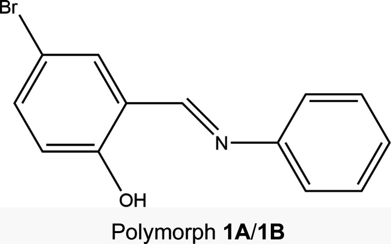
Experimental
Synthesis and crystallization
(E)-4-Bromo-2-[(phenylimino)methyl]phenol was synthesized by direct condensation of 5-bromosalicylaldehyde and aniline in ethanol. The two materials (0.005 mol of each, 1.000 g of 5-bromosalicylaldehyde and 0.466 g of aniline) were dissolved separately in ethanol (25 ml). The resultant solutions were combined and refluxed with stirring for 4 h. After removal of any precipitate, the solution was rotary evaporated until further precipitate formed, the solid filtered off, rinsed with ethanol and left to dry, giving a yield of 94% (1.304 g, 0.0047 mol). Yellow single crystals (of 1B) crashed out of the crude reaction mixture and orange single crystals (of 1A) were produced by recrystallization from ethanol.
Refinement
Crystal data, data collection and structure refinement details are summarized in Table 1 ▸. All H atoms, apart from the OH hydrogen involved in the intramolecular hydrogen bonding with the imine N atom, were positioned geometrically and refined using a riding model. The H atoms involved in the intramolecular hydrogen bond were located in the Fourier difference map wherever feasible. In 1A, the O—H distance was restrained to 0.86 (1) Å.
Table 1. Experimental details.
For both structures: C13H10BrNO, M r = 276.13, Z = 4. Experiments were carried out with Mo Kα radiation using an Oxford Diffraction Xcalibur (Sapphire3, Gemini ultra) diffractometer. An analytical absorption correction [CrysAlis PRO (Oxford Diffraction, 2010 ▸), based on expressions derived by Clark & Reid (1995 ▸)] was used. Refinement was with 2 restraints. H atoms were treated by a mixture of independent and constrained refinement.
| Polymorph 1A | Polymorph 1B | |
|---|---|---|
| Crystal data | ||
| Crystal system, space group | Orthorhombic, P c a21 | Monoclinic, C c |
| Temperature (K) | 120 | 150 |
| a, b, c (Å) | 12.2768 (3), 4.4829 (1), 19.6694 (4) | 25.8944 (13), 6.9439 (4), 6.1499 (4) |
| α, β, γ (°) | 90, 90, 90 | 90, 91.381 (5), 90 |
| V (Å3) | 1082.52 (4) | 1105.48 (11) |
| μ (mm−1) | 3.77 | 3.69 |
| Crystal size (mm) | 0.46 × 0.20 × 0.05 | 0.58 × 0.49 × 0.22 |
| Data collection | ||
| T min, T max | 0.383, 0.847 | 0.190, 0.585 |
| No. of measured, independent and observed [I > 2σ(I)] reflections | 13133, 2215, 2133 | 7049, 2254, 2142 |
| R int | 0.043 | 0.051 |
| (sin θ/λ)max (Å−1) | 0.625 | 0.625 |
| Refinement | ||
| R[F 2 > 2σ(F 2)], wR(F 2), S | 0.022, 0.053, 1.05 | 0.039, 0.100, 1.05 |
| No. of reflections | 2215 | 2254 |
| No. of parameters | 149 | 148 |
| Δρmax, Δρmin (e Å−3) | 0.39, −0.23 | 0.95, −0.34 |
| Absolute structure | Flack x determined using 993 quotients [(I +) − (I −)]/[(I +) + (I −)] (Parsons et al., 2013 ▸) | Flack x determined using 1007 quotients [(I +) − (I −)]/[(I +) + (I −)] (Parsons et al., 2013 ▸) |
| Absolute structure parameter | −0.006 (8) | −0.010 (19) |
Results and discussion
The structures of polymorphs 1A and 1B are shown in Fig. 1 ▸. The structure of 1A at 120 K was consistent with the previously published structure at room temperature (Yan et al., 2014 ▸). The structure of 1A was obtained in the orthorhombic space group Pca21, while 1B was obtained in the monoclinic space group Cc. The compound consists of a hydroxy-substituted phenyl ring linked via an imine group to a second unsubstituted phenyl group. In both polymorphs, the structures were found to exist in the enol form, with C7=N1 bond lengths of 1.282 (4) Å for 1A and 1.284 (10) Å for 1B, indicating a double bond, and C1—O1 bond lengths of 1.350 (5) Å for 1A and 1.351 (9) Å for 1B, indicating a single bond. The structures showed quite different dihedral angles, with 1A having Φ = 1.8 (2)° at 120 K and 1B having Φ = 45.6 (1)° at 150 K. Upon cooling, the structures were both found to display some degree of thermochromism with 1A changing from orange at room temperature to yellow at 120 K and 1B, which was yellow at room temperature, becoming slightly paler at 150 K (Fig. 2 ▸). The differences in the thermochromic behaviour of the two polymorphs are consistent with literature suggestions that a larger dihedral angle increases the overlap of the π-system reducing the nitrogen basicity, disfavouring the keto form and thus also reducing the thermochromism of the compound.
Figure 1.

Illustration of the structures of (a) 1A and (b) 1B at 120 (2) and 150 (2) K, respectively, with the atomic numbering schemes depicted. Anisotropic displacement parameters are shown at the 50% probability level.
Figure 2.

Illustration of the colour change observed upon cooling (a) 1A and (b) 1B.
An intramolecular O1—H1⋯N1 hydrogen bond, involving the phenol OH group and imine N atom, was identified in the structures of both polymorphs and creates an S(6) ring. The hydrogen-bonding parameters were almost identical in the two structures, with a donor–acceptor distance of ∼2.59 Å and a hydrogen-bond angle of ∼150° (Tables 2 ▸ and 3 ▸.). The packing of the two polymorphs was unsurprisingly significantly different given the large difference in the dihedral angles. In polymorph 1A, the molecules are essentially planar and orientated diagonally such that the plane of the molecule is perpendicular to the bc plane and, as a result of the 21 screw axis, the diagonal slant of alternate molecules along the a-axis direction essentially align in opposite directions (Fig. 3 ▸
a). It was also noted that there were short π-type contacts between the C=N group and the phenol ring in the 0 1 direction, with a centroid-to-centroid (C=N) distance of 3.326 (1) Å. These can be seen on the Hirshfeld surface of 1A as red dots (Fig. 4 ▸
a). In polymorph 1B, although the molecules themselves are twisted, the molecules are orientated relative to each other such that they create planes parallel to the ac plane direction (see Fig. 3 ▸
b).
1 direction, with a centroid-to-centroid (C=N) distance of 3.326 (1) Å. These can be seen on the Hirshfeld surface of 1A as red dots (Fig. 4 ▸
a). In polymorph 1B, although the molecules themselves are twisted, the molecules are orientated relative to each other such that they create planes parallel to the ac plane direction (see Fig. 3 ▸
b).
Table 2. Hydrogen-bond geometry (Å, °) for polymorph 1A .
| D—H⋯A | D—H | H⋯A | D⋯A | D—H⋯A |
|---|---|---|---|---|
| O1—H1⋯N1 | 0.86 (1) | 1.82 (3) | 2.593 (4) | 150 (5) |
Table 3. Hydrogen-bond geometry (Å, °) for polymorph 1B .
| D—H⋯A | D—H | H⋯A | D⋯A | D—H⋯A |
|---|---|---|---|---|
| O1—H1⋯N1 | 0.86 (11) | 1.82 (11) | 2.590 (10) | 148 (10) |
Figure 3.

(a) Illustration of the packing for 1A, looking down the a axis; molecules in blue are in-plane behind those in element colours. (b) View of polymorph 1B, looking down the c axis.
Figure 4.

The Hirshfeld surface plot (top) and fingerprint plot (bottom) for (a) 1A and (b) 1B.
Examining the Hirshfeld fingerprint plots (Turner et al., 2017 ▸) for the two structures highlights the differences in the two structures, not least in the shapes of the two plots (Fig. 4 ▸). For 1A, the O⋯H and Br⋯H contacts are quite obvious, while in 1B the H⋯H and C⋯H contacts are significantly more pronounced, slightly masking the O⋯H and Br⋯H contacts. These differences are very apparent on the Hirshfeld surface for both compounds with a greater number of red spots on the surface of 1A that are more noticeable than for 1B, showing that 1A has more short contacts.
The two polymorphs of (E)-4-bromo-2-[(phenylimino)methyl]phenol reported herein are particularly interesting as part of a study into N-salicylideneanilines because they show significantly different molecular conformations and colours at room temperature. In line with the literature, the extent of the thermochromism was found to be linked to the dihedral angle, with 1A [Φ = 1.8 (2)°] showing a greater colour change upon cooling than observed for 1B [Φ = 45.6 (1)°].
Supplementary Material
Crystal structure: contains datablock(s) 1A, 1B, global. DOI: 10.1107/S2053229620011560/yd3009sup1.cif
Structure factors: contains datablock(s) 1A. DOI: 10.1107/S2053229620011560/yd30091Asup2.hkl
Structure factors: contains datablock(s) 1B. DOI: 10.1107/S2053229620011560/yd30091Bsup3.hkl
Acknowledgments
The authors gratefully acknowledge funding for HEM from the EPSRC and from Durham University, and useful discussions with Professor Jonathan Steed of Durham University.
References
- Bregman, J., Leiserowitz, L. & Schmidt, G. M. J. (1964). J. Chem. Soc. pp. 2068–2085.
- Clark, R. C. & Reid, J. S. (1995). Acta Cryst. A51, 887–897.
- Cohen, M. D. & Schmidt, G. M. J. (1962). J. Phys. Chem. 66, 2442–2446.
- Cohen, M. D., Schmidt, G. M. J. & Flavian, S. (1964). J. Chem. Soc. pp. 2041–2051.
- Dolomanov, O. V., Bourhis, L. J., Gildea, R. J., Howard, J. A. K. & Puschmann, H. (2009). J. Appl. Cryst. 42, 339–341.
- Fujiwara, T., Harada, J. & Ogawa, K. (2004). J. Phys. Chem. B, 108, 4035–4038.
- Fujiwara, T., Harada, J. & Ogawa, K. (2009). J. Phys. Chem. A, 113, 1822–1826. [DOI] [PubMed]
- Hadjoudis, E. & Mavridis, I. M. (2004). Chem. Soc. Rev. 33, 579–588. [DOI] [PubMed]
- Johmoto, K., Ishida, T., Sekine, A., Uekusa, H. & Ohashi, Y. (2012). Acta Cryst. B68, 297–304. [DOI] [PubMed]
- Ogawa, K., Harada, J., Fujiwara, T. & Yoshida, S. (2001). J. Phys. Chem. A, 105, 3425–3427.
- Ogawa, K., Kasahara, Y., Ohtani, Y. & Harada, J. (1998). J. Am. Chem. Soc. 120, 7107–7108.
- Oxford Diffraction (2010). CrysAlis PRO. Oxford Diffraction Ltd, Abingdon, Oxfordshire, England.
- Özek, A., Albayrak, C., Odabaşoğlu, M. & Büyükgüngör, O. (2007). Acta Cryst. C63, o177–o180. [DOI] [PubMed]
- Parsons, S., Flack, H. D. & Wagner, T. (2013). Acta Cryst. B69, 249–259. [DOI] [PMC free article] [PubMed]
- Robert, F., Naik, A. D., Tinant, B., Robiette, R. & Garcia, Y. (2009). Chem. Eur. J. 15, 4327–4342. [DOI] [PubMed]
- Sheldrick, G. M. (2008). Acta Cryst. A64, 112–122. [DOI] [PubMed]
- Sheldrick, G. M. (2015). Acta Cryst. C71, 3–8.
- Swetha, G., Ida Malarselvi, R., Ramachandra Raja, C., Thiruvalluvar, A. & Priscilla, J. (2017). IUCrData, 2, x171671.
- Swetha, G., Ida Malarselvi, R., Ramachandra Raja, C., Thiruvalluvar, A. & Priscilla, J. (2019). IUCrData, 4, x190788.
- Turner, M. J., McKinnon, J. J., Wolff, S. K., Grimwood, D. J., Spackman, P. R., Jayatilaka, D. & Spackman, M. A. (2017). CrystalExplorer17. University of Western Australia. https://crystalexplorer.scb.uwa.edu.au/.
- Yan, X.-X., Lu, L. & Zhu, M. (2014). Acta Cryst. E70, o853. [DOI] [PMC free article] [PubMed]
Associated Data
This section collects any data citations, data availability statements, or supplementary materials included in this article.
Supplementary Materials
Crystal structure: contains datablock(s) 1A, 1B, global. DOI: 10.1107/S2053229620011560/yd3009sup1.cif
Structure factors: contains datablock(s) 1A. DOI: 10.1107/S2053229620011560/yd30091Asup2.hkl
Structure factors: contains datablock(s) 1B. DOI: 10.1107/S2053229620011560/yd30091Bsup3.hkl