

Abstract
Currently, due to the lack of long-term postoperative follow-up outcomes of the congenital divided eyelid nevus, we described our surgical approaches and presented the functional and cosmetic results of 13 patients with an average of 5-year follow-up. Based on the surgical treatments and the follow-ups, the selection of total or subtotal excision depends on the lesion location and the use of blepharoplasty approaches is determined by defect size. The CO2 laser may be a useful second-stage procedure to ablate remnant lesions, but long-term monitoring is required.
Keywords: divided eyelid nevus, pedicled flaps, grafts
Abstract
Étant donné le peu de suivi postopératoire à long terme sur les résultats cliniques de la réparation du nævus palpébral congénital en miroir, les chercheurs décrivent les méthodes chirurgicales utilisées et présentent les résultats fonctionnels et esthétiques chez 13 patients, suivis sur une période moyenne de cinq ans. D’après les traitements chirurgicaux et les suivis, le choix d’une excision totale ou partielle dépend du foyer de la lésion, et le mode de blépharoplastie repose sur la dimension de l’anomalie. Le laser CO2 peut être utilisé en deuxième phase pour faire disparaître les vestiges des lésions, mais une surveillance à long terme s’impose.
Introduction
Divided eyelid nevus is a type of congenital melanocytic nevus on both upper and lower eyelids of one eye. The nevus appears as one entity when the eye closed.
Based on size, divided eyelid nevus can be classified as small (<1.5 cm), medium (1.5-20 cm), and large (>20 cm).1 Current literature indicates that the size of congenital divided eyelid nevus varied from small to medium.2-4 The lesion is more common in Asians and whites, especially prevalent in females.5 Because of potential malignant transformation and cosmetic concern, prophylactic procedures including lesion excision and blepharoplasty are the main and effective approaches.
In the past decades, 18 patients from the rural area of Gansu province, China, were treated in our department. Based on our experience, we describe treatment techniques and follow-up outcomes.
Patients and Methods
During January 2006 to August 2017, 18 patients with unilateral divided eyelid nevus were enrolled (4 male and 14 female patients). The average age was 15 years (range, 2-35 years). The diseased regions included full eyelids (n = 5), central eyelid (n = 10), lid margin involvement (n = 6), palpebral conjunctiva involvement (n = 3), bulbar conjunctiva involvement (n = 1), and cheek extension (n = 1) (Table 1).
Table 1.
Patient Demographics.
| Patient | Sex | Age (years) | Location | Size (cm) | Treatment | Postoperative complications (during the most recent follow-up) |
|---|---|---|---|---|---|---|
| 1 | F | 5 | Right, lateral | Upper eyelid 3.0 × 1.5; lower eyelid 1.5 × 0.5 | Incomplete excision and full-thickness skin graft | Lid margin remnants |
| 2 | F | 17 | Entire eyelids | Entire lesion 8.0 × 5.0 | Incomplete excision and full-thickness skin graft | Lid margin remnants |
| 3 | F | 11 | Right, medial | Upper eyelid 0.3 × 0.5; lower eyelid 1.5 × 1 | Complete excision and orbicularis oculi pedicled flap | None |
| 4 | M | 19 | Left, central | Upper eyelid 1.5 × 1; lower eyelid × 1.5 × 1 | Incomplete excision, nasolabial island flap, and full-thickness skin graft | Lid margin remnants |
| 5 | M | 21 | Left, central | Entire lesion 6.0 × 1.5 | Incomplete excision and full-thickness skin graft | Lid margin remnants |
| 6 | M | 11 | Right, lateral | Upper eyelid 2 × 1.5; lower eyelid 2 × 1.8 | Incomplete excision and full-thickness skin graft | Lid margin remnants |
| 7 | F | 2 | Left, lateral | Upper eyelid 1.5 × 2.5; lower eyelid 1.7 × 3.0 | Incomplete excision and full-thickness skin graft with CO2 laser | Lid margin remnants and partial eyelash loss |
| 8 | F | 32 | Right, central | Upper eyelid 1.1 × 1.5; lower eyelid 0.9 × 1.3 | Complete excision and subcutaneous pedicle flap | Partial eyelash loss |
| 9 | F | 20 | Left, lateral | Upper eyelid 0.5 × 0.5; lower eyelid 1.5 × 2.0 | Complete excision and orbicularis oculi pedicled flap | None |
| 10 | F | 24 | Right, medial | Upper eyelid 1.0 × 1.0; lower eyelid 1.5 × 1.0 | Incomplete excision and full-thickness skin graft | Lid margin remnants |
| 11 | F | 19 | Left, lateral | Upper eyelid 2 × 1.5; lower eyelid 2 × 1.5 | Complete excision and full-thickness skin graft | None |
| 12 | F | 40 | Right, lateral | Upper eyelid 0.4 × 1; lower eyelid 1.7 × 1.2 | Complete excision and orbicularis oculi pedicled flap | None |
| 13 | F | 38 | Right, central | Upper eyelid 2.6 × 1.5; lower eyelid 1.8 × 1.8 | Incomplete excision and full-thickness skin graft | Lid margin remnants |
Abbreviations: F, female; M, male.
Surgical procedures were selected depending on lesion location, size, and extension. In general, the upper and lower eyelids were reconstructed at 1-stage surgery. Lesions were excised with 1 mm of normal surrounding tissue, and the eyelids were reconstructed by flaps or skin grafts. Partial excision was adopted that affected palpebral margin, and lacrimal punctum were left untreated. Repair options were in relation to the defective area that local flaps were used to cover a medium-sized nevus (<1.5 cm in diameter). Additionally, when the local flap had limited coverage, a nasolabial island flap could be transported to reconstruct the inferior lesion. When more than two-thirds of the eyelid was involved (>1.5 cm in diameter), a full-thickness skin graft harvested from the medial upper arm or abdomen was employed to repair eyelid, and the tie-over dressing was applied to fix the graft.
The oral mucosal graft was for the repair of the palpebral conjunctiva. To promote healing, the eye ointment was applied to the conjunctival sac postoperatively.
The second-stage surgery was designed by using CO2 laser (HGTECH super-pulsed, 10 600 nm, 15-second pulse width, 3-4 W energy) to ablate nevus residuals for at least 3 months after suture removal. Eyelash hair follicles are maximally preserved during the treatment. Follow-ups were focused on cosmetic results, eyelids function, and malignant transformation.
Results
Of the 18 cases, 8 patients underwent full-thickness skin grafts repair, 5 advanced flaps, 2 nasolabial flaps and skin grafts, and 1 oral mucosal graft. One patient received subtotal remnant ablation by CO2 laser after graft repair. Histologically, 6 cases were compound type and 12 were intradermal type. Postoperative complications included lesion residues (n = 9) and eyelash sacrifice (n = 3). Thirteen patients had an average of 5-year follow-up (range, 1-9 years), and 5 patients refused any follow-up or lost to follow-up. During the follow-up period, no malignant transformation was observed. Lesion recurrence was observed in 2 patients.
Case 1
A 5-year-old girl who had divided nevus of the right eyelid was admitted to our department. The lesion had a diameter of 3.0 cm × 1.5 cm and 1.5 cm × 0.5 cm on upper and lower eyelids, respectively (Figures 1 and 2). The nevus was subtotally excised, sparing upper and lower palpebral margins. Two size-matched full-thickness abdominal skin grafts were obtained and trimmed to cover the defects respectively. Moreover, a double-eyelid line was formed at the superior graft to prevent lagophthalmos and graft shrinkage. Then the grafts were sutured with 3-0 silk suture. Postoperatively, the graft survived well and no complications were noted (Figure 3). Histologically, the 2 defects were compound type. The patient parents preferred follow-up without second-stage treatment. After 9 years of the blepharoplasty, the results showed acceptable appearance and good eyelid function without malignant change, although the recurrent nevus was noted (Figure 4).
Figure 1.

A 5-year-old girl with right eyelid divided nevus and hair bearing observed on upper eyelid.
Figure 2.

The upper and lower nevi were excised and repaired with skin grafts.
Figure 3.

Postoperative 1 year with the good appearance.
Figure 4.

Postoperative 9 years without lesion recurrence or malignant transformation.
Case 2
A 17-year-old girl was observed with a giant hairy congenital divided nevus affecting both right upper and lower eyelids, right eyebrow, and bulbar conjunctiva, extending to right temporal region and right cheek (Figures 5 and 6). The entire lesion measured approximately 8.0 cm × 5.0 cm. Under general anesthesia, the nevus was partially excised underlying superficial fascia, sparing the lid margin and bulbar conjunctiva. A full-thickness abdominal skin graft sized 8.0 cm × 7.0 cm was harvested. To prevent graft contracture, the graft was horizontally divided into 2 parts and then covered the defect (Figure 7). Postoperatively, scar softening cream was applied on sutures. Histology examination revealed an intradermal type of nevus. The patient preferred no further treatment of the remnants after informed risk of malignant transformation. At 5-year follow-up, the appearance was acceptable, and the eyelids were functionally good without any complications (Figure 8).
Figure 5.

A 17-year-old girl with right eyelid divided nevus involving bulbar conjunctiva and extending to the zygomatic and temporal regions (lateral).
Figure 6.

A 17-year-old girl with right eyelid divided nevus involving bulbar conjunctiva and extending to the zygomatic and temporal regions (front).
Figure 7.

The lesion was repaired with a zoned skin graft, and the sutures were removed.
Figure 8.
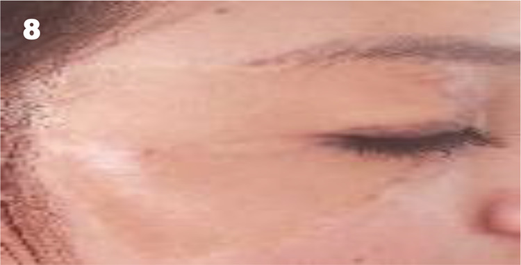
Postoperative 5 years, the appearance was acceptable without (complications).
Case 3
An 11-year-old girl presented with divided nevus on her right eyelid. Upon physical examination, the lesions located on the medial eyelid measured 0.3 cm × 0.5 cm and 1.5 cm × 1.0 cm on upper and lower eyelids, respectively (Figure 9). The lid margin and palpebral conjunctiva were not involved. Under local anesthesia, the complete excision was performed underlying superficial fascia (Figures 10 and 11). Then, at the upper eyelid, the two pedicled z-plasty flaps were designed and interchanged to correct lagophthalmos and Asian epicanthus. A pedicled orbicularis oculi flap measured 3.5 cm × 1.2 cm was prepared from the right lower eyelid and advanced to cover the inferior defect. Histology revealed a compound type of nevus. Three years after the operation, the eyelid was functionally and cosmetically excellent without complications (Figure 12).
Figure 9.

An 11-year-old girl with right eyelid divided nevus on her medial eyelids.
Figure 10.

Postoperative suture removal at day 7 (with right eye closed).
Figure 11.

Postoperative suture removal at day 7.
Figure 12.

Postoperative 3 years, the appearance and function of right eyelid were excellent.
Discussion
After entering puberty, the risk of developing lifetime melanoma from a single small-sized or medium-sized congenital nevus was estimated less than 1%.5 Moreover, only 1 case of untreated divided eyelid nevus malignant transformation was reported.6 However, we prefer surgical approaches because of aesthetic concerns and potential malignant changes. In our series, all patients sought treatments due to cosmetic complaints and psychological stress. Individualized treatment plans were designed based on the patient’s age, lesion size, and location. Although complete lesion resection was described,4 we prioritize eyelid functional and aesthetic integrity: When the palpebral margin is involved, partial excision is performed to prevent lagophthalmos and protect natural curve of the palpebral fissure. Local flaps were recommended to repair eyelid defects for Asian patients that the flaps are the best match in regard to color, texture, and sufficient blood supply.4
Because of the extremely thin skin and abundant blood vascularity of the eyelid, pedicle flaps could survive well. In patient 3, the flaps and donor sites healed well with inconspicuous scarring. However, according to our research, the local flap coverage capability is limited when the lesion occupies more than half horizontal length of the palpebral fissure and more than 1 cm of the vertical height of the eyelid. Although the tissue expander was used to provide enough local flaps,7,8 the radical multistaged surgical treatment is not suitable for young children.
Skin grafts are a simple and effective reconstructive approach for larger defects. Nevertheless, one of the disadvantages of skin grafts is visible color disparity that in patient 2, 5 years after the surgery, the skin graft had the difference in color compared with adjacent skin. Due to the giant pigmented nevus in patient 2, the skin graft was separated into two grafts horizontally during the surgery to minimize postoperative graft shrinkage, inhibit scar hyperplasia, and conceal scars.
In our series, 1 child patient had remnants incompletely ablated by CO2 laser after full-thickness graft reconstruction. During the treatment, the patient had little compliance that the lesion recurrence was observed at 2-year follow-up. The CO2 laser may not be the optimal treatment for children that eyelid ectropion and double eyelid height asymmetry were observed.9 For the small-sized divided eyelid nevus, the CO2 laser was used in an adult patient with satisfactory results.10 However, in consideration of remnant malignant change or recurrence, the long-term follow-ups are needed for all patients especially with lid residual lesion.
Footnotes
Level of Evidence: Level 5, Therapeutic
Authors’ Note: All procedures performed in studies involving human participants were in accordance with the ethical standards of the institutional and/or national research committee and with the 1964 Helsinki declaration and its later amendments or comparable ethical standards. Informed consent was obtained from all individual participants included in the study.
Declaration of Conflicting Interests: The author(s) declared no potential conflicts of interest with respect to the research, authorship, and/or publication of this article.
Funding: The author(s) received no financial support for the research, authorship, and/or publication of this article.
ORCID iD: Xianying Zhang, MD  https://orcid.org/0000-0001-5740-2199
References
- 1. McDonnell PJ, Mayou BJ. Congenital divided naevus of the eyelids. Br J Ophthalmol. 1988;72(1):198–201. [DOI] [PMC free article] [PubMed] [Google Scholar]
- 2. Jia R, Zhu H, Lin M, et al. Clinicopathological characteristics and surgical outcomes of divided nevus of the eyelids: a decade’s experience on 73 cases. Ann Plast Surg. 2012;68(2):166–170. [DOI] [PubMed] [Google Scholar]
- 3. Alfano C, Chiummariello S, De Gado F, et al. Divided nevus of the eyelids: three case studies. In Vivo. 2007;21(1):137–139. [PubMed] [Google Scholar]
- 4. Lu R, Li Q, Quan Y, et al. Staged surgery with total excision and lamellar reconstructive for medium-sized divided nevus of the eyelids. Plast Reconstr Surg Glob Open. 2015;3(6):7. [DOI] [PMC free article] [PubMed] [Google Scholar]
- 5. Price HN. Congenital melanocytic nevi: update in genetics and management. Curr Opin Pediatr. 2016; 28(4):476–482. [DOI] [PubMed] [Google Scholar]
- 6. Kharel RS, Bhatta S, Shrestha GB, et al. Malignant transformation of kissing nevus-a rare entity. Nepal J Ophthalmol. 2012;4(2):329–332. [DOI] [PubMed] [Google Scholar]
- 7. Yamamichi K, Kosaka M. A novel reconstructive procedure for the divided nevus of the eyelids using a tissue expander. Plast Reconstr Surg Glob Open. 2016;4(12):7. [DOI] [PMC free article] [PubMed] [Google Scholar]
- 8. Bayramiçli M, Ersoy B, Sirinoglu H. Surgical management of a congenital panda nevus with preexpanded triple forehead flaps and temporal island flap. J Craniofac Surg. 2012;23(5):1396–1398. [DOI] [PubMed] [Google Scholar]
- 9. Zeng Y. Divided nevus of the eyelid: successful treatment with CO2 laser. J Dermatol Treat. 2014;25(4):358–359. [DOI] [PubMed] [Google Scholar]
- 10. Zhu L, Jia Y, Wang X. Treatment of eyelid nevus with CO2 laser: a double-edged sword. J Dermatol Treat. 2015;26(3):257–258. [DOI] [PubMed] [Google Scholar]