

Abstract
A microdeletion within human chromosome 5q14.3 has been associated with the occurrence of neurodevelopmental disorders, such as autism and intellectual disability, and MEF2C haploinsufficiency was identified as main cause. Here, we report that a brain‐enriched long non‐coding RNA, NDIME, is located near the MEF2C locus and is required for normal neural differentiation of mouse embryonic stem cells (mESCs). NDIME interacts with EZH2, the major component of polycomb repressive complex 2 (PRC2), and blocks EZH2‐mediated trimethylation of histone H3 lysine 27 (H3K27me3) at the Mef2c promoter, promoting MEF2C transcription. Moreover, the expression levels of both NDIME and MEF2C were strongly downregulated in the hippocampus of a mouse model of autism, and the adeno‐associated virus (AAV)‐mediated expression of NDIME in the hippocampus of these mice significantly increased MEF2C expression and ameliorated autism‐like behaviors. The results of this study reveal an epigenetic mechanism by which NDIME regulates MEF2C transcription and neural differentiation and suggest potential effects and therapeutic approaches of the NDIME/ MEF2C axis in autism.
Keywords: autism, epigenetic regulation, LncRNA, MEF2C, neural differentiation
Subject Categories: Chromatin, Epigenetics, Genomics & Functional Genomics; Neuroscience; RNA Biology
The lncRNA NDIME is required for proper neural differentiation of mESCs. Viral‐mediated expression of NDIME in the hippocampus of a mouse model of autism ameliorates autism‐like behaviors.
Introduction
Autism spectrum disorder (ASD) is a neurodevelopmental condition of complex etiology characterized by social and cognitive impairments and repetitive behaviors. Although the pathogenesis of autism is complex, disturbance of the excitatory and inhibitory (E/I) balance has been strongly implicated in ASD etiology (Rubenstein & Merzenich, 2003). Attenuated GABAergic signals and enhanced glutamatergic signals have been reported to alter neural network connectivity and probably cause autism‐like behaviors (Sohal & Rubenstein, 2019). An increasing number of factors affecting early neurodevelopment have been identified, including gene mutations and environmental elements, and recent studies have suggested that epigenetic factors including non‐coding RNAs also play important roles in the pathogenesis of autism (Gabel et al, 2015; Valluy et al, 2015). However, the exact pathogenesis of autism remains unclear.
Strongly disease‐associated variants in the chromosome 5q14.3 microdeletions that cause autism in children have been identified by genome‐wide association studies (GWASs) as well as by genome sequencing. These studies have pointed to myocyte enhancer factor 2C (MEF2C) haploinsufficiency as the cause (Zweier et al, 2010; Saitsu et al, 2011). Further, neural stem cell (NSC)‐conditional knockout of MEF2C in mice resulted in abnormal neurons and behavioral deficits similar to findings reported for Rett syndrome, classified as an ASD (Li et al, 2008a). Additionally, MEF2C heterozygous mice exhibit both MEF2C haploinsufficiency syndrome (MCHS)‐like and autism‐like behaviors associated with an increase in hippocampal network activity, suggesting an altered hippocampal E/I balance (Tu et al, 2017). The Mef2c gene is surrounded by multiple non‐coding RNAs in the chromosome 5q14.3 and chromosome conformation capture (3C) assays reveal a high order chromatin at the Mef2c locus (Mitchell et al, 2018). Moreover, breakpoints of balanced chromosomal abnormalities (BCAs) in subjects with congenital anomalies altered a single topologically associated domain (TAD) encompassing MEF2C, resulting in decreased MEF2C expression (Redin et al, 2017). It remains unclear, however, just how the expression of MEF2C is regulated and how MEF2C affects the pathogenesis of autism.
Long non‐coding RNAs (lncRNAs) are defined as transcripts longer than 200 nucleotides (nt) with little or no protein‐coding potential (Marques & Ponting, 2009), and they are emerging in research as important regulators in embryogenesis and in developmental processes including neural development (Guttman et al, 2011; Ramos et al, 2015; Sarropoulos et al, 2019). Increasing evidence indicates that lncRNAs can regulate the expression of their adjacent genes through cis‐acting via co‐expression in a tissue‐specific pattern with adjacent genes forming transcription units synergistically regulating the life activities and functions of cells (Chalei et al, 2014; Engreitz et al, 2016; Kotzin et al, 2016; Joung et al, 2017; Guo et al, 2018; Sallam et al, 2018; Sarropoulos et al, 2019). Earlier research on hotspots in central nervous system (CNS) diseases mainly focused on protein‐coding genes. Recent studies, however, have shown that the dysregulation of lncRNAs is linked to neurodevelopmental and neurodegenerative diseases (Gandal et al, 2018), and that most genetic variations in these cases occur in non‐coding regions (Nott et al, 2019; Zhou et al, 2019). Studies have shown that the expression levels of many lncRNAs in the peripheral blood of autistic patients are significantly different from those in normal controls (Wang et al, 2015; Sayad et al, 2019). Certain lncRNAs are associated with the risk of ASD, including IFNG‐AS1 (Fallah et al, 2020), RPS10P2‐AS1 (Bilinovich et al, 2019), HOTAIR (Safari et al, 2020), PTCHD1‐AS (Ross et al, 2020), lnc‐NR2F1 (Ang et al, 2019), and MSNP1AS (DeWitt et al, 2016). The studies have all suggested that lncRNAs may play crucial roles in ASD, and yet our knowledge of lncRNAs that function in the pathogenesis of autism remains limited, and a further understanding of the molecular mechanism that they regulate is lacking.
Here we report a homologue of linc00461 in mice, lincRNA C130071C03Rik, and identify its role in Neural Differentiation Initiation of MEF2C Expression (NDIME). NDIME is located near the Mef2c locus and was highly expressed in neuronal cells as a critical regulator of MEF2C expression and neural differentiation. We also found that the expression levels of both NDIME and MEF2C were strongly downregulated in the hippocampus of a mouse model of autism, and the viral‐mediated expression of NDIME in the hippocampus of these mice ameliorated autism‐like behaviors.
Results
The lncRNA NDIME is highly expressed in Sox1‐positive neuronal cells during neural differentiation
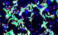
In order to study the role of the lncRNA NDIME in mESCs differentiation, we first analyzed the histone marks trimethylation of histone H3 lysine 4 (H3K4me3) and H3K27me3 in embryonic tissue by chromatin immunoprecipitation‐sequencing (ChIP‐seq) with ENCODE 3 (UCSD/Ren) obtained from the UCSC Genome Browser and found that NDIME is specifically expressed in the CNS (Appendix Fig S1A). Using quantitative real‐time PCR (qRT–PCR), we found that the expression of NDIME in the mouse brain was higher than that in other mouse tissues and mESCs (Appendix Fig S1B). We then induced differentiation of mESCs, with green fluorescent protein (GFP) knocked‐in at the locus of Sox1, a neural‐progenitor‐specific gene (Sox1‐GFP cells), toward a neural cell fate and revealed the upregulation of NDIME during neural differentiation (Fig 1A and B). Furthermore, expression of this lncRNA was strongly detected in the Sox1‐GFP+ population but not in the Sox1‐GFP− population or in mESCs (Fig 1C). Upon neural differentiation, the expression of NDIME was upregulated during the first 7 days of serum‐free culture of embryoid body‐like quick aggregates (SFEBq; Eiraku et al, 2008; Kamiya et al, 2011), whereas its expression was markedly reduced by the addition of fetal bovine serum (FBS), a potent inhibitor of neural differentiation (Kawasaki et al, 2000) (Fig 1D and E). Using subcellular fractionation and RNA fluorescence in situ hybridization (RNA‐FISH) experiments, we confirmed the nuclear localization of NDIME (Fig 1F and Appendix Fig S1C).
Figure 1. LncRNA NDIME is required for proper neural differentiation.

-
AThe production of Sox1‐GFP+ neural‐lineage cells during 7 days of neural differentiation, as shown by microscopy. Scale bars, 100 μm.
-
BqRT–PCR analysis of NDIME expression during 7 days of neural differentiation (D3, P = 0.068; D5, P = 0.0049; D7, P = 0.0003).
-
CRelative expression level of NDIME mRNA in the Sox1‐GFP− population and Sox1‐GFP+ population at day 7 of neural differentiation (P = 0.0118).
-
DmESCs were cultured in differentiation medium alone, or with 10% FBS, at day 5 of neural differentiation as shown by microscopy. Scale bars, 100 μm.
-
ETemporal expression profile of NDIME by qRT–PCR. mESCs were cultured in differentiation medium alone or with 10% FBS (D3, P = 0.074; D5, P = 0.0048; D7, P = 0.0034).
-
FDistribution of NDIME in the cytoplasm and nucleus at day 7 of neural differentiation. The XIST RNA and GAPDH mRNA were considered as the positive controls for the nuclear and cytosolic fractions, respectively. Data are represented as mean, and error bars represent SEM in all panels. n = 3.
-
GVerification of the efficiency of knockout of exon 3 of NDIME in mESCs via qRT–PCR.
-
H, IKnockout of NDIME strongly inhibited neural differentiation, as shown by microscopy (H) and FACS (I) analyses during 7 days of neural differentiation. Scale bars, 100 μm.
-
JKnockout of NDIME inhibits the expression of neural‐lineage genes at day 5 of neural differentiation.
-
KNESTIN and GFP immunostaining of the cells in (H) at day 5 of neural differentiation. Nuclei were stained with Hoechst 33342 (blue). Scale bars, 100 μm.
-
LNESTIN and GFP immunostaining of Ctrl, NDIME −/−, and NDIME −/−+Rs26‐NDIME cells at day 5 of neural differentiation. Scale bars, 100 μm.
-
MExpression of neural‐lineage genes of Ctrl, NDIME −/−, and NDIME −/−+Rs26‐NDIME cells at day 5 of neural differentiation.
-
NRelative expression of Emx1, Brn2, Sox5, and Zeb2 mRNA in different differentiation stages of Ctrl and NDIME‐knockout cells (compared with control cells).
NDIME is required for proper neural differentiation
Next, in neural differentiation of mESCs, we used a clustered regularly interspaced short palindromic repeats (CRISPR)/Cas9‐mediated genome‐editing approach to inactivate NDIME in 46C cells (mESCs line) through knockout of exon 3 of NDIME, which contains a conserved sequence in mammals (Appendix Fig S1D). Genomic depletion was validated by genomic DNA PCR and qRT–PCR (Fig 1G and Appendix Fig S1E). As observed, knockout of NDIME did not affect the morphology of mESCs colonies compared to that of control cells (Fig 1H). Importantly, upon the induction of neural differentiation, knockout of NDIME strongly inhibited the generation of Sox1‐GFP+ neural‐lineage cells (Fig 1H). Consistent with this, fluorescence‐activated cell sorting (FACS) assay showed that knockout of NDIME markedly decreased the percentages of Sox1‐GFP+ populations in SFEBq‐cultured mESCs (Fig 1I). The expression levels of neural markers Sox1, Zfp521, and Pax6, as well as N‐cadherin (N‐cad) but not Zic2 (Fig 1J), were also reduced in NDIME‐knockout mESCs, as was the percentage of Sox1‐GFP+/NESTIN+ neural progenitor cells (Fig 1K).
To further clarify the role of NDIME in neural differentiation, we then knocked full‐length NDIME into the ROSA26 locus (used for constitutive gene expression) of NDIME‐knockout mESCs (Appendix Fig S1G). As expected, the percentages of Sox1‐GFP+ populations and the percentage of Sox1‐GFP+/NESTIN+ neural progenitor cells were rescued by overexpression of NDIME (Fig 1L, Appendix Fig S1G and H). qRT–PCR analysis further confirmed that the expression of Sox1, Zfp521, Pax6, and N‐cad were also rescued (Fig 1M).
We further sought to decrease NDIME expression through the expression of small hairpin RNAs (shRNAs) to achieve ~80% knockdown of NDIME expression in 46C cells (Appendix Fig S2A). In consistence with the previous data, FACS, immunostaining, and qRT–PCR assays showed that knockdown of NDIME also strongly inhibited the neural differentiation of mESCs (Appendix Fig S2B–E). We further used shRNAs to decrease the expression of lncRNA NDIME in another mESCs line (E14 cells). Immunostaining and qRT–PCR assays showed that knockdown of NDIME also strongly inhibited the neural differentiation of E14 cells (Appendix Fig S2F and G). These results indicate that NDIME is essential for the early neural differentiation stages of mESCs.
To further clarify the role of NDIME in differentiation timing, the SFEBq‐induced neural progenitor cells were differentiated into neurons (Eiraku et al, 2008). We found that the expression of neuronal genes Emx1, Brn2 (Pou3f2), Sox5, and Zeb2 were strongly downregulated in NDIME‐knockout cells at D7, by D16 the NDIME‐knockout cells and control cells displayed similar expression of these genes (Fig 1N). Knockout of NDIME led to increased expression of non‐neural ectodermal marker gene Keratin14 (Krt14) at D12 and D16 and decreased expression of early mesodermal marker gene Mest (Peg1) at D7, D12, and D16 (Appendix Fig S1I). Thus, knockout of NDIME might be involved in the delaying neural differentiation, activating non‐neural ectodermal genes’ expression, and repressing mesodermal genes’ expression.
NDIME functions upstream of its neighbor gene Mef2c
MEF2C is located near the lncRNA NDIME and is a critical regulator of neural differentiation (Li et al, 2008a,b; Cho et al, 2011). Strikingly, the protein and mRNA levels of MEF2C were markedly decreased in both the NDIME‐knockout cells (Fig 2A and B) and the NDIME‐knockdown cells (Appendix Fig S3A and B). To test whether NDIME functions upstream of MEF2C, we next knocked the coding sequence of MEF2C into the ROSA26 locus in NDIME‐knockout cells (Fig 2C). Notably, MEF2C overexpression in the NDIME‐knockout cells led to an increase in the percentages of Sox1‐GFP+ populations and the percentage of Sox1‐GFP+/NESTIN+ neural progenitor cells compared with the NDIME‐knockout cells (Fig 2D–F). qRT–PCR analysis further confirmed that the expression of Sox1, Zfp521, Pax6, and N‐cad was rescued by MEF2C overexpression in NDIME‐knockdown cells (Fig 2G). In contrast, MEF2C overexpression in NDIME‐knockout cells did not change the expression of Zic2 (Fig 2G). In consistence, MEF2C overexpression in the NDIME‐knockdown cells also restored the neural differentiation of mESCs (Appendix Fig S3C–G).
Figure 2. NDIME functions upstream of its neighbor gene Mef2c.

-
A, BExpression levels of MEF2C (A) (D0, P = 0.0020; D3, P = 0.0025; D5, P = 0.0003; D7, P = 0.0142) of Ctrl and NDIME −/− cells during 7 days of neural differentiation, and protein level of MEF2C (B) following knockout of NDIME at day 5 of neural differentiation.
-
CWestern blot analysis of the effects of MEF2C overexpression in NDIME‐knockout cells at day 5 of neural differentiation.
-
D, EOverexpression of MEF2C in the NDIME‐knockout cells rescued neural differentiation defects at day 5 of neural differentiation, as shown by microscopy (D) and FACS (E). Scale bars, 100 μm.
-
FNESTIN and GFP immunostaining of the cells in (D) at day 5 of neural differentiation. Scale bars, 100 μm.
-
GExpression of neural‐lineage genes in Ctrl, NDIME −/−, and NDIME −/−+Mef2c cells during 7 days of neural differentiation.
We next sought to decrease MEF2C expression through the expression of small hairpin RNAs (shRNAs) against MEF2C. We observed that the protein level of MEF2C was markedly decreased in the MEF2C‐knockdown cells (Appendix Fig S4A). Importantly, knockdown of Mef2c significantly inhibited the neural differentiation of mESCs, with decreased Sox1‐GFP+ populations, Sox1‐GFP+/NESTIN+ neural progenitor cells, and neural‐specific gene expression but not Zic2 expression (Appendix Fig S4B–E). Thus, our study indicates that NDIME regulates the expression of MEF2C during neural differentiation and that MEF2C may be a downstream target gene of NDIME in this process.
NDIME physically interacts with the PRC2 complex
Histone H3K27 methylation is a critical epigenetic mechanism for the transient silencing and activation of genes during development (Kouzarides, 2002; Silva et al, 2003). ChIP coupled to a quantitative real‐time PCR (ChIP‐qPCR) assay showed that knockout of NDIME led to a significant increase in H3K27me3 level at the Mef2c promoter (Fig 3A). The enrichment of RNA polymerase II (RNA Pol II) at the Mef2c promoter was consistently and significantly decreased in the NDIME‐knockout cells (Fig 3B). Thus, knockout of NDIME may affect MEF2C expression in part by altering the chromatin state affecting the assembly of the transcription initiation complex machinery at the Mef2c promoter. Further, using an in vivo RNA immunoprecipitation (RIP) assay, we found that NDIME binds the PRC2 complex, a complex that catalyzes H3K27me3 and mediates transcriptional repression (Surface et al, 2010) (Fig 3C). To test whether NDIME physically binds PRC2, we co‐transfected NDIME with Flag‐Ezh2 or Flag‐Suz12 into HEK293T cells. EZH2 and SUZ12 proteins were immunoprecipitated and tested for associated RNAs by qRT–PCR. The immunoprecipitation of EZH2 and SUZ12 did specifically retrieve NDIME (Fig 3D and E). We therefore hypothesized that NDIME may regulate the expression of MEF2C by preventing the binding of PRC2 at the Mef2c promoter.
Figure 3. NDIME blocks PRC2 binding at the Mef2c promoter.

-
A, BEnrichment of H3K27me3 (A) and RNA polymerase II (B) at the Mef2c promoter in the NDIME‐knockout cells at day 5 of neural differentiation compared with control cells.
-
CNDIME interacts with EZH2 (P = 0.0043) and SUZ12 (P = 0.0003), but not EED (P = 0.6720) and EZH1 (P = 0.0951) compared with IgG, as determined by RIP using EZH2, SUZ12, EED, and EZH1 antibodies at day 5 of neural differentiation. Nuclear RNA U1 as a negative control. The presence of NDIME and U1 was measured by qRT–PCR with or without reverse transcriptase (RT).
-
D, ENDIME levels following EZH2‐RIP (P = 0.0028) (D) or SUZ12‐RIP (P = 0.0015) (E) in HEK293T cells compared with pcDNA3.1. Nuclear RNA U1 was used as a negative control. The presence of NDIME and U1 was measured by qRT–PCR with or without reverse transcriptase (RT).
-
F, GEnrichment of EZH2 (F) and SUZ12 (G) at the Mef2c promoter in NDIME‐knockout cells at day 5 of neural differentiation compared with control cells.
-
HExpression of neural‐lineage genes at day 5 of neural differentiation in the NDIME‐knockout cells after adding the EZH2 inhibitor GSK126 (7.5 μM) partially rescued neural differentiation defects, as shown by qRT–PCR; the wild‐type and NDIME‐knockout cells treated with DMSO.
To test this hypothesis, we performed a ChIP‐qPCR assay for the PRC2 subunits EZH2 and SUZ12 in the NDIME‐knockout cells and control cells. We found that the levels of EZH2 and SUZ12 remained high at the Mef2c promoter in the NDIME‐knockout cells (Fig 3F and G). Moreover, after adding the inhibitor GSK126 (7.5 μM), which inhibits Ezh2‐dependent H3K27me3 formation, the expression of MEF2C and neural markers was partially rescued, suggesting that the H3K27me3 level was indeed an important cause for the changed expression of genes in the NDIME‐knockout cells (Fig 3H). These results suggest that NDIME blocks the binding of PRC2 at the Mef2c promoter and ensures that mESCs progress properly toward neural differentiation.
Exons 1–3 of NDIME are responsible for PRC2 binding
In an effort to gain further molecular and mechanistic insights into NDIME, NDIME RNA secondary structures were predicted based on the minimum free energy (MFE) and partition function through the RNAfold WebServer (Fig 4A; left panel). The thermodynamic ensemble of RNA structures showed that the sequence at 5′ end of NDIME forms a stable structure, suggesting that the 5′ end of NDIME might play a key role in its function (Fig 4A; right panel). We next constructed vectors expressing either exons 1–3 or exon 4 of NDIME and co‐transfected them with Flag‐Ezh2 into HEK293T cells. Interestingly, immunoprecipitation of EZH2 showed that EZH2 predominantly bound exons 1–3 but not exon 4 of NDIME (Fig 4B). Additionally, we knocked exons 1‐3 or exon 4 of NDIME into the ROSA26 locus of NDIME‐knockout mESCs (Fig 4C and Appendix Fig S1D). As expected, the introduction of exons 1–3 but not exon 4 of NDIME, significantly increased the percentages of Sox1‐GFP+ populations and the percentage of Sox1‐GFP+/NESTIN+ neural progenitor cells (Fig 4D, F, and G). qRT–PCR analysis proved that the expression levels of Sox1, Zfp521, Pax6, and N‐cad and the MEF2C protein were rescued by the introduction of exons 1‐3 but not exon 4 of NDIME (Fig 4E and I). ChIP‐qPCR analysis further confirmed that the H3K27me3 level at the Mef2c promoter was rescued by the introduction of exons 1–3 but not exon 4 of NDIME (Fig 4H). Taken together, these results indicate that exons 1–3 of NDIME are sufficient to block the binding of PRC2 at the Mef2c promoter.
Figure 4. Exons 1–3 of NDIME antagonize EZH2‐mediated epigenetic silencing of Mef2c.

-
ARNA secondary structure was predicted based on MFE and partition function through RNAfold WebServer (left panel, and the color scale represents positional entropy). The thermodynamic ensemble of RNA structures (right panel).
-
BRIP analysis using EZH2 antibody for the co‐transfection of Flag‐EZH2 and NDIME exons 1–3 (P = 0.0177) or exon 4 (P = 0.4335) in HEK293T extracts compared with IgG.
-
CVerification of the efficiency of overexpression of exons 1–3 or exon 4 of NDIME in NDIME‐knockout cells at day 5 of neural differentiation via qRT–PCR. Three different primers P1, P2, and P3 were used to analyze NDIME or exons of NDIME expression in Ctrl, NDIME −/−, NDIME −/−+Rs26‐Exon123, and NDIME −/−+Rs26‐Exon4 cells (compared with control cells). The position of primers P1, P2, and P3 are shown in the schematic diagram.
-
D, EFACS (D), expression of neural‐lineage genes (E), in Ctrl, NDIME −/−, NDIME −/−+Rs26‐Exon123, NDIME −/−+Rs26‐Exon4 cells at day 5 of neural differentiation.
-
FMicroscopy of the control, NDIME −/−, NDIME −/−+Rs26‐Exon123, and NDIME −/−+Rs26‐Exon4 cells at day 5 of neural differentiation. Scale bars, 100 μm.
-
GNESTIN and GFP immunostaining of the cells in (F) at day 5 of neural differentiation. Scale bars, 100 μm.
-
HEnrichment of H3K27me3 at the Mef2c promoter of the cells in (F) at day 5 of neural differentiation.
-
IWestern blot analysis of expression of MEF2C in the cells in (F) at day 5 of neural differentiation.
NDIME/MEF2C axis promotes the neural gene expression programs and regulates numerous ASD risk genes expression
To test whether NDIME and MEF2C function in a similar pathway, we next performed RNA‐sequencing (RNA‐seq) analysis and compared the differentially expressed genes (DEGs) in the NDIME‐knockout cells and the MEF2C‐knockdown cells compared to control at day 5. Notably, a 2‐fold cutoff analysis of the DEGs showed that 1,151 genes were upregulated and 1,102 genes were downregulated in the NDIME‐knockout cells compared to control cells (Fig 5A). The downregulated DEGs were mainly enriched for multicellular organism development, nervous system development, and cell differentiation (Fig 5B). Correspondingly, the upregulated DEGs were mainly associated with the meiotic cell cycle, multicellular organism development, and spermatogenesis (Appendix Fig S6A). A 2‐fold cutoff analysis of the DEGs further showed that 1,229 genes were upregulated and 1,529 genes were downregulated in the MEF2C knockdown cells compared to control cells (Fig 5C). As expected, the downregulated DEGs were mainly enriched for nervous system development, multicellular organism development, and neuron migration (Fig 5D). Correspondingly, the upregulated DEGs were mainly enriched for anterior/posterior pattern specification, multicellular organism development, and spermatogenesis (Appendix Fig S6B). Importantly, Kyoto Encyclopedia of Genes and Genomes (KEGG) pathway analysis revealed that DEGs in both the NDIME‐knockout cells and the MEF2C‐knockdown cells were strongly related to axon guidance, cell adhesion molecules (CAMs), and ECM‐receptor interactions (Fig 5E and F). To gain further insights, we compared the DEGs in the NDIME‐knockout cells and the MEF2C‐knockdown cells and found that 412 genes were co‐upregulated and 410 genes were co‐downregulated (Fig 5G). Gene ontology (GO) term analysis showed that the overlapping co‐downregulated DEGs were mainly enriched for multicellular organism development, nervous system development, cell differentiation, neuron differentiation, and neuron migration (Fig 5H; left panel). The overlapping co‐upregulated DEGs were mainly enriched for cell cycle and stem cell maintenance (Fig 5H; right panel). Furthermore, heatmaps revealed the neural‐related genes and the enrichment of mESCs genes in the overlapping DEGs in the NDIME‐knockout cells and the MEF2C‐knockdown cells, which were confirmed by qRT–PCR (Fig 5I and J).
Figure 5. The NDIME/ MEF2C axis promotes the neural gene expression programs and regulates numerous ASD risk genes expression.

- Summary of the DEGs in the NDIME‐knockout cells compared with control cells at day 5 of neural differentiation.
- Analysis of significant GO terms for genes that were downregulated by at least two‐fold in the NDIME‐knockout cells at day 5 of neural differentiation (hypergeometric test, P < 0.05).
- Summary of the DEGs in the MEF2C‐knockdown cells compared with control cells at day 5 of neural differentiation.
- Analysis of significant GO terms for genes that were downregulated by at least 2‐fold in the MEF2C‐knockdown cells compared with control cells at day 5 of neural differentiation (hypergeometric test, P < 0.05).
- Pathway enrichment analysis for the DEGs in the NDIME‐knockout cells compared with control cells at day 5 of neural differentiation.
- Pathway enrichment analysis for the DEGs in the Mef2c‐knockdown cells compared with control cells at day 5 of neural differentiation.
- Overlap of DEGs between the NDIME‐knockout cells and the MEF2C‐knockdown cells.
- Analysis of significant GO terms for genes that were dysregulated in (G) (hypergeometric test, P < 0.05).
- Heatmap illustrating the expression of selected neurectoderm genes and mESCs‐specific genes that were shown as log2 FPKM in Ctrl, NDIME −/−, shCtrl and shMef2c mESCs at day 5 of neural differentiation. Each lane corresponds to an independent biological sample.
- qRT–PCR analysis of representative co‐upregulated and co‐downregulated genes in (G) at day 5 of neural differentiation.
- Overlapping between co‐regulated genes in (G) and gene sets of interest (hypergeometric test, P = 0.0174).
- Relative expression of selectively downregulated ASD risk genes from the DEGs of the NDIME‐knockout cells compared with control cells at day 5 and from the DEGs of the MEF2C‐knockdown cells compared with control cells at day 5.
To test whether knockout of NDIME or knockdown of MEF2C in mESCs is compromised for neural differentiation at day 0, we performed RNA‐Seq and compared the DEGs in the NDIME‐knockout cells and the MEF2C‐knockdown cells compared to the control at day 0. DEGs showed that 479 genes were upregulated and 443 genes were downregulated in the NDIME‐knockout cells compared to control cells (Appendix Fig S6C). GO and KEGG pathway analysis revealed that the DEGs were mainly related to multicellular organism development, cell differentiation, RNA transport, and Rap1 signaling pathway (Appendix Fig S6D and G). DEGs showed that 92 genes were upregulated and 41 genes were downregulated in the MEF2C‐knockdown cells compared to control cells (Appendix Fig S6E). GO and KEGG pathway analysis revealed that the DEGs were mainly related to positive regulation of gene expression, mRNA surveillance pathway, and RNA transport (Appendix Fig S6F and H). The results suggest that the knockout of NDIME or knockdown of MEF2C in mESCs might not compromise for neural differentiation at day 0 and NDIME/MEF2C might function as important regulators during the process of neural differentiation.
In humans, linc00461 is the homologue of NDIME, and bioinformatics predictions have suggested that linc00461 is a potential autism risk gene (Cogill & Wang, 2016; Cogill et al, 2018). Mutation or deletion of Mef2c leads to the occurrence of neurological diseases such as autism, mental retardation, and epilepsy (Novara et al, 2010). Thus, we compared the overlapping DEGs at day 5 in the NDIME‐knockout cells and the MEF2C‐knockdown cells with ASD risk genes annotated by the AutDB platform (http://autism.mindspec.org/autdb/Welcome.do; Fig 5K). The results were confirmed by qRT–PCR (Fig 5L). Overall, these data suggest that the NDIME/MEF2C axis promotes the neural gene expression programs and regulates numerous ASD risk genes expression.
NDIME is associated with autism
Co‐expression network analysis in the developing human brain revealed that lncRNAs are potential associated with ASD, with linc00461 being one of the prioritized lncRNA genes grouped into a module that shows overlap in biological processes for transcription and molecular function for DNA binding (Cogill et al, 2018). Transcriptome‐wide isoform‐level dysregulation in three major psychiatric disorders identified that 944 ncRNAs exhibit differential expression pattern in the disorders. One of these linc00461 is grouped into the astrocyte module that shows enrichment for genes in pericytes, oligodendrocytes, interneurons, and astrocytes, and the expression of linc00461 is downregulated in schizophrenia (SCZ) with trends in ASD and bipolar disorder (Gandal et al, 2018). In addition, the expression of MEF2C is downregulated in the ASD cortex (Parikshak et al, 2016). Whole‐genome deep‐learning analysis of de novo non‐coding mutations in ASD recently revealed that de novo mutations are associated with ASD, among the mutations, including linc00461 and Mef2c locus (Zhou et al, 2019). These findings point to unknown functions and mechanisms of linc00461 in neural differentiation and autism.
MEF2C is the first member of the MEF2 family able to be detected in the CNS at embryonic day 11.5 (E11.5), and it is highly expressed in the neocortex and dentate gyrus (Lyons et al, 1995). The expression levels of NDIME and MEF2C in whole brain tissues were strongly upregulated from E12.5 to E18.5 (Appendix Fig S7A). RNA‐FISH analysis showed that lncRNA NDIME was expressed in the cerebral cortex and hippocampus at E14.5 and E18.5 (Appendix Fig S7B and C). MEF2C and EZH2 were co‐expressed with lncRNA NDIME in the cerebral cortex and hippocampus at E14.5 and E18.5 (Appendix Fig S7B and C). These results indicate that lncRNA NDIME may be involved in mouse embryonic neurogenesis and subsequent biological activity.
To further study the relationship between NDIME and autism, we used valproic acid (VPA), which has been reported to cause birth defects (including ASD) in humans, to induce a mouse model of autism (Kataoka et al, 2013). In ICR mice, VPA (500 mg/kg) was injected intraperitoneally (i.p.) at E12.5, and the control group mice were treated with equal volumes of 0.9% saline. Four weeks after birth, the developmental behaviors were tested in the VPA‐treated and the saline‐treated mice. We found that the mice injected with VPA exhibited enhanced center activity but not total activity in the open field test, anxiety‐like behaviors, sociability deficits, and reduced adult hippocampal neurogenesis compared with the saline‐treated mice, confirming that they phenotypically resembled the VPA‐induced mouse model of autism (Juliandi et al, 2015; Appendix Fig S8A–D).
Next, we found that the expression levels of both NDIME and MEF2C were strongly decreased in the hippocampus of the VPA‐treated mice (Fig 6A). Notably, we found that the enrichment of EZH2 in the Mef2c promoter was increased in the VPA‐treated mice, as determined by the ChIP‐qPCR assay (Fig 6B). Western blot and immunostaining assays further confirmed that MEF2C expression was significantly decreased in the hippocampus (Fig 6C–F) and cortex (Appendix Fig S8E–H) of the VPA‐treated mice than saline‐treated mice. Therefore, we considered whether increasing NDIME expression in the hippocampus of the VPA‐treated mice would provide a means of attenuating autism‐like behaviors in the VPA‐treated mice. To test this, we randomly divided 4‐week‐old autism model mice into two groups. One group of mice was injected with adeno‐associated virus (AAV) to express NDIME in the hippocampus, and the other group as well as saline‐treated control mice were injected with equal volumes of AAV‐GFP virus (Fig 6G). Immunostaining assays showed the restoration of the MEF2C protein in the hippocampus of the VPA‐treated mice overexpressing NDIME (Fig 6H). Importantly, overexpressing NDIME in the VPA‐treated mice decreased center activity in the open field test, decreased anxiety‐like behaviors, and increased sociability compared with the VPA‐treated mice injected control virus, indicating that the autism‐like behaviors were ameliorated (Fig 6I–K). In addition, the expression levels of ASD risk genes co‐regulated by NDIME and MEF2C were also significantly rescued in the VPA‐treated mice overexpressing of NDIME (Appendix Fig S9A). We also found that the mRNA levels of the inhibitory presynaptic marker genes Ccl9 and VGAT and the excitatory synapse marker gene VGLUT2 were significantly rescued in the VPA‐treated mice overexpressing NDIME (Appendix Fig S9B). Besides, neurogenesis was partially rescued in these mice (Appendix Fig S9C–F).
Figure 6. Overexpression of NDIME in the hippocampus rescues autism‐like social deficits in mice.

-
AqRT–PCR analysis of NDIME (P = 0.0051) and MEF2C (P = 0.0062) in VPA‐treated mice and saline‐treated mice; n = 9 per group.
-
BEnrichment of EZH2 at the Mef2c promoter in VPA‐treated mice and saline‐treated mice (compared with saline‐treated mice); n = 9 per group.
-
CWestern blot analysis of the protein levels of MEF2C and GAPDH in the hippocampus of VPA‐treated mice (n = 9) and saline‐treated mice (n = 10).
-
DImmunofluorescence images for MEF2C in the hippocampus of VPA‐treated mice and saline‐treated mice. Nuclei were stained with Hoechst 33342 (blue); n = 9 per group. Scale bars, 50 μm.
-
EStatistical analysis of the grayscale levels of MEF2C in (C) by ImageJ (P = 0.0074); saline‐treated mice (n = 10), VPA‐treated mice (n = 9).
-
FStatistical analysis of the Mean MEF2C staining intensity/unit area in (D) by ImageJ (P = 0.0096); n = 9 per group.
-
GImmunofluorescence images for GFP in the hippocampus of saline‐treated mice, VPA‐treated mice, and VPA‐treated mice with viral‐mediated expression of NDIME; n = 9 per group. Scale bars, 100 μm.
-
HImmunofluorescence images for MEF2C of mice in (G); n = 9 per group. Scale bars, 50 μm.
-
I–KAdeno‐associated virus overexpression of NDIME in the dentate gyrus (DG) of the dorsal hippocampus of VPA‐treated mice rescued autism‐like social deficits. Open field test (I), three‐chamber test (J), and elevated‐plus maze (EPM) test (K).
To explore the mechanism governing the VPA‐induced regulation of NDIME and MEF2C expression, we cultured 46C cells and treated them with different VPA concentrations at the early differentiation stages. VPA exposure inhibited the neural differentiation in a dose‐dependent manner (Appendix Fig S10A). VPA exposure also decreased the expression of NDIME and MEF2C in a dose‐dependent manner (Appendix Fig S10B). In addition, the enrichment of EZH2 at the Mef2c promoter was increased in the VPA‐treated cells compared with the control cells, as determined by the ChIP‐qPCR assay (Appendix Fig S10C). Taken together, these results suggest that deregulation of NDIME by VPA may be involved in the pathogenesis of VPA‐induced mouse model of autism.
Discussion
The incidence of autism in children and adolescents has reportedly increased year by year, yet the pathogenesis of autism is not well understood. High‐throughput sequencing has revealed that many lncRNAs are dysregulated in autistic patients (Ziats & Rennert, 2013), but the exact role of lncRNAs in autism remains unclear. In the present study, we discovered that lncRNA NDIME, located near the autism risk gene Mef2c, regulates MEF2C expression, and through it the progression of mESCs toward neural differentiation and the development of autism.
LncRNAs can act in a cis‐ or trans‐acting manner to regulate the expression of neighboring genes, such as Xist, lncOb, and Morrbid, regulating the expression of adjacent genes through cis‐acting (Zhao et al, 2008; Kotzin et al, 2016; Dallner et al, 2019), while HOTAIR and linc‐p21 regulate gene expression through trans‐acting (Rinn et al, 2007; Huarte et al, 2010). Interestingly, lncRNAs such as NeST and Jpx regulate the expression of adjacent genes through either cis‐ or trans‐acting (Tian et al, 2010; Gomez et al, 2013). In this study, we found that NDIME regulates the expression of MEF2C in its vicinity and acts on the same gene regulatory network to synergistically regulate the differentiation of mESCs. Overexpression of exons 1‐3 of NDIME in the NDIME‐knockout cells rescued the neural differentiation defects and MEF2C expression, indicating that NDIME regulates the expression of MEF2C through trans‐acting. In contrast to this regulation model, a previous study showed that linc‐MD1 can act as a competing endogenous RNA (ceRNA) to adsorb miR‐133 and miR‐135 and hence regulate the expression of MAML1 and MEF2C in muscle differentiation (Cesana et al, 2011). Thus, we discovered a new long non‐coding RNA and new mechanism that regulates the expression of MEF2C during neural differentiation.
Previous studies have shown that some lncRNAs, such as Xist and HOTAIR, recruit a complex that mediates the binding of chromatin with PRC2, which plays an important inhibitory role in development, thereby silencing genes expression (Tsai et al, 2010; Simon et al, 2013). In contrast, lncRNAs such as Bvht (Klattenhoff et al, 2013), Chaer (Wang et al, 2016), yylncT (Frank et al, 2019), and lnc‐ob1 (Sun et al, 2019) impair the binding of inhibitory epigenetic factors on chromatin. A recent study has shown that EZH2, the catalytic component of PRC2, is necessary for the repression of MEF2C (Asp et al, 2011; Delgado‐Olguin et al, 2012). In this study, we showed that NDIME directly interacted with PRC2 through EZH2 and SUZ12, core components of the complex, regulating the transcription of Mef2c during neural differentiation. Notably, EZH2 and SUZ12 and the associated modified H3K27me3 were enriched at the Mef2c promoter in the NDIME‐knockout cells. These findings suggest that NDIME blocks the binding of the inhibitory epigenetic factor PRC2 at the Mef2c promoter. Recent studies have shown that lncRNAs contain critical modular domains and motifs for protein binding and other functions. For example, the 5′ end of the lncRNA Chaer contains a bi‐tetra‐loop and is necessary and sufficient for EZH2 binding. We found that exons 1–3 of NDIME can bind EZH2. Interestingly, there is a 99 nt region in exon 3 of NDIME with a secondary structure of two connected stem‐loops (Appendix Fig S5A and B), which a similar to the structure of a previously validated EZH2‐interacting motif in the lncRNA Chaer (Wang et al, 2016). In addition, our results showed that the knockout of exon 3 of NDIME significantly inhibited neural differentiation. These results point to a critical role of this 99 nt region in exon 3 of NDIME in the NDIME‐PRC2 interaction and the regulation of MEF2C expression.
Children born to mothers who are treated with the antiepileptic drug VPA during pregnancy exhibit increased incidence of neurodevelopmental delay, cognitive deficits, and autism (Jentink et al, 2010; Bromley et al, 2013). Rodents prenatally expose to VPA at E12.5 display behavioral anomalies resembling ASD symptoms once born and are widely used as an animal model of autism (Rinaldi et al, 2007; Kataoka et al, 2013). We found that prenatal VPA exposure at E12.5 decreased the expression of the lncRNA NDIME and MEF2C at postnatal day 28 (PND28). Viral‐mediated expression of NDIME in the hippocampus of VPA‐treated mice ameliorated autism‐like behaviors and partially rescued neurogenesis. Knockout and knockdown of NDIME in mESCs have shown that NDIME is required for early neural differentiation. According to Eiraku et al (2008), the neural induction peak is around day 10 and induced neural progenitors were dissociated on day 12 and differentiated into neurons expressing characteristic cortical markers, which recapitulates key features of early cortical and forebrain development. Importantly, NDIME is critical for the regulation of the timing of neuronal differentiation during in vitro corticogenesis (Fig 1N). Interestingly, VPA treatment at the early differentiation stages inhibited the neural differentiation and decreased the expression of NDIME and MEF2C in a dose‐dependent manner. Thus, downregulation of NDIME by VPA may provide a novel molecular mechanism for VPA‐induced mouse model of fetal brain development and autism.
We show that NDIME binds the PRC2 complex to regulate the expression of MEF2C through epigenetic regulation, thereby regulating the differentiation of mESCs and the occurrence of a mouse model of autism, which indicates a new layer of complex regulatory networks between mESCs differentiation and disease development. As this lncRNA is present in humans and dysregulated in patients with SCZ, a detailed understanding of how NDIME regulates MEF2C expression may provide a novel therapeutic approach for neurodevelopmental diseases.
Materials and Methods
Mice and VPA treatment
ICR mice (6–8 weeks) and C57BL/6 mice (6–8 weeks) were purchased from the Shanghai SLAC Laboratory Animal Co. Ltd. After weaning at PND21, all mice were group‐housed by the same sex in standard plastic cages (5 per cage) with food and water available ad libitum. Mice were maintained in a temperature and humidity‐controlled room (22 ± 1°C and 50–60% relative humidity) under a standard 12‐h light/dark cycle. All procedures involving animals were approved by the Laboratory Animal Care Committee of Tongji University under the Guide for the Care and Use of Laboratory Animals (NIH Guide). All efforts were made to minimize animal suffering and to reduce the number of animals used.
The ICR mice were mated. Sperm plugs were visually detected and the day of detection was labeled gestational day 0. The pregnant mice were injected with 500 mg/kg VPA (i.p.) on E12.5 and the control group mice were treated with equal volumes of 0.9% saline. The VPA was dissolved in isotonic 0.9% saline, and the volume of injection was 10 ml/kg. All the animals were returned to their home cages immediately after the injection and left undisturbed until weaning of the offspring. The offspring pups were weaned, sexed, and caged in groups of 5 mice of the same sex at PND21, and were then subjected to behavioral and biochemical analyses at PND28.
Vector
The cDNAs for the Ezh2 (NM_007971) and Suz12 (NM_199196) were cloned into the pcDNA3.1 vector (Addgene). The cDNA fragments corresponding to the full NDIME (NR_015561.2), NDIME exons 1–3, and NDIME exon 4 were cloned into the Fuw vector (Addgene). All primer sequences used in this study are listed in Appendix Table S2.
Behavioral tests
Behavioral tests, namely, the open field, three‐chamber, and elevated‐plus maze tests were conducted in a soundproof room with a neutral environment. After each test, all the mice were returned to their home cages and given at least 1 or 2 days of rest before the next test. All the tests were conducted during the light phase of the light/dark cycle. Mice were given a half‐hour or 1‐hour habituation period after transportation to the behavioral room before the test. The test device was cleaned with 75 percent ethanol after a mouse finished a test. Behavioral tests were videotaped with a video camera, and the recorded video files were analyzed.
Open field test
The open field test was performed to analyze the locomotor activity and anxiety‐like behaviors of the rodents in a novel environment. The open field apparatus was a transparent plexiglass box (30 cm × 30 cm × 21 cm) placed in a soundproof box with controlled lighting (about 200 lx). Mice were placed along the edge of the arena and allowed to freely explore for 5 min. The total distance traveled, distance traveled in the central zone (10 cm × 10 cm square area), and time spent in the central zone were recorded by Activity Monitor software (Med Associates, Inc.).
Three‐chamber test
The three‐chamber social apparatus was a (90 cm × 50 cm × 30 cm) clear plexiglass box, which was divided into three equal‐size compartments by two transparent partitions. At the floor level of each partition, there was a hole (5 cm × 5 cm) located in the center, allowing mice to freely access each chamber. Two small, round wire cages were put in the diagonal corner of the apparatus to enclose stranger mice. After a 10‐min habituation period, a novel mouse was placed into one wire cage. Then, the test mouse was placed in the center, the partitions were removed, and the mice were allowed to freely explore the chamber for 10 min. Immediately, after the sociability phase, another stranger mouse was placed into the other wire cage, and the test mouse was allowed to freely explore the chamber for another 10 min. Thus, the test mouse would now have the choice between the mouse that the test mouse had already encountered and a new stranger mouse (preference for social novelty phase). Interaction time was scored when the test mouse was sniffing toward the cages. All the stranger mice used in the test were age‐ and gender‐matched ICR mice that were not exposed to the test mouse before.
Elevated‐plus maze (EPM) test
This test was conducted to assess the anxiety‐like behavior of the rodents (Moy et al, 2007). The apparatus used consisted of a plus‐shaped maze, having two enclosed arms (30 cm × 5 cm, with 15 cm high nontransparent walls) and two open arms (30 cm × 5 cm). The maze was elevated 40 cm above the ground. The test was conducted in a testing room with standard lighting (about 100 lx) with a camera overhead for tracking the movements of the animals. Each mouse was placed in the center (5 cm × 5 cm) of the maze facing one of the enclosed arms and allowed to freely explore for 10 min. The amount of time spent in the open arms by each mouse was measured.
Generation and use of AAV‐NDIME‐WPRE‐CAG‐GFP
AAV viruses with CMV promoter‐driven gene expression were used (VPK‐410, Cell Biolabs). The NDIME vector contained the full NDIME (NR_015561.2) and WPRE and CAG promoter sequence and a GFP fluorescent reporter. The resulting custom plasmids (AAV‐NDIME‐WPRE‐CAG‐GFP) with pAAV‐RC and pHelper (ratio of vectors at 1:1:1) were packaged into 293AAV cells. The control vector contained the CMV promoter driving the expression of GFP. Mice were injected with 20 mg/ml of anesthetic Avertin (Aladdin) (i.p.) after being weighed and then received 2 μl of AAV vector/site (0.3 μl/min) into 2 sites per hippocampus by means of an automated stereotaxic injection system (Stoelting Quintessential) equipped with a 5 μl glass syringe (Hamilton 7633‐01) and bevel‐tip needle (Hamilton, 30°, 30 gauge, 0.5”). The coordinates for the dentate gyrus (DG) of the dorsal hippocampus injections were determined as follows: AP 1.75 mm; ML ± 1.75 mm; DV −2.16 mm slowly up to −2.06 mm. Waiting for 10 min after the viruses injected. The mice were returned to their home cages following the injections.
Cell culture and differentiation
mESCs line 46C (Watanabe et al, 2005) was cultured on irradiated mouse embryonic fibroblasts (MEFs), and mESCs line E14 was cultured on 0.1% gelatin‐coated plates in mESCs medium in Dulbecco's modified eagle medium (DMEM) medium (Gibco, USA) containing 15% fetal bovine serum (FBS, Gibco), 0.1 mmol/l β‐mercaptoethanol (Sigma, USA), 2 mmol/l glutamine (Invitrogen, USA), 2 mmol/l nonessential amino acids (NEAA) (Invitrogen), 1 mmol/l sodium pyruvate (Invitrogen), and 20 ng/ml homemade leukemia inhibitory factor (LIF) at 37°C in a humidified incubator with 5% CO2. HEK293T cells were cultured in Dulbecco's modified eagle medium (DMEM) medium (Gibco, USA) containing 10% fetal bovine serum (FBS, Gibco) at 37°C in a humidified incubator with 5% CO2. The plasmids were transfected into the HEK293T cells with Lipofectamine 3000 (Invitrogen, CA) according to the manufacturer's instructions. All the sources of cell lines were identified and tested for mycoplasma contamination.
The differentiation Medium was G‐MEM (Gibco) supplemented with 8% knockout serum replacement (KOSR, Gibco), 0.1 mmol/l β‐mercaptoethanol, 2 mmol/l glutamine, 2 mmol/l nonessential amino acids, and 1 mmol/l sodium pyruvate. For the SFEBq culture, mESCs were dissociated to single cells with 0.25% trypsin‐EDTA (Invitrogen) and suspended to form aggregates in the differentiation medium (1.8 × 10⁵ cells per 4 ml per dish) using low‐cell‐adhesion 6 cm dishes (Fisher Scientific, catalog number: FB0875713A). On day 7, cell aggregates were transferred to a 10 cm low‐cell‐adhesion dish (Fisher Scientific, catalog number: FB0875712) in N2 medium (DMEM/F12 (Gibco) supplemented with 1% N2 (Invitrogen), 1% NEAA (Invitrogen)). On day 12, cell aggregates were dissociated using accutase (Gibco) and plated on poly‐L‐ornithine (Sigma)‐ and laminin (Sigma, 10 ng/ml)‐coated plates in seeding medium (DMEM/F12 supplemented with N2, 1% NEAA). On day 14, medium was changed to the differentiation neuronal culture medium (Neurobasal (Gibco) supplemented with 2% B27 supplement (Invitrogen), 1% NEAA).
Gene knockdown and overexpression in ESCs
mESCs stably expressing NDIME shRNA and Mef2c shRNA were generated by infection with lentiviral pLKO.1 vector (Addgene) and then selected with puromycin for 48 h. The coding sequence of Mef2c (NM_001170537.1) and cDNA fragments corresponding to the full NDIME (NR_015561.2), NDIME exons 1–3, and NDIME exon 4 were cloned into a vector driven by the CAG promoter and then were knocked into the Rosa26 locus via electroporation (Lonza) using engineered zinc‐finger nucleases, as described previously (Perez‐Pinera et al, 2012). All the primers used for cloning are listed in Appendix Table S2.
Generation of mESCs NDIME‐depleted lines with CRISPR/Cas9
A pair of single‐guide RNAs (sgRNAs) flanking exon 3 of NDIME were designed using an online CRISPR design website (http://crispr.mit.edu/) and cloned as previously described (Aparicio‐Prat et al, 2015). 5 μg of each of the sgRNAs, 5 μg of Cas9 plasmids, and 1 μg of puromycin plasmids were mixed in P3 Primary Cell Solution via electroporation (Lonza), and then cultured on irradiated MEFs. Genomic DNA was then purified using the TIANamp Genomic DNA Kit (DP304‐03). The genomic integration was validated by PCR. The primer sequences used in this study are listed in Appendix Tables S2 and S4.
RNA extraction and qRT–PCR analysis
Total RNA was isolated from tissues or cells with RNAiso (Takara). 500 ng of total RNA was used to synthesize complementary DNA (cDNA) using the PrimeScript™ RT reagent kit (Takara, RR037A). The resulting cDNA was diluted in ddH2O by 20 times, and 2 μl was assayed by qRT–PCR. qRT–PCR was conducted on a Mx3000 instrument (Agilent) using SYBR®_Premix Ex Taq™ (Takara, RR420A). The levels of mRNAs were normalized to the levels of GAPDH mRNA. Relative expression levels were calculated using the 2−ΔΔCt method (Muller et al, 2002). The primer sequences used in this study are listed in Appendix Table S1.
Fluorescent in situ hybridization
Day 5 SFEBs were dissociated into single cells and plated onto 12‐mm coverslips coated with 2% (v/v) Matrigel (BD Biosciences). FISH probes of NDIME (LNC1100575, RiboBio) were designed and synthesized by RiboBio Co., Ltd. Mouse U6 FISH probes (LNC110103, RiboBio) were used as a nuclear RNA. Mouse 18S FISH probes (LNC110104, RiboBio) were used as a cytoplasmic RNA. Images were captured by confocal microscope (Nikon A1R).
Western blot
Cells were washed in ice‐cold PBS and incubated in 1 × SDS lysis buffer with 1 × complete protease inhibitor tablet (Roche). For fresh brain tissue, the tissue was washed twice with ice‐cold PBS and harvested in protein lysis buffer and 1 × complete protease inhibitor tablet (Roche). Lysates were centrifuged at 13,800 g for 15 min at 4°C, and equal amounts of cell lysates were separated by SDS–PAGE and transferred onto PVDF membranes (Millipore). Blots were blocked in 3% non‐fat dry milk and then incubated with primary antibody overnight at 4°C. Blots were washed three times with TBST, and then incubated with horseradish peroxidase (HRP)‐conjugated secondary antibodies for 1 h at room temperature. Blots were washed three times with TBST and then viewed under the SuperSignal West Pico Chemiluminescent Substrate (Thermo Scientific). GAPDH was used as the loading control. ImageJ (https://imagej.en.softonic.com) was used for western blot grayscale analysis. The antibodies are listed in Appendix Table S5.
Immunostaining
For cell immunostaining, day 5 SFEBs were dissociated into single cells and plated onto 12‐mm coverslips coated with 2% (v/v) Matrigel (BD Biosciences) and fixed with 4% paraformaldehyde (PFA) at room temperature for 20 min. The fixed cells were permeabilized with 0.1% Triton X‐100 at room temperature for 8 min. The cells were then blocked with PBS containing 10% FBS for 1 h at room temperature, before being incubated with primary antibody overnight at 4°C. For brain section immunostaining, the brains were dissected from sacrificed mice, post‐fixed overnight in 4% PFA at 4°C. The brains were then washed three times with PBS and embedded in Tissue‐Tek O.C.T. compound. Next, the brains were cryoprotected using 20% sucrose/PBS solution for 8 h and 30% sucrose/PBS solution overnight at 4°C, and then sectioned at 35 μm using a Leica CM3050 S cryostat. The brain sections were stored in the brain freezing medium which contains 30% sucrose and 30% ethanediol, 11.36% Na2HPO4, and 2.4% NaH2PO4. The sections were washed three times with PBS and then incubated with PBS containing 0.1% Triton X‐100 and 10% FBS for 1 h at room temperature. Subsequently, the sections were incubated with the appropriate dilutions of primary antibodies in PBS with 0.1% Triton X‐100 and 5% FBS overnight at 4°C. Finally, all immunofluorescence‐labeled images were captured by confocal microscope (Nikon A1R). For endogenous GFP analysis, the cell aggregates were spun to the center of dishes and captured by fluorescence microscopy (Nikon ECLIPSE Ti‐S). The antibodies are listed in Appendix Table S5.
FACS analysis
SFEBs were dissociated into single cells using 0.05% trypsin‐EDTA at 37°C for 3 min, then washed with PBS and filtered with a 200 mesh strainer, and analyzed with a BD FACSVerse and FlowJo software. To sort the Sox1‐GFP+ and Sox1‐GFP− cells, SFEBs were dissociated into single cells for sorting using BD FACS AriaII.
Chromatin immunoprecipitation
The ChIP protocol was previously described (Creyghton et al, 2008). For cultured cells ChIP, 1 × 10⁷ cells were crosslinked with 37% formaldehyde to final concentration of 1% on a rotator for 10 min and quenched with 1.25 M glycine to a final concentration of 0.125 M on a rotator for 5 min at room temperature. For fresh brain tissue ChIP, 50 mg tissue was homogenized and crosslinked with formaldehyde and quenched with glycine. Cells were lysed with cell lysis buffer on a rotator for 15 min at 4°C and with nuclei lysis buffer on a rotator for 30 min at 4°C, then the samples were sonicated using M220 Focused‐ultrasonicator (Covaris) to generate 200–1,000 bp fragments. The samples were added to 3 mg of antibody on a rotator overnight at 4°C. The immunoprecipitation samples were mixed with magnetic beads (ChIP‐Grade Protein G beads; Cat# 9006s; Cell Signaling Technology) on a rotator for 1.5 h at 4°C. The beads were washed and reversed crosslinked overnight at 65°C (at least 6 h). The samples were treated with RNase A and Proteinase K and then purified with phenol–chloroform (Sangon Biotech). The immunoprecipitated DNA was diluted in ddH2O by 5 times, and input DNA was diluted in ddH2O by 10 times. 2 μl of resulting DNA was assayed as templates for qRT–PCR and normalized to the input DNA. The primers used for ChIP‐qPCR are listed in Appendix Table S3. The antibodies are listed in Appendix Table S5.
RNA immunoprecipitation
The RIP protocol was performed as described (Rinn et al, 2007). 5 × 10⁶ cells were collected by trypsinization and lysed with RIP buffer for 30 min on ice. 3 mg of antibodies were used for immunoprecipitation in each RIP assay. For the exogenous RIP assay, antibodies were used to bind the RNA after Flag‐EZH2 or Flag‐SUZ12 overexpressing plasmids were transfected into HEK293T cells. For the native RIP assay, anti‐EZH2 (Cat# 5246; RRID: AB_10694683), anti‐SUZ12 (Cat# 3737; RRID: AB_2196850), anti‐EED (Cat# 05‐1320; RRID: AB_1586999), and anti‐EZH1 (Cat# 42088; RRID: AB_2799212) antibodies were used to pull down the RNAs associated with the corresponding proteins. Co‐immunoprecipitated RNAs were isolated using RNAiso (Takara), and equal volumes RNA was used to synthesize cDNA with the PrimeScript™ RT reagent Kit (Takara, RR037A). The resulting cDNA was diluted in ddH2O by 10 times, and 2.5 μl was assayed by qRT–PCR for U1 (used as a nuclear RNA) and NDIME using SYBR®_Premix Ex Taq™ (Takara, RR420A) and normalized to input RNA. The antibodies are listed in Appendix Table S5.
RNA secondary structures prediction and protein‐RNA interaction
RNA secondary structures were predicted by the RNAfold WebServer (http://rna.tbi.univie.ac.at/cgi-bin/RNAfold.cgi) based on minimum free energy (MFE) and partition function. The interaction propensity between EZH2 and RNA fragments was predicted using catRAPID (http://s.tartaglialab.com/page/catrapid_group).
RNA‐seq and data analysis
Total RNA was isolated from the four independent samples (Ctrl, NDIME−/−, shCtrl, shMef2c) using TRIzol (Invitrogen, Carlsbad, CA, USA). RNA quality validation was conducted using the Agilent 2100 Bioanalyzer (Thermo Fisher Scientific, MA, USA), and then, samples were paired‐end sequenced using BGISEQ‐500 (BGI, Shenzhen, China). Differentially expressed genes (fold change difference ≥ 2) were selected using a false discovery rate of < 0.05 (Limma). Gene ontology analysis was performed using DAVID V6.8 (https://david.ncifcrf.gov/). DAVID adjusted P‐values were used for further evaluation. Differentially expressed genes were cross‐referenced with ASD risk genes annotated by the AutDB platform (http://autism.mindspec.org/autdb/Welcome.do). The number of genes present in both datasets was determined, and a hypergeometric test was employed (statistical significance was accepted at P < 0.05).
Statistical analysis
Data are presented as means ± SEM from at least three independent biological replicates. Data were analyzed using GraphPad Prism software. For two groups’ comparisons, unpaired two‐tailed Student's t‐test would be employed. For multiple groups’ comparisons, one‐way analysis of variance (ANOVA) with Bonferroni post hoc test would be employed. Statistical significance was accepted at P < 0.05. NS—non‐significant. *,# P < 0.05, **,## P < 0.01, ***,### P < 0.001.
Author contributions
MB and JK conceived the study; MB involved in data curation, formal analysis, and visualization and wrote the original draft of the manuscript; MB, DY, XG, JX, and J.K. designed the methodology; MB, NL, YW, GL, and ZJ investigated the study; MB, XG, JX, DY, WJ, GW, WC, and JK wrote, reviewed, and edited the manuscript; and JK involved in funding acquisition and project administration, provided the resources, and supervised the study.
Conflict of interest
The authors declare that they have no conflict of interest.
Supporting information
Appendix
Source Data for Figure 2
Source Data for Appendix
Review Process File
Acknowledgments
This work was supported by grants obtained from the Ministry of Science and Technology (grant 2016YFA0101300), National Natural Science Foundation of China (grants 31800863, 81530042, 31830059, 31871495, and 31721003), and Fundamental Research Funds for the Central Universities (22120200411, 8100141376, and 22120200105).
EMBO Reports (2020) 21: e50283
Data availability
The datasets produced in this study are available in the following databases: Gene Expression Omnibus accession number GSE143606 at (http://www.ncbi.nlm.nih.gov/geo/query/acc.cgi?acc=GSE143606).
References
- Ang CE, Ma Q, Wapinski OL, Fan S, Flynn RA, Lee QY, Coe B, Onoguchi M, Olmos VH, Do BT et al (2019) The novel lncRNA lnc‐NR2F1 is pro‐neurogenic and mutated in human neurodevelopmental disorders. eLife 8: e41770 [DOI] [PMC free article] [PubMed] [Google Scholar]
- Aparicio‐Prat E, Arnan C, Sala I, Bosch N, Guigo R, Johnson R (2015) DECKO: single‐oligo, dual‐CRISPR deletion of genomic elements including long non‐coding RNAs. BMC Genom 16: 846 [DOI] [PMC free article] [PubMed] [Google Scholar]
- Asp P, Blum R, Vethantham V, Parisi F, Micsinai M, Cheng J, Bowman C, Kluger Y, Dynlacht BD (2011) Genome‐wide remodeling of the epigenetic landscape during myogenic differentiation. Proc Natl Acad Sci USA 108: E149–E158 21551099 [Google Scholar]
- Bilinovich SM, Lewis K, Grepo N, Campbell DB (2019) The long noncoding RNA RPS10P2‐AS1 is implicated in autism spectrum disorder risk and modulates gene expression in human neuronal progenitor cells. Front Genet 10: 970 [DOI] [PMC free article] [PubMed] [Google Scholar]
- Bromley RL, Mawer GE, Briggs M, Cheyne C, Clayton‐Smith J, Garcia‐Finana M, Kneen R, Lucas SB, Shallcross R, Baker GA et al (2013) The prevalence of neurodevelopmental disorders in children prenatally exposed to antiepileptic drugs. J Neurol Neurosurg Psychiatry 84: 637–643 [DOI] [PMC free article] [PubMed] [Google Scholar]
- Cesana M, Cacchiarelli D, Legnini I, Santini T, Sthandier O, Chinappi M, Tramontano A, Bozzoni I (2011) A long noncoding RNA controls muscle differentiation by functioning as a competing endogenous RNA. Cell 147: 358–369 [DOI] [PMC free article] [PubMed] [Google Scholar]
- Chalei V, Sansom SN, Kong L, Lee S, Montiel JF, Vance KW, Ponting CP (2014) The long non‐coding RNA Dali is an epigenetic regulator of neural differentiation. eLife 3: e04530 [DOI] [PMC free article] [PubMed] [Google Scholar]
- Cho EG, Zaremba JD, McKercher SR, Talantova M, Tu S, Masliah E, Chan SF, Nakanishi N, Terskikh A, Lipton SA (2011) MEF2C enhances dopaminergic neuron differentiation of human embryonic stem cells in a parkinsonian rat model. PLoS ONE 6: e24027 [DOI] [PMC free article] [PubMed] [Google Scholar]
- Cogill S, Wang L (2016) Support vector machine model of developmental brain gene expression data for prioritization of Autism risk gene candidates. Bioinformatics 32: 3611–3618 [DOI] [PubMed] [Google Scholar]
- Cogill SB, Srivastava AK, Yang MQ, Wang L (2018) Co‐expression of long non‐coding RNAs and autism risk genes in the developing human brain. BMC Syst Biol 12: 91 [DOI] [PMC free article] [PubMed] [Google Scholar]
- Creyghton MP, Markoulaki S, Levine SS, Hanna J, Lodato MA, Sha K, Young RA, Jaenisch R, Boyer LA (2008) H2AZ is enriched at polycomb complex target genes in ES cells and is necessary for lineage commitment. Cell 135: 649–661 [DOI] [PMC free article] [PubMed] [Google Scholar]
- Dallner OS, Marinis JM, Lu YH, Birsoy K, Werner E, Fayzikhodjaeva G, Dill BD, Molina H, Moscati A, Kutalik Z et al (2019) Dysregulation of a long noncoding RNA reduces leptin leading to a leptin‐responsive form of obesity. Nat Med 25: 507–516 [DOI] [PubMed] [Google Scholar]
- Delgado‐Olguin P, Huang Y, Li X, Christodoulou D, Seidman CE, Seidman JG, Tarakhovsky A, Bruneau BG (2012) Epigenetic repression of cardiac progenitor gene expression by Ezh2 is required for postnatal cardiac homeostasis. Nat Genet 44: 343–347 [DOI] [PMC free article] [PubMed] [Google Scholar]
- DeWitt JJ, Grepo N, Wilkinson B, Evgrafov OV, Knowles JA, Campbell DB (2016) Impact of the autism‐associated long noncoding RNA MSNP1AS on neuronal architecture and gene expression in human neural progenitor cells. Genes 7: 76 [DOI] [PMC free article] [PubMed] [Google Scholar]
- Eiraku M, Watanabe K, Matsuo‐Takasaki M, Kawada M, Yonemura S, Matsumura M, Wataya T, Nishiyama A, Muguruma K, Sasai Y (2008) Self‐organized formation of polarized cortical tissues from ESCs and its active manipulation by extrinsic signals. Cell Stem Cell 3: 519–532 [DOI] [PubMed] [Google Scholar]
- Engreitz JM, Haines JE, Perez EM, Munson G, Chen J, Kane M, McDonel PE, Guttman M, Lander ES (2016) Local regulation of gene expression by lncRNA promoters, transcription and splicing. Nature 539: 452–455 [DOI] [PMC free article] [PubMed] [Google Scholar]
- Fallah H, Sayad A, Ranjbaran F, Talebian F, Ghafouri‐Fard S, Taheri M (2020) IFNG/IFNG‐AS1 expression level balance: implications for autism spectrum disorder. Metab Brain Dis 35: 327–333 [DOI] [PubMed] [Google Scholar]
- Frank S, Ahuja G, Bartsch D, Russ N, Yao W, Kuo JC, Derks JP, Akhade VS, Kargapolova Y, Georgomanolis T et al (2019) yylncT defines a class of divergently transcribed lncRNAs and safeguards the T‐mediated mesodermal commitment of human PSCs. Cell Stem Cell 24: 318–327 [DOI] [PubMed] [Google Scholar]
- Gabel HW, Kinde B, Stroud H, Gilbert CS, Harmin DA, Kastan NR, Hemberg M, Ebert DH, Greenberg ME (2015) Disruption of DNA‐methylation‐dependent long gene repression in Rett syndrome. Nature 522: 89–93 [DOI] [PMC free article] [PubMed] [Google Scholar]
- Gandal MJ, Zhang P, Hadjimichael E, Walker RL, Chen C, Liu S, Won H, van Bakel H, Varghese M, Wang Y et al (2018) Transcriptome‐wide isoform‐level dysregulation in ASD, schizophrenia, and bipolar disorder. Science 362: eaat8127 [DOI] [PMC free article] [PubMed] [Google Scholar]
- Gomez JA, Wapinski OL, Yang YW, Bureau JF, Gopinath S, Monack DM, Chang HY, Brahic M, Kirkegaard K (2013) The NeST long ncRNA controls microbial susceptibility and epigenetic activation of the interferon‐gamma locus. Cell 152: 743–754 [DOI] [PMC free article] [PubMed] [Google Scholar]
- Guo X, Xu Y, Wang Z, Wu Y, Chen J, Wang G, Lu C, Jia W, Xi J, Zhu S et al (2018) A Linc1405/Eomes complex promotes cardiac mesoderm specification and cardiogenesis. Cell Stem Cell 22: 893–908 [DOI] [PubMed] [Google Scholar]
- Guttman M, Donaghey J, Carey BW, Garber M, Grenier JK, Munson G, Young G, Lucas AB, Ach R, Bruhn L et al (2011) lincRNAs act in the circuitry controlling pluripotency and differentiation. Nature 477: 295–300 [DOI] [PMC free article] [PubMed] [Google Scholar]
- Huarte M, Guttman M, Feldser D, Garber M, Koziol MJ, Kenzelmann‐Broz D, Khalil AM, Zuk O, Amit I, Rabani M et al (2010) A large intergenic noncoding RNA induced by p53 mediates global gene repression in the p53 response. Cell 142: 409–419 [DOI] [PMC free article] [PubMed] [Google Scholar]
- Jentink J, Loane MA, Dolk H, Barisic I, Garne E, Morris JK, de Jong‐van den Berg LT, Group EASW (2010) Valproic acid monotherapy in pregnancy and major congenital malformations. N Engl J Med 362: 2185–2193 [DOI] [PubMed] [Google Scholar]
- Joung J, Engreitz JM, Konermann S, Abudayyeh OO, Verdine VK, Aguet F, Gootenberg JS, Sanjana NE, Wright JB, Fulco CP et al (2017) Genome‐scale activation screen identifies a lncRNA locus regulating a gene neighbourhood. Nature 548: 343–346 [DOI] [PMC free article] [PubMed] [Google Scholar]
- Juliandi B, Tanemura K, Igarashi K, Tominaga T, Furukawa Y, Otsuka M, Moriyama N, Ikegami D, Abematsu M, Sanosaka T et al (2015) Reduced adult hippocampal neurogenesis and cognitive impairments following prenatal treatment of the antiepileptic drug valproic acid. Stem Cell Reports 5: 996–1009 [DOI] [PMC free article] [PubMed] [Google Scholar]
- Kamiya D, Banno S, Sasai N, Ohgushi M, Inomata H, Watanabe K, Kawada M, Yakura R, Kiyonari H, Nakao K et al (2011) Intrinsic transition of embryonic stem‐cell differentiation into neural progenitors. Nature 470: 503–509 [DOI] [PubMed] [Google Scholar]
- Kataoka S, Takuma K, Hara Y, Maeda Y, Ago Y, Matsuda T (2013) Autism‐like behaviours with transient histone hyperacetylation in mice treated prenatally with valproic acid. Int J Neuropsychopharmacol 16: 91–103 [DOI] [PubMed] [Google Scholar]
- Kawasaki H, Mizuseki K, Nishikawa S, Kaneko S, Kuwana Y, Nakanishi S, Nishikawa SI, Sasai Y (2000) Induction of midbrain dopaminergic neurons from ES cells by stromal cell‐derived inducing activity. Neuron 28: 31–40 [DOI] [PubMed] [Google Scholar]
- Klattenhoff CA, Scheuermann JC, Surface LE, Bradley RK, Fields PA, Steinhauser ML, Ding H, Butty VL, Torrey L, Haas S et al (2013) Braveheart, a long noncoding RNA required for cardiovascular lineage commitment. Cell 152: 570–583 [DOI] [PMC free article] [PubMed] [Google Scholar]
- Kotzin JJ, Spencer SP, McCright SJ, Kumar DBU, Collet MA, Mowel WK, Elliott EN, Uyar A, Makiya MA, Dunagin MC et al (2016) The long non‐coding RNA Morrbid regulates Bim and short‐lived myeloid cell lifespan. Nature 537: 239–243 [DOI] [PMC free article] [PubMed] [Google Scholar]
- Kouzarides T (2002) Histone methylation in transcriptional control. Curr Opin Genet Dev 12: 198–209 [DOI] [PubMed] [Google Scholar]
- Li H, Radford JC, Ragusa MJ, Shea KL, McKercher SR, Zaremba JD, Soussou W, Nie Z, Kang YJ, Nakanishi N et al (2008a) Transcription factor MEF2C influences neural stem/progenitor cell differentiation and maturation in vivo . Proc Natl Acad Sci USA 105: 9397–9402 [DOI] [PMC free article] [PubMed] [Google Scholar]
- Li Z, McKercher SR, Cui J, Nie Z, Soussou W, Roberts AJ, Sallmen T, Lipton JH, Talantova M, Okamoto S et al (2008b) Myocyte enhancer factor 2C as a neurogenic and antiapoptotic transcription factor in murine embryonic stem cells. J Neurosci 28: 6557–6568 [DOI] [PMC free article] [PubMed] [Google Scholar]
- Lyons GE, Micales BK, Schwarz J, Martin JF, Olson EN (1995) Expression of mef2 genes in the mouse central nervous system suggests a role in neuronal maturation. J Neurosci 15: 5727–5738 [DOI] [PMC free article] [PubMed] [Google Scholar]
- Marques AC, Ponting CP (2009) Catalogues of mammalian long noncoding RNAs: modest conservation and incompleteness. Genome Biol 10: R124 [DOI] [PMC free article] [PubMed] [Google Scholar]
- Mitchell AC, Javidfar B, Pothula V, Ibi D, Shen EY, Peter CJ, Bicks LK, Fehr T, Jiang Y, Brennand KJ et al (2018) MEF2C transcription factor is associated with the genetic and epigenetic risk architecture of schizophrenia and improves cognition in mice. Mol Psychiatry 23: 123–132 [DOI] [PMC free article] [PubMed] [Google Scholar]
- Moy SS, Nadler JJ, Young NB, Perez A, Holloway LP, Barbaro RP, Barbaro JR, Wilson LM, Threadgill DW, Lauder JM et al (2007) Mouse behavioral tasks relevant to autism: phenotypes of 10 inbred strains. Behav Brain Res 176: 4–20 [DOI] [PMC free article] [PubMed] [Google Scholar]
- Muller PY, Janovjak H, Miserez AR, Dobbie Z (2002) Processing of gene expression data generated by quantitative real‐time RT‐PCR. Biotechniques 32: 1372–1374, 1376, 1378–1379 [PubMed] [Google Scholar]
- Nott A, Holtman IR, Coufal NG, Schlachetzki JCM, Yu M, Hu R, Han CZ, Pena M, Xiao J, Wu Y et al (2019) Brain cell type‐specific enhancer‐promoter interactome maps and disease‐risk association. Science 366: 1134–1139 [DOI] [PMC free article] [PubMed] [Google Scholar]
- Novara F, Beri S, Giorda R, Ortibus E, Nageshappa S, Darra F, Dalla Bernardina B, Zuffardi O, Van Esch H (2010) Refining the phenotype associated with MEF2C haploinsufficiency. Clin Genet 78: 471–477 [DOI] [PubMed] [Google Scholar]
- Parikshak NN, Swarup V, Belgard TG, Irimia M, Ramaswami G, Gandal MJ, Hartl C, Leppa V, Ubieta LT, Huang J et al (2016) Genome‐wide changes in lncRNA, splicing, and regional gene expression patterns in autism. Nature 540: 423–427 [DOI] [PMC free article] [PubMed] [Google Scholar]
- Perez‐Pinera P, Ousterout DG, Brown MT, Gersbach CA (2012) Gene targeting to the ROSA26 locus directed by engineered zinc finger nucleases. Nucleic Acids Res 40: 3741–3752 [DOI] [PMC free article] [PubMed] [Google Scholar]
- Ramos AD, Andersen RE, Liu SJ, Nowakowski TJ, Hong SJ, Gertz C, Salinas RD, Zarabi H, Kriegstein AR, Lim DA (2015) The long noncoding RNA Pnky regulates neuronal differentiation of embryonic and postnatal neural stem cells. Cell Stem Cell 16: 439–447 [DOI] [PMC free article] [PubMed] [Google Scholar]
- Redin C, Brand H, Collins RL, Kammin T, Mitchell E, Hodge JC, Hanscom C, Pillalamarri V, Seabra CM, Abbott MA et al (2017) The genomic landscape of balanced cytogenetic abnormalities associated with human congenital anomalies. Nat Genet 49: 36–45 [DOI] [PMC free article] [PubMed] [Google Scholar]
- Rinaldi T, Kulangara K, Antoniello K, Markram H (2007) Elevated NMDA receptor levels and enhanced postsynaptic long‐term potentiation induced by prenatal exposure to valproic acid. Proc Natl Acad Sci USA 104: 13501–13506 [DOI] [PMC free article] [PubMed] [Google Scholar]
- Rinn JL, Kertesz M, Wang JK, Squazzo SL, Xu X, Brugmann SA, Goodnough LH, Helms JA, Farnham PJ, Segal E et al (2007) Functional demarcation of active and silent chromatin domains in human HOX loci by noncoding RNAs. Cell 129: 1311–1323 [DOI] [PMC free article] [PubMed] [Google Scholar]
- Ross PJ, Zhang WB, Mok RSF, Zaslavsky K, Deneault E, D'Abate L, Rodrigues DC, Yuen RKC, Faheem M, Mufteev M et al (2020) Synaptic Dysfunction in Human Neurons With Autism‐Associated Deletions in PTCHD1‐AS. Biol Psychiat 87: 139–149 [DOI] [PMC free article] [PubMed] [Google Scholar]
- Rubenstein JL, Merzenich MM (2003) Model of autism: increased ratio of excitation/inhibition in key neural systems. Genes Brain Behav 2: 255–267 [DOI] [PMC free article] [PubMed] [Google Scholar]
- Safari M, Noroozi R, Taheri M, Ghafouri‐Fard S (2020) The rs12826786 in HOTAIR lncRNA Is Associated with Risk of Autism Spectrum Disorder. J Mol Neurosci 70: 175–179 [DOI] [PubMed] [Google Scholar]
- Saitsu H, Igarashi N, Kato M, Okada I, Kosho T, Shimokawa O, Sasaki Y, Nishiyama K, Tsurusaki Y, Doi H et al (2011) De novo 5q14.3 translocation 121.5‐kb upstream of MEF2C in a patient with severe intellectual disability and early‐onset epileptic encephalopathy. Am J Med Genet A 155A: 2879–2884 [DOI] [PubMed] [Google Scholar]
- Sallam T, Jones M, Thomas BJ, Wu X, Gilliland T, Qian K, Eskin A, Casero D, Zhang Z, Sandhu J et al (2018) Transcriptional regulation of macrophage cholesterol efflux and atherogenesis by a long noncoding RNA. Nat Med 24: 304–312 [DOI] [PMC free article] [PubMed] [Google Scholar]
- Sarropoulos I, Marin R, Cardoso‐Moreira M, Kaessmann H (2019) Developmental dynamics of lncRNAs across mammalian organs and species. Nature 571: 510–514 [DOI] [PMC free article] [PubMed] [Google Scholar]
- Sayad A, Omrani MD, Fallah H, Taheri M, Ghafouri‐Fard S (2019) Aberrant expression of long non‐coding RNAs in peripheral blood of autistic patients. J Mol Neurosci 67: 276–281 [DOI] [PubMed] [Google Scholar]
- Silva J, Mak W, Zvetkova I, Appanah R, Nesterova TB, Webster Z, Peters AH, Jenuwein T, Otte AP, Brockdorff N (2003) Establishment of histone h3 methylation on the inactive X chromosome requires transient recruitment of Eed‐Enx1 polycomb group complexes. Dev Cell 4: 481–495 [DOI] [PubMed] [Google Scholar]
- Simon MD, Pinter SF, Fang R, Sarma K, Rutenberg‐Schoenberg M, Bowman SK, Kesner BA, Maier VK, Kingston RE, Lee JT (2013) High‐resolution Xist binding maps reveal two‐step spreading during X‐chromosome inactivation. Nature 504: 465–469 [DOI] [PMC free article] [PubMed] [Google Scholar]
- Sohal VS, Rubenstein JLR (2019) Excitation‐inhibition balance as a framework for investigating mechanisms in neuropsychiatric disorders. Mol Psychiatry 24: 1248–1257 [DOI] [PMC free article] [PubMed] [Google Scholar]
- Sun Y, Cai M, Zhong J, Yang L, Xiao J, Jin F, Xue H, Liu X, Liu H, Zhang Y et al (2019) The long noncoding RNA lnc‐ob1 facilitates bone formation by upregulating Osterix in osteoblasts. Nat Metab 1: 485–496 [DOI] [PubMed] [Google Scholar]
- Surface LE, Thornton SR, Boyer LA (2010) Polycomb group proteins set the stage for early lineage commitment. Cell Stem Cell 7: 288–298 [DOI] [PubMed] [Google Scholar]
- Tian D, Sun S, Lee JT (2010) The long noncoding RNA, Jpx, is a molecular switch for X chromosome inactivation. Cell 143: 390–403 [DOI] [PMC free article] [PubMed] [Google Scholar]
- Tsai MC, Manor O, Wan Y, Mosammaparast N, Wang JK, Lan F, Shi Y, Segal E, Chang HY (2010) Long noncoding RNA as modular scaffold of histone modification complexes. Science 329: 689–693 [DOI] [PMC free article] [PubMed] [Google Scholar]
- Tu S, Akhtar MW, Escorihuela RM, Amador‐Arjona A, Swarup V, Parker J, Zaremba JD, Holland T, Bansal N, Holohan DR et al (2017) NitroSynapsin therapy for a mouse MEF2C haploinsufficiency model of human autism. Nat Commun 8: 1488 [DOI] [PMC free article] [PubMed] [Google Scholar]
- Valluy J, Bicker S, Aksoy‐Aksel A, Lackinger M, Sumer S, Fiore R, Wust T, Seffer D, Metge F, Dieterich C et al (2015) A coding‐independent function of an alternative Ube3a transcript during neuronal development. Nat Neurosci 18: 666–673 [DOI] [PubMed] [Google Scholar]
- Wang Y, Zhao X, Ju W, Flory M, Zhong J, Jiang S, Wang P, Dong X, Tao X, Chen Q et al (2015) Genome‐wide differential expression of synaptic long noncoding RNAs in autism spectrum disorder. Transl Psychiatry 5: e660 [DOI] [PMC free article] [PubMed] [Google Scholar]
- Wang Z, Zhang XJ, Ji YX, Zhang P, Deng KQ, Gong J, Ren S, Wang X, Chen I, Wang H et al (2016) The long noncoding RNA Chaer defines an epigenetic checkpoint in cardiac hypertrophy. Nat Med 22: 1131–1139 [DOI] [PMC free article] [PubMed] [Google Scholar]
- Watanabe K, Kamiya D, Nishiyama A, Katayama T, Nozaki S, Kawasaki H, Watanabe Y, Mizuseki K, Sasai Y (2005) Directed differentiation of telencephalic precursors from embryonic stem cells. Nat Neurosci 8: 288–296 [DOI] [PubMed] [Google Scholar]
- Zhao J, Sun BK, Erwin JA, Song JJ, Lee JT (2008) Polycomb proteins targeted by a short repeat RNA to the mouse X chromosome. Science 322: 750–756 [DOI] [PMC free article] [PubMed] [Google Scholar]
- Zhou J, Park CY, Theesfeld CL, Wong AK, Yuan Y, Scheckel C, Fak JJ, Funk J, Yao K, Tajima Y et al (2019) Whole‐genome deep‐learning analysis identifies contribution of noncoding mutations to autism risk. Nat Genet 51: 973–980 [DOI] [PMC free article] [PubMed] [Google Scholar]
- Ziats MN, Rennert OM (2013) Aberrant expression of long noncoding RNAs in autistic brain. J Mol Neurosci 49: 589–593 [DOI] [PMC free article] [PubMed] [Google Scholar]
- Zweier M, Gregor A, Zweier C, Engels H, Sticht H, Wohlleber E, Bijlsma EK, Holder SE, Zenker M, Rossier E et al (2010) Mutations in MEF2C from the 5q14.3q15 microdeletion syndrome region are a frequent cause of severe mental retardation and diminish MECP2 and CDKL5 expression. Hum Mutat 31: 722–733 [DOI] [PubMed] [Google Scholar]
Associated Data
This section collects any data citations, data availability statements, or supplementary materials included in this article.
Supplementary Materials
Appendix
Source Data for Figure 2
Source Data for Appendix
Review Process File
Data Availability Statement
The datasets produced in this study are available in the following databases: Gene Expression Omnibus accession number GSE143606 at (http://www.ncbi.nlm.nih.gov/geo/query/acc.cgi?acc=GSE143606).