Abstract
In this issue of Blood, Margraf et al selectively delete integrin linked kinase (ILK) in myeloid cells of mice to show that this integrin-binding protein suppresses chemokine-induced neutrophil extravasation and ischemia-induced reperfusion injury.1
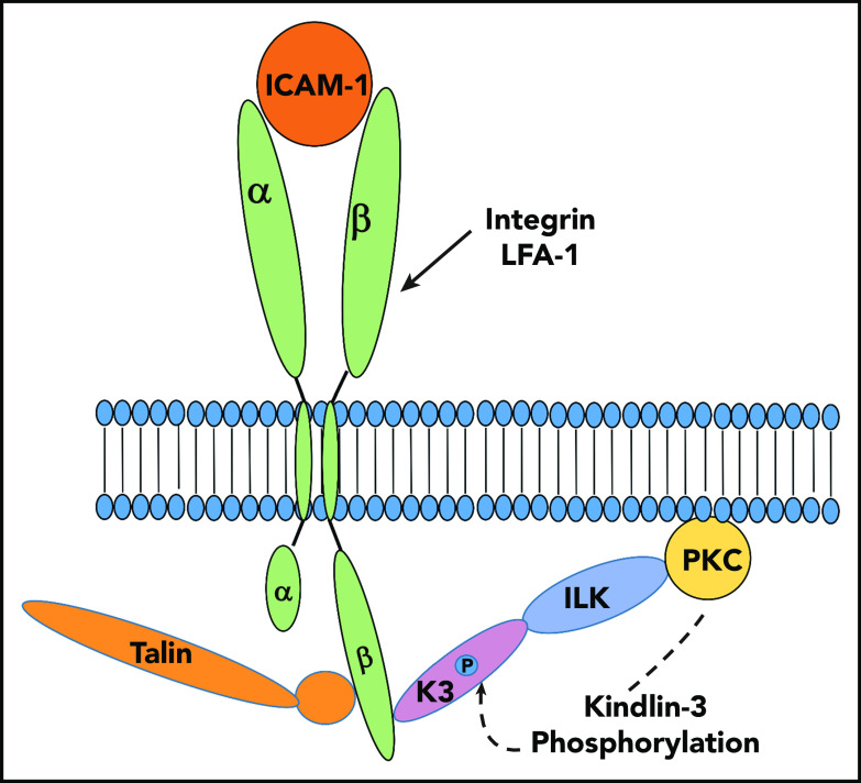
A new ILK-dependent pathway of LFA-1 activation on neutrophils. ILK facilitates recruitment of PCK-α to the cell membrane where it supports phosphorylation of a specific residue in kindlin-3. This modification enhances the capacity of kindlin-3 to cooperate with talin-1 to activate integrin LFA-1 and its recognition of ICAM-1. Illustration by Katarzyna Bialkowska.
The mechanism underlying these blunted responses in the ILK-deficient animals is a consequence of failure of the cells to activate the leukocyte integrin lymphocyte function-associated antigen-1 (LFA-1) (αLβ2, CD11a/CD18), such that it can recognize ICAM-1 on endothelial cells. ILK localizes in adherent complexes on the plasma membrane and is well-recognized to be a key regulator of integrin function. Indeed, ILK was first identified as an integrin β1 subunit binding partner in a yeast 2 hybrid screen.2 ILK associates tightly with 2 other proteins—particularly interesting new cysteine-histidine rich protein (PINCH) and parvin—to form the ILK-PINCH-parvin (IPP) complex, which serves as an adaptor from many binding partners and provides a linkage between integrins and the actin cytoskeleton.3 Global knockout of ILK is lethal in mice, fish, and flies,3,4 and tissue-specific knockouts of ILK have been associated with a wide range of organ-specific defects.
In the study by Margraf et al, the central role of ILK relates to its regulation of LFA-1 (αLβ2, CD11a/CD18), 1 of the 4 β2 leukocyte integrins that is composed of a non-covalent heterodimer of the transmembrane αL and β2 subunits (see figure). On unstimulated leukocytes, LFA-1 exists in a quiescent state in which it exhibits low capacity to bind ICAM-1. Upon cytokine stimulation of leukocytes, LFA-1 transforms to a high-affinity and high-avidity state in which it can bind to ICAM-1 expressed from endothelial cells. This interaction can lead to arrest and extravasation of leukocytes, including neutrophils. This transition of the integrin, referred to as activation, is not only necessary for LFA-1 to efficiently recognize ICAM-1 but is also necessary for productive engagement of many ligands by many different integrins. Integrin activation is induced by association of the cytoplasmic tail of the β subunit with specific binding partners. These interactions trigger an inside-out signal that traverses the transmembrane segments and ultimately optimizes the relative positioning of the ligand-binding motifs within the extracellular domains of the α and β subunits to accommodate ligands such as ICAM-1. Talins, which are large cytoskeletal proteins, and kindlins, a 3-member family of cytoplasmic proteins, are both critical for integrin activation.5,6 Both talin and the kindlins contain a 4.1-ezrin-radixin-moesin (FERM) domain that regulates integrin activation by binding to the cytoplasmic tail of the integrin β subunit, although they bind to different sites in the β subunit. In leukocytes, it is kindlin-3 that binds to integrin β subunits and regulates activation of LFA-1.5,6
The study by Margraf et al establishes a pivotal role of ILK in activating LFA-1 and its recognition of ICAM-1 for neutrophil recruitment into tissues and also defines a mechanism for the ILK–LFA-1 collaboration in mounting an inflammatory response. The authors show that ILK is required for phosphorylation of kindlin-3 on a single serine residue, and this posttranslational modification enhances its ability to promote activation of LFA-1. Such phosphorylation of kindlin-3 has been shown to enhance activation of β3 integrins.7 If ILK were a kinase, the pathway would be simple and direct because ILK and kindlins bind to one another.8 However, although still somewhat controversial, the preponderance of recent evidence suggests that ILK is not a true kinase but is instead a pseudokinase.3 Therefore, it is likely that the ability of ILK to support kindlin-3 phosphorylation is indirect. Margraf et al demonstrate that kindlin-3 phosphorylation depends on the capacity of ILK to recruit PKC-α to the cell membrane, where its kinase activity leads (directly or indirectly) to kindlin-3 phosphorylation. Thus, by using a PKC-α inhibitor or bone marrow chimera deficient in PKC-α, LFA-1–dependent leukocyte arrest is significantly blunted, and a mutant kindlin-3 that could not be phosphorylated reduced LFA-1–mediated interaction with ICAM-1.
To summarize, the Margraf et al study suggests a pathway leading to LFA-1 activation as depicted in the figure. Cytokine stimulation supports recruitment and extravasation of neutrophils during inflammation by activation of LFA-1. This activation is dependent upon ILK, which recruits PKC-α to the cell membrane where it encourages the phosphorylation of kindlin-3, which enhances LFA-1 activation in cooperation with talin. Thus, optimal alignment of talin, kindlin, and PKC-α by their interaction with membrane lipids is likely to be critical for maximal integrin activation.
One interesting facet of the Margraf et al study is that although ILK is involved in ICAM-1 recognition by LFA-1, fibrinogen recognition by a second leukocyte integrin, Mac-1 (αMβ2, CD11b/CD18) (which contains the same β2 subunit and therefore the same binding sites for ILK, kindlin-3, and talin-1), does not show the same dependence on ILK for its activation. There are several possible explanations. One interesting possibility is that the mechanism of activation may be distinct for the 2 integrins. Intracellular signaling induced by fibrinogen binding to Mac-1 is particularly dependent on integrin clustering9 whereas LFA-1 activation is more dependent on conformational changes in the integrin.10 Both LFA-1 and Mac-1 interact with ICAM-1, but distinct sites in ICAM-1 are recognized by the 2 integrins, and the induction of different downstream signaling events raises the possibility of biased agonism/antagonism to control specific inflammatory responses mediated by each integrin.
Footnotes
Conflict-of-interest disclosure: The authors declare no competing financial interests.
REFERENCES
- 1.Margraf A, Germena G, Drexler HCA, et al. The integrin-linked kinase is required for chemokine-triggered high-affinity conformation of the neutrophil β2-integrin LFA1. Blood. 2020;136(19):2200-2205. [DOI] [PubMed] [Google Scholar]
- 2.Hannigan GE, Leung-Hagesteijn C, Fitz-Gibbon L, et al. Regulation of cell adhesion and anchorage-dependent growth by a new beta 1-integrin-linked protein kinase. Nature. 1996;379(6560):91-96. [DOI] [PubMed] [Google Scholar]
- 3.Qin J, Wu C. ILK: a pseudokinase in the center stage of cell-matrix adhesion and signaling. Curr Opin Cell Biol. 2012;24(5):607-613. [DOI] [PMC free article] [PubMed] [Google Scholar]
- 4.Legate KR, Montañez E, Kudlacek O, Fässler R. ILK, PINCH and parvin: the tIPP of integrin signalling. Nat Rev Mol Cell Biol. 2006;7(1):20-31. [DOI] [PubMed] [Google Scholar]
- 5.Calderwood DA, Campbell ID, Critchley DR. Talins and kindlins: partners in integrin-mediated adhesion. Nat Rev Mol Cell Biol. 2013;14(8):503-517. [DOI] [PMC free article] [PubMed] [Google Scholar]
- 6.Plow EF, Qin J. The kindlin family of adapter proteins. Circ Res. 2019;124(2):202-204. [DOI] [PMC free article] [PubMed] [Google Scholar]
- 7.Bialkowska K, Sossey-Alaoui K, Pluskota E, et al. Site-specific phosphorylation regulates the function of kindlin-3 in a variety of cells. Life Sci Alliance. 2020;3(3):e201900594. [DOI] [PMC free article] [PubMed] [Google Scholar]
- 8.Fukuda K, Bledzka K, Yang J, Perera HD, Plow EF, Qin J. Molecular basis of kindlin-2 binding to integrin-linked kinase pseudokinase for regulating cell adhesion. J Biol Chem. 2014;289(41):28363-28375. [DOI] [PMC free article] [PubMed] [Google Scholar]
- 9.Shi C, Zhang X, Chen Z, et al. Integrin engagement regulates monocyte differentiation through the forkhead transcription factor Foxp1. J Clin Invest. 2004;114(3):408-418. [DOI] [PMC free article] [PubMed] [Google Scholar]
- 10.Lefort CT, Rossaint J, Moser M, et al. Distinct roles for talin-1 and kindlin-3 in LFA-1 extension and affinity regulation. Blood. 2012;119(18):4275-4282. [DOI] [PMC free article] [PubMed] [Google Scholar]


